
BIOL 252-ANATOMY AND PHYSIOLOGY II W,LAB- MODULE 4 EXAM-digestive system proctored exam
BIOL 252-ANATOMY AND PHYSIOLOGY II W,LAB- MODULE 4 EXAM-digestive system proctored exam
Total Questions : 51
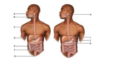
Showing 10 questions Sign up for moreSelect the letter of the organ with the following function: Muscular propulsion of materials into the esophagus.
Explanation
The pharynxis a muscular tube located behind the nasal cavity and mouth, connecting to the esophagus. Its primary role in digestion is to propel swallowed food (bolus) from the oral cavity into the esophagus through coordinated muscular contractions. This process is part of deglutition (swallowing), which involves voluntary initiation in the mouth followed by involuntary reflexes in the pharynx. The pharyngeal muscles contract in sequence, pushing the bolus downward while preventing entry into the respiratory tract.
Which muscles of the tongue are responsible for changing its shape for speech and swallowing?
Explanation
A. Circumvallate muscles:These are not muscles but rather large, circular papillae located in a V-shaped row at the posterior aspect of the tongue. They contain numerous gustatory receptors but lack contractile properties for lingual movement. They do not influence the organ's shape.
B. Extrinsic muscles:These muscles, including the genioglossus and hyoglossus, originate from structures outside the tongue and insert into its substance. They are primarily responsible for the gross movement and positioning of the tongue, such as protrusion and retraction. They move the tongue as a whole.
C. Salivary muscles:There is no anatomical classification for muscles specifically designated as salivary muscles in the human body. Salivary secretion is controlled by the autonomic nervous system acting on glandular epithelial cells, not by specialized lingual muscles. This is not a valid anatomical term.
D. Submandibular muscles:This term typically refers to the muscles of the floor of the mouth, such as the mylohyoid, which support the tongue and hyoid bone. While they assist in the elevation of the tongue during swallowing, they do not reside within the tongue. They do not change its internal shape.
E. Intrinsic muscles:These muscle fibers are located entirely within the tongue and are not attached to bone. They are arranged in longitudinal, transverse, and vertical planes, allowing the tongue to curl, flatten, or thicken for complex articulation and deglutition. They are the primary effectors for changing tongue shape.
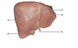
The letter D is pointing to the right lobe of the liver.
Explanation
False:The liver is divided into two primary lobes by the falciform ligament (labeled C). The right lobeis the significantly larger portion (labeled A). The left lobeis the smaller portion located on the opposite side of the ligament, which is where letter D is pointing.
Starting at the pyloric sphincter, which sequence represents the correct path food follows?
Explanation
A. Pyloric sphincter → Jejunum → Ileum → Transverse colon:This sequence incorrectly bypasses the duodenum, which is the immediate proximal segment of the small intestine. Chyme must traverse the 25 cm C-shaped duodenal loop before reaching the jejunum. The transverse colon is located much further distally.
B. Pyloric sphincter → Duodenum → Jejunum → Ileum:This represents the physiological anterograde flow of chyme through the small intestine. The duodenum receives gastric contents, followed by the highly vascularized jejunum and the lymphoid-rich ileum. This anatomical order ensures sequential nutrient absorption and enzymatic processing.
C. Pyloric sphincter → Duodenum → Ileum → Jejunum → Ascending colon:This choice incorrectly transposes the positions of the ileum and jejunum. The jejunum constitutes the proximal 40% of the post-duodenal small bowel, while the ileum forms the distal 60%. Proper transit requires the jejunum to precede the ileum.
D. Pyloric sphincter → Duodenum → Cecum → Sigmoid colon:This path omits the majority of the small intestine, specifically the jejunum and ileum. The cecum is the commencement of the large intestine, separated from the duodenum by several meters of bowel. It represents an incomplete anatomical progression.
E. Pyloric sphincter → Ileum → Jejunum → Cecum:This sequence proposes a retrograde or inverted order for the small intestinal segments. The ileum is the most distal part of the small intestine and connects to the cecum at the ileocecal valve. It cannot precede the jejunum.
Which of the following doesn't belong?
Explanation
A. Transverse colon:This structure is a major segment of the large intestine involved in water resorption and electrolyte balance. Unlike the other choices, it contains haustra and taeniae coli. It is not part of the small intestinal tract.
B. Ileum:This is the terminal section of the small intestine characterized by Peyer's patches and increased goblet cell density. It belongs to the same functional and anatomical group as the duodenum and jejunum. It facilitates vitamin B12 absorption.
C. Duodenum:This short, fixed segment is the first part of the small intestine where most chemical digestion occurs. It shares the histological features of the small bowel, such as villi, with the ileum and jejunum. It belongs to the small intestine.
D. Jejunum:This middle segment of the small intestine is the primary site for nutrient absorption. It possesses long villi and prominent plicae circulares, matching the anatomical classification of the duodenum and ileum. It is not a colonic structure.
True or False
The pancreas is intraperitoneal and lies transverse at the level of L1 and L2 vertebrae.
Explanation
False:The pancreas is primarily a retroperitoneal organ, with the exception of the tail which may be intraperitoneal. It is situated behind the lesser sac of the omentum. Its anatomical position at L1 and L2 is correct, but its peritoneal status is not.
True or False
Fungiform papillae contain about 50% of the taste buds and are located at the posterior of the tongue.
Explanation
False:Fungiform papillae are concentrated at the tip and lateral margins of the tongue, not the posterior. They contain only a small fraction of total gustatory receptors. The vallate papillae at the posterior contain the majority of taste buds.
A patient comes in complaining of sensitivity when biting into cold foods. Upon examination, you notice a chisel-shaped tooth appears slightly cracked. What type of tooth is this?
Explanation
A. Incisor:These anterior teeth possess a sharp, chisel-shaped incisal edge designed for shearing and cutting food. They have a single root and are the most proximal teeth to the midline. Their morphology matches the clinical description provided.
B. Canine:These teeth are characterized by a single, pointed cusp or "fang" shape intended for tearing and piercing. They are not chisel-shaped and possess the longest roots in the human dentition. They are located lateral to the incisors.
C. Premolar:Also known as bicuspids, these teeth have two prominent cusps on their occlusal surface for crushing and grinding. They do not have a chisel-like edge. They are located between the canines and the molar teeth.
D. Molar:These posterior teeth have large, broad occlusal surfaces with 4 to 5 cusps for heavy mastication. They are designed for grinding boluses into smaller particles. Their flat, multi-cusped morphology is entirely different from a chisel shape.
Bile is produced by hepatocytes, and eventually moves into the portal triad's bile ducts, which make it way into the:
Explanation
A. Pancreatic duct:This duct transports exocrine enzymes from the acinar cells to the duodenum. While it often joins the common bile duct at the ampulla of Vater, it is not the primary conduit for biliary drainage. It does not carry bile.
B. Common bile duct:This structure is formed by the union of the common hepatic duct and the cystic duct. It serves as the definitive pathway for bile to reach the second part of the duodenum. It is the direct continuation of biliary flow.
C. Hepatic vein:These vessels are responsible for draining deoxygenated, filtered blood from the liver into the inferior vena cava. They are part of the systemic venous circulation. They do not participate in the transport of biliary secretions.
D. Gallbladder:This organ serves as a secondary storage site where bile is concentrated and held until needed for digestion. While bile enters it via the cystic duct, the ultimate destination from the portal triads is the ductal system.
Peyer's patches, which house lymphocytes, are found in the
Explanation
A. Esophagus:The esophageal mucosa consists of stratified squamous epithelium designed for protection against mechanical abrasion. It contains mucous glands but lacks organized lymphoid aggregates like Peyer's patches. It does not have a specialized immune role in the bowel.
B. Stomach:The gastric environment is highly acidic, which provides a non-specific barrier against pathogens. While it contains some diffuse lymphoid tissue, it does not possess the organized nodular Peyer's patches. These are absent in the proximal upper gastrointestinal tract.
C. Distal ileum:This region of the small intestine contains large, visible clusters of lymphatic nodules within the lamina propria and submucosa. These Peyer's patches are critical for immunosurveillance of the intestinal microbiome. They are the hallmark histological feature of the ileum.
D. Jejunum:Although the jejunum has diffuse lymphoid tissue, Peyer's patches are generally absent or extremely rare in this segment. The jejunum is primarily specialized for nutrient absorption rather than immune aggregation. Its histology is characterized by long villi instead.
You just viewed 10 questions out of the 51 questions on the BIOL 252-ANATOMY AND PHYSIOLOGY II W,LAB- MODULE 4 EXAM-digestive system proctored exam Exam. Subscribe to our Premium Package to obtain access on all the questions and have unlimited access on all Exams. Subscribe Now

