





 Video Library


Please set your exam date
Acquired Cardiovascular Disorders: Congestive Heart Failure
Study Questions
Practice Excercise 1
A nurse is teaching a group of nursing students about the pathophysiological changes that occur during pediatric congestive heart failure. Which statement by a student indicates a correct understanding of the body's compensatory mechanisms?
Explanation
Pediatric congestive heart failuretriggers systemic neurohormonal pathways to maintain tissue perfusion when cardiac output declines. Decreased renal perfusion activates the renin-angiotensin-aldosteronesystem,forcing peripheral vasoconstriction,increasing systemic vascular resistance, and promoting sodium and water retention to increaseblood volumeand maintain target arterial perfusion pressure.
Rationale for correct answer:
3. Activation of the renin-angiotensin-aldosterone cascadeserves as a vital neurohormonal compensatory mechanismduring heart failure. Angiotensin II induces widespread vasoconstriction to maintain systemic blood pressure. Concurrently, aldosterone promotes renal fluid conservation to expand circulatory volume and optimize cardiac output.
Rationale for incorrect answers:
1. The kidneys retain rather than excrete water and sodiumduring heart failure compensation. Diminished renal blood flow triggers mechanisms that maximizefluid preservationto restore low circulating volumes. This pathological response unintentionally increases preload and worsens systemic venous congestion over time.
2. The sympathetic nervous system increases rather than decreases the heart rateduring cardiac decompensation. Adrenergic stimulation releases catecholamines to trigger compensatory tachycardiaand increase myocardial contractility. This response attempts to sustain peripheral perfusion but elevatesmyocardial oxygen consumptiondangerously.
4. The myocardium undergoes hypertrophic enlargementrather than cellular atrophy under chronic structural strain. Ventricular walls experience myocardial hypertrophyto counter high afterload and increase pumping force. This structural remodeling eventually reduces ventricular compliance and accelerates progressive cardiac dysfunction.
Test-taking strategy:
- Analyze the scenario/question:The nurse is assessing nursing students' knowledge regarding the compensatory mechanisms activated during pediatric congestive heart failure. The objective is to identify the statement that accurately describes these physiological responses.
- Evaluate neurohormonal compensation pathways:
- Map the physiological cascade: Low cardiac output decreases organ perfusion, which immediately activates hormonal and autonomic survival responses.
- Contrast protective actions versus destructive outcomes: Short-term volume retention and vasoconstriction maintain pressure but cause long-term damage.
- Rule in Choice 3because it correctly identifies the vasoconstrictive and fluid-retaining actions of the renin-aldosterone axis.
- Rule out Choice 1:Heart failure causes renal conservation of sodium, not excretion, which increases preload.
- Rule out Choice 2:The sympathetic nervous system drives tachycardia rather than bradycardia to maintain systemic flow.
- Rule out Choice 4:Chronic cardiac workload leads to ventricular hypertrophy rather than tissue atrophy.
Take home points
- Decreased cardiac output stimulates the renin-angiotensin-aldosterone system to preserve arterial blood pressure through systemic vasoconstriction.
- Aldosterone release causes the kidneys to retain sodium and water, which increases circulating blood volume but elevates preload.
- Sympathetic nervous system activation releases catecholamines that cause tachycardia and increased contractility to boost cardiac output.
- Chronic neurohormonal stimulation leads to maladaptive myocardial hypertrophy and remodeling, which ultimately worsens pediatric heart failure.
An infant with a history of a large ventricular septal defect (VSD) is admitted with acute heart failure. The nurse notes cold, pale extremities, a capillary refill of 4 seconds, and a heart rate of 172 beats per minute. The nurse understands these findings are a direct result of which compensatory response?
Explanation
2. Sympathetic nervous system stimulationreleases epinephrine and norepinephrine to counter falling systemic cardiac output. Adrenergic activation induces peripheral vasoconstrictionto divert blood toward vital organs, causing cold, pale extremities and prolonged capillary refill.It concurrently drives compensatory tachycardia to maximize forward blood flow.
Rationale for incorrect answers:
1. Parasympathetic nervous system withdrawaloccurs during acute heart failure to allow sympathetic dominance and elevate the heart rate. However, simple vagal suppressiondoes not actively trigger the intense alpha-adrenergic peripheral vasoconstriction that causes pale, cold extremities and a four-second capillary refilltime.
3. Systemic vascular resistance increasesdrastically rather than experiencing systemic vasodilation during acute cardiac failure. Vasodilation would cause decreased afterloadand lower blood pressure, which contradicts the body's compensatory framework of using intense vasoconstriction to maintain an adequate perfusion pressure.
4. Myocardial tissue undergoes compensatory hypertrophy and stretchingrather than wall thinning during volume overload states. This mechanical change increases myocardial contractilityvia the Frank-Starling law, which significantly elevates, rather than decreases, the total myocardial oxygen consumption.
Test-taking strategy:
- Analyze the scenario/question:An infant with a large ventricular septal defect presents with cold, pale extremities, a capillary refill time of 4 seconds, and a heart rate of 172 beats per minute. The nurse must identify the specific compensatory mechanism responsible for these findings.
- Link assessment data to physiology:
- Cold, pale skin and prolonged capillary refill indicate poor peripheral perfusion driven by vasoconstriction.
- A heart rate of 172 beats per minute represents tachycardia in an infant.
- Connect both findings to a single cause: Alpha-adrenergic and beta-adrenergic stimulation from the autonomic nervous system.
- Rule in Choice 2because sympathetic activation directly causes both vasoconstriction and tachycardia.
- Rule out Choice 1:Vagal withdrawal assists in raising heart rate but does not drive peripheral vasoconstriction.
- Rule out Choice 3:Heart failure triggers vasoconstriction to maintain pressure, which increases afterload rather than decreasing it.
- Rule out Choice 4:Chronic volume overload causes myocardial hypertrophy and increases oxygen demand.
Take home points
- Decreased systemic perfusion from a large ventricular septal defect triggers immediate sympathetic nervous system activation.
- Sympathetic catecholamine release causes peripheral vasoconstriction, leading to cold extremities, pallor, and a prolonged capillary refill time greater than 2 seconds.
- Compensatory tachycardia develops simultaneously to maintain adequate cardiac output in the presence of a large left-to-right shunt.
- Prolonged sympathetic overactivation increases myocardial workload and oxygen consumption, eventually accelerating clinical heart failure.
Practice Excercise 2
A pediatric client presents with tachycardia, edema, dyspnea, orthopnea, and crackles. A nurse performs a physical assessment of the client and notifies a physician immediately. Which condition does the nurse most likely suspect?
Explanation
Left-sided heart failureimpairs systemic ventricular emptying,forcing backward hydrostatic pressure into the pulmonary vasculature. This fluid shift causes interstitial pulmonary edema,which presents as severe respiratory distress and impaired gas exchange in pediatric populations.
Rationale for correct answer:
4. Left-sidedheart failureis characterized by fluid backup into the pulmonary bed. The accumulation of alveolar fluid causes clinical manifestations including dyspnea, orthopnea, and cracklesduring lung auscultation. The heart rates elevate concurrently to compensate for the reduction in forward systemic cardiac output.
Rationale for incorrect answers:
1. Right-sidedheart failureleads to systemic venous congestion rather than primary pulmonary edema. The clinical presentation involves hepatomegaly,jugular venous distension, and peripheral dependent edema rather than acute adventitious lung sounds. It does not match the predominantly respiratory cluster of symptoms detailed in this pediatric scenario.
2. Rheumaticfeveris an inflammatory disease that develops following an untreated group A streptococcal infection. It manifests clinically with polyarthritis,chorea, erythema marginatum, and subcutaneous nodules along with systemic carditis. It does not cause sudden, isolated left-ventricular fluid backing into the pulmonary parenchyma.
3. Kawasakidiseaseis an acute febrile vasculitis that primarily targets the coronary arteries. The diagnostic criteria require a prolonged high fever paired with conjunctivitis,strawberry tongue, truncal rash, and desquamation of hands and feet. It does not present primarily with immediate pulmonary hydrostatic congestion signs.
Test-taking strategy:
- Analyze the scenario/question:The pediatric patient presents with a combination of tachycardia, edema, dyspnea, orthopnea, and crackles. The nurse needs to identify the specific underlying condition based on these clinical assessment findings.
- Differentiate cardiac hemodynamics:
- Group the symptoms by system: Dyspnea, orthopnea, and crackles indicate fluid collection within the pulmonary system.
- Trace the fluid backup: Pulmonary congestion originates from a failure of the left side of the heart to pump blood forward.
- Rule in Choice 4because left-ventricular failure directly explains the fluid accumulation in the alveoli.
- Rule out Choice 1:Right-sided failure causes systemic systemic signs like hepatomegaly rather than pulmonary crackles.
- Rule out Choice 2:Rheumatic fever presents with distinct inflammatory criteria like migratory arthritis and carditis.
- Rule out Choice 3:Kawasaki disease involves specific vasculitis features like mucosal changes and desquamation.
Take home points
- Left-sided heart failure causes blood to back up into the left atrium and pulmonary veins, causing pulmonary congestion and respiratory distress.
- Right-sided heart failure leads to systemic venous engorgement, causing systemic edema, hepatomegaly, and jugular venous distension.
- Pediatric indicators of left-ventricular failure include tachypnea, grunting, retractions, orthopnea, and crackles upon auscultation.
- Immediate nursing priorities for pulmonary congestion include elevating the head of the bed, administering oxygen, and providing rapid-acting diuretics.
A nurse is assessing a child with heart failure. Which of the following findings should the nurse expect to find?
Explanation
Pediatric heart failure involves myocardial dysfunctionleading to inadequate systemic perfusion.This reduction in systemic cardiac output triggers intense compensatory neural-hormonal cascades, leading to systemic and pulmonary vascular congestion. It causes tachycardia, tachypnea, cardiomegaly, hepatomegaly,and impaired growth due to metabolic demands.
Rationale for correct answer:
3.A gallop rhythmreflects ventricular fluid overload. This abnormal S3 or S4 sound indicates rapid ventricular filling against increased pressure. It signifies compensatory cardiac dilatationin pediatric heart failure. The myocardium fails to pump efficiently.
Rationale for incorrect answers:
1.Tachycardia, not bradycardia, is an early compensatory mechanism. The sympathetic nervous system accelerates the heart rate to maintain cardiac output. This counters reduced stroke volumein failing hearts. Bradycardia indicates impending cardiovascular collapse.
2.Tachypnea occurs instead of a decreased respiratory rate.Pulmonary congestion triggers rapid, shallow breathing to improve gas exchange. The infant exhibits intercostal retractionsand nasal flaring. Respiratory depression suggests a different, non-cardiac pathology.
4.Pulses are typically weak and thready rather than strong, bounding pulses. Low cardiac output diminishes peripheral perfusion significantly. Bounding pulses are characteristic of a patent ductus arteriosusor severe anemia instead. Weak pulses reflect poor systemic stroke volume.
Test-taking strategy:
- Analyze the scenario/question:The nurse is assessing a pediatric client for clinical manifestations of heart failure, which requires identifying signs of impaired myocardial contractilityand volume overload.
- Apply clinical assessment principles:
- Analyze Choice 3:This option is correct because an extra heart sound indicates severe ventricular stress during rapid passive filling.
- Rule out Choice 1:Tachycardia is the expected sympathetic response to preserve cardiac output, making bradycardia an incorrect assessment finding.
- Rule out Choice 2:Pulmonary venous congestioncauses tachypnea, so a decreased respiratory rate is an inappropriate selection.
- Rule out Choice 4:Reduced stroke volume results in weak, thready peripheral pulses rather than strong, bounding pulsations.
- This evaluation strategy relies on separating sympathetic compensatory mechanisms from signs of systemic venous congestion.
Take home points
- An S3 or S4 gallop murmur is a classic sign of pediatric ventricular volume overload.
- Tachycardia and tachypnea are early compensatory mechanisms for decreased cardiac output.
- Peripheral pulses in pediatric heart failure are typically weak, thready, and diminished.
- Hepatomegaly and poor feeding are common systemic signs of right-sided venous congestion.
An emergency room nurse is caring for a pediatric client in heart failure. Which symptom is consistent with a diagnosis of left-sided heart failure?
Explanation
Pediatric heart failure is clinically divided into left-sided and right-sided dysfunction based on the specific vascular circuit experiencing fluid backup and congestion. Pediatric left-sided heart failurecauses pulmonary congestiondue to backpressure from the left ventricle into the pulmonary vasculature. It leads to impaired gas exchange,increased work of breathing, and interstitial edema.
Rationale for correct answer:
4.Tachypnea and dyspnearepresent classic signs of pulmonary fluid accumulation. Elevated pressures back up into the pulmonary veins, flooding alveoli. This creates significant ventricular preloadstress and respiratory distress. The child exhibits rapid breathing to compensate.
Rationale for incorrect answers:
1.Weight gainis typically associated with systemic venous congestion in right-sided failure. Fluid accumulation occurs in tissues and abdominal organs rather than the pulmonary circuit. This reflects systemic fluid retentionfrom right ventricular dysfunction. Left-sided failure causes poor feeding instead.
2.Peripheral edemaoccurs when the right ventricle fails to pump forward efficiently. Fluid backs up into the systemic venous circulation, causing dependent swelling. This indicates right-sided heart failurerather than isolated left ventricular dysfunction. Children manifest this less prominently than adults.
3.Neck vein distentionis a classic sign of elevated right atrial pressure. Jugular venous pressure rises when systemic return is impeded. It points to systemic venous engorgementfrom right-sided heart failure. Infants rarely show this sign due to short necks.
Test-taking strategy:
- Analyze the scenario/question:The nurse is assessing an emergency room pediatric client specifically for manifestations of left-sided heart failure,which requires separating pulmonary symptoms from systemic congestion signs.
- Apply pathophysiological principles:
- Analyze Choice 4:Left ventricular failure elevates pulmonary pressures, directly causing pulmonary edemawhich presents as tachypnea and dyspnea.
- Rule out Choice 1:Rapid weight gain points to systemic venous volume overload, which is an expected finding in right-sided heart failure.
- Rule out Choice 2:Dependent peripheral edema indicates systemic capillary fluid shift, confirming a right-sided pathologyrather than left-sided failure.
- Rule out Choice 3:Jugular venous engorgement reflects elevated right atrial pressures, which helps rule out isolated left ventricularfailure.
- This evaluation strategy depends on localizing the fluid backup to either the pulmonary circuit or the systemic venous circuit.
Take home points
- Left-sided heart failure predominantly causes respiratory symptoms due to pulmonary venous congestion.
- Right-sided heart failure manifests as systemic venous congestion including hepatomegaly and peripheral edema.
- Pediatric clients with left-sided failure frequently present with orthopnea, grunting, and intercostal retractions.
- Chronic left-sided heart failure eventually increases pulmonary vascular resistance and causes secondary right-sided failure.
A nurse is educating a group of nurses on congestive heart failure (CHF). Which physiological changes occur as a result of hypoxemia in CHF?
Explanation
Pediatric congestive heart failurecomplicated by chronic hypoxemia triggers profound compensatory hematological and tissue changes due to tissue hypoxia.In response to low arterial oxygen saturation, the kidneys accelerate erythropoietin production, stimulating the bone marrow to dramatically increase red blood cell synthesis to maximize oxygen-carrying capacity.Concurrently, prolonged tissue hypoxemia alters peripheral capillary beds, stimulating endothelial proliferation and vascular endothelial growth factor release within the digits, which causes characteristic digital clubbingover time.
Rationale for correct answer:
1.Polycythemia and clubbingare the direct physiological consequences of chronic hypoxemia. The body counters low oxygen levels by manufacturing excess erythrocytes, resulting in therapeutic polycythemia to boost oxygen delivery. Over months, unremitting capillary hypoxia induces soft tissue proliferation and hypertrophic changes at the nail beds, manifesting as structural fingertip clubbing.These signs are hallmark indicators of long-standing cyanotic cardiac diseaseor advanced heart failure.
Rationale for incorrect answers:
2.Anemia and barrel chestdo not result from hypoxemia in this context. Hypoxemia drives an elevation in red blood cells, which directly contradicts the erythrocyte deficit seen in anemia. A barrel chest is a structural musculoskeletal deformation caused by chronic alveolar hyperinflationand air trapping, which is typical of chronic obstructive pulmonary disease or severe asthma rather than heart failure.
3.Increased white blood cells and low plateletsare inflammatory and hematological markers that are not triggered by hypoxemia. An elevated leukocyte count points to active microbial infectionor systemic inflammation, while thrombocytopenia indicates platelet destruction or sequestration. Neither of these cellular changes serves as a physiological compensation for low arterial oxygenation.
4.Elevated erythrocyte sedimentation rate and peripheral edemais an incorrect grouping because hypoxemia does not elevate this inflammatory marker. In fact, severe polycythemia increases blood viscosity, which characteristically slows the descent of erythrocytes, leading to a decreased or low sedimentation rate. While peripheral edema occurs in right-sided heart failure, it is caused by hydrostatic systemic venous congestion,not hypoxemia.
Test-taking strategy:
- Analyze the scenario/question:The nurse is identifying the specific physiological alterations that develop as a direct compensatory response to long-term hypoxemiain heart failure.
- Evaluate compensatory mechanisms:
- Analyze Choice 1:Chronic hypoxemia triggers erythropoietin-driven erythrocyte multiplicationand peripheral vascular changes, leading directly to polycythemia and clubbing.
- Rule out Choice 2:Low oxygen tension stimulates bone marrow production, making a state of red blood cell deficiencyan inappropriate selection.
- Rule out Choice 3:White blood cell and platelet fluctuations reflect immune responses and coagulation cascades rather than respiratory gas imbalances.
- Rule out Choice 4:High blood viscosity from polycythemia slows cell suspension settling, which predictably lowers rather than raises the sedimentation rate.
- This evaluation strategy relies on linking chronic tissue hypoxia to its specific hematological and peripheral structural adaptations.
Take home points
- Chronic hypoxemia stimulates renal erythropoietin secretion, resulting in compensatory polycythemia.
- Digital clubbing is a structural change in the nail beds caused by chronic peripheral capillary hypoxia.
- Polycythemia significantly increases blood viscosity, which elevates the risk of thromboembolic events.
- An elevated red blood cell mass in hypoxemic cardiac states causes a characteristically low erythrocyte sedimentation rate.
A nurse is assessing a 4-month-old infant with congestive heart failure. The nurse notes that the infant is sweating profusely on the forehead during feedings, takes 45 minutes to consume 2 ounces of milk, and has a respiratory rate of 68 breaths per minute. How should the nurse document and interpret these clinical findings?
Explanation
Infants with left-sided heart failureexhibit pronounced pulmonary venous congestion, which severely compromises their respiratory mechanics and metabolic reserves. The fluid backup increases alveolar pressure, causing pulmonary congestionand a rapid respiratory rate, which leads to feeding intoleranceas the infant expends excessive energy trying to coordinate sucking, swallowing, and breathing.
Rationale for correct answer:
3. Left-sided heart failure leads to pulmonary fluid backup,which manifests clinically as tachypnea and respiratory distress. The infant's feeding intolerance,characterized by taking 45 minutes to consume a small volume and sweating profusely, reflects sympathetic nervous system overactivation as the baby expends maximum metabolic energy during feeds.
Rationale for incorrect answers:
1. Respiratorysyncytial viruscauses acute bronchiolitis, which presents with rhinorrhea, wheezing, coughing, and a low-grade fever. Although it induces tachypnea, it does not explain the chronic pattern of diaphoresis during feeds or the structural history of congestive heart failure detailed in this scenario.
2. Profusediaphoresisand taking 45 minutes to finish 2 ounces of milk are pathologically abnormal for a 4-month-old infant. Standard feeding should take under 30 minutes, so these findings represent clear, severe cardiac strain rather than normal physiologicalfeeding workloads.
4. Right-sidedheart failurecauses systemic venous engorgement rather than primary pulmonary symptoms. The typical clinical manifestations include hepatomegaly, jugular venous distension, and peripheral dependent edema, rather than acute pulmonary crackles, tachypnea, and gastric refluxtriggers.
Test-taking strategy:
- Analyze the scenario/question:The nurse is assessing a 4-month-old infant with congestive heart failure who presents with profuse forehead diaphoresis during feeding, severe feeding fatigue, and a respiratory rate of 68 breaths per minute. The goal is to identify the correct clinical interpretation and documentation of these findings.
- Correlate symptoms with cardiac pathophysiology:
- Cluster the clinical signs: Tachypnea, feeding fatigue, and diaphoresis are classic indicators of high metabolic demand and respiratory compromise.
- Trace the mechanism: Left-ventricular dysfunction backs blood up into the pulmonary circulation, causing a high respiratory rate and making feeding an exhausting physical chore.
- Rule in Choice 3because left-sided failure directly results in pulmonary congestion and marked feeding intolerance.
- Rule out Choice 1:Acute viral infections present with infectious signs like a fever and coryza rather than isolated feeding-induced diaphoresis.
- Rule out Choice 2:These findings represent clear milestones of distress and are never considered standard feeding responses.
- Rule out Choice 4:Right-sided failure involves systemic venous organs, causing hepatomegaly rather than primary respiratory feeding fatigue.
Take home points
- Profuse diaphoresis on the forehead during feedings is a classic sign of sympathetic nervous system overactivation in infants with heart failure.
- Infant feeding sessions lasting longer than 30 minutes indicate severe feeding intolerance and excessive metabolic energy expenditure.
- Left-sided heart failure characteristically presents with respiratory distress, tachypnea, and pulmonary congestion due to backward fluid pressures.
- Nursing interventions for feeding fatigue include offering smaller, more frequent meals and using high-calorie formulas to minimize workload.
The nurse is reviewing lab values for an adolescent client admitted with suspected acute viral myocarditis and congestive heart failure. Which laboratory value is most specific in supporting a diagnosis of heart failure?
Explanation
Acute viral myocarditistriggers severe myocardial inflammation, compromising ventricular compliance and elevating intraventricular filling pressures. The resulting stretching of the ventricular myocytesstimulates the synthesis and release of brain natriuretic peptide,a diagnostic biomarker that helps differentiate heart failurefrom primary pulmonary disorders by reflecting localized cardiac wall stress.
Rationale for correct answer:
3. Elevated natriuretic peptide concentrationsconfirm ventricular wall stretch and high filling pressures, serving as a highly sensitive biomarker for cardiac dysfunction. Although pulmonary disorders cause dyspnea and cough, they do not elevate this cardiac-specific neurohormone, making it the premier metric to isolate myocardial failure.
Rationale for incorrect answers:
1. An elevatedleukocyte countindicates an active systemic inflammatory or infectious process, such as viral myocarditis or acute pneumonia. Because it rises in response to numerous generalized bodily infections, it lacks the necessary diagnostic specificityto confirm the presence of ventricular failure.
2. A lowhemoglobin concentrationidentifies systemic anemia, which can exacerbate pre-existing heart failure by reducing tissue oxygen delivery and forcing a compensatory increase in cardiac output. However, anemia is an independent hematologic condition that does not directly confirm an acute cardiacetiology.
4. Maintaining apotassium concentrationwithin standard reference parameters is essential to preserve regular myocardial electrical conduction and prevent life-threatening dysrhythmias. While crucial for therapeutic safety, a normal electrolyte level does not provide active diagnostic evidence regarding congestive failure.
Test-taking strategy:
- Analyze the scenario/question:The nurse is analyzing laboratory values for an adolescent with suspected acute viral myocarditis and congestive heart failure. The goal is to identify the laboratory biomarker that is most specific for diagnosing heart failure over a respiratory infection.
- Evaluate diagnostic specificity:
- Identify the physiological origin of each marker: White blood cells respond to general infection, hemoglobin measures oxygen-carrying capacity, and potassium measures electrolyte balance.
- Target the cardiac-specific mechanism: Brain natriuretic peptide is released directly from ventricular walls in response to increased volume and pressure stretch.
- Rule in Choice 3because natriuretic peptide rises specifically due to cardiac wall stress, differentiating it from pulmonary disease.
- Rule out Choice 1:Leukocytosis occurs in both respiratory tract infections and myocarditis, making it non-specific.
- Rule out Choice 2:Anemia alters hemodynamic workloads but is not a definitive diagnostic indicator of heart muscle failure.
- Rule out Choice 4:Normal potassium levels indicate electrolyte stability but hold no diagnostic value for identifying heart failure.
Take home points
- Brain natriuretic peptide is a specific biomarker released by the ventricles in response to increased myocardial wall stretch and pressure overload.
- Measuring natriuretic peptide levels allows clinicians to differentiate between cardiac-driven dyspnea and primary respiratory infections or disorders.
- Leukocytosis and elevated inflammatory markers accompany viral myocarditis but lack the diagnostic specificity to confirm ventricular failure.
- Comprehensive heart failure diagnostic screening requires pairing specific cardiac biomarkers with echocardiography to evaluate the ventricular ejection fraction.
Practice Excercise 3
A nurse is caring for an infant with heart failure. Which intervention is most appropriate when caring for this client?
Explanation
Pediatric heart failureinduces profound myocardial metabolic demandsthat rapidly deplete the infant's limited cardiac reserve. This state of energy expenditurecauses severe growth failure and fatigue, making absolute energy conservation a primary therapeutic goal. Interventions focus on optimizing oxygenation, maintaining tissue perfusion, and structuring care to provide uninterrupted rest periods.
Rationale for correct answer:
3.To cluster nursing activitiesis a vital strategy to conserve the infant's energy. Grouping assessments, diaper changes, and medication administration minimizes disruptions and maximizes uninterrupted rest periods. This significantly reduces oxygen consumptionand cardiac workload in a failing heart. It prevents exhaustion during essential care routines.
Rationale for incorrect answers:
1.To limit fluid intakestrictly via fluid restriction is rarely done in infants unless severe fluid overload is present. Infants have high metabolic turnover and are at extreme risk for dehydration and electrolyte imbalances. Instead, fluid management focuses on utilizing concentrated, high-calorie formulasto maintain nutrition.
2.To avoid using infant seatsis incorrect because an semi-upright position actually improves respiratory mechanics. Placing the infant in an infant seat or elevating the head of the bed to a 45-degree angle reduces diaphragmatic pressurefrom abdominal organs. This optimizes lung expansion and decreases pulmonary venous congestion.
4.To place the infant prone or supineis contraindicated as flat positioning exacerbates orthopnea and respiratory distress. Flat positioning increases venous return to a failing heart, causing acute pulmonary congestionand worsening gas exchange. Semi-Fowler positioning is the preferred standard for pediatric clients.
Test-taking strategy:
- Analyze the scenario/question:The nurse is planning interventions for an infant with heart failure, requiring the selection of a care measure that balances metabolic demandswith respiratory and nutritional support.
- Apply pediatric care principles:
- Analyze Choice 3:Organizing care to allow for consolidated rest periods directly targets the core problem of cardiac fatigueand high metabolic demands.
- Rule out Choice 1:Restricting absolute fluid volume can compromise renal perfusion and cause dehydration; managing fluid density via caloric concentrationis preferred.
- Rule out Choice 2:Avoiding upright positioning removes a beneficial non-pharmacological method for reducing pulmonary venous congestionand work of breathing.
- Rule out Choice 4:Using fully flat horizontal positioning increases central blood volume, which exacerbates volume overload and worsens interstitial edema.
- This evaluation strategy centers on identifying non-invasive methods that decrease myocardial workload while optimizing tissue oxygenation.
Take home points
- Clustering nursing care is the gold standard intervention for reducing myocardial oxygen demands in infants.
- Semi-Fowler positioning in an infant seat or elevated bed reduces venous return and eases respiratory distress.
- Infant fluid management relies on high-calorie density formulas rather than strict, standard fluid restrictions.
- Feeding sessions should be limited to 30 minutes maximum to prevent excessive physical exhaustion and caloric loss.
A nurse is caring for an infant with heart failure. Which diet plan is recommended for this client?
Explanation
Pediatric heart failureinduces a profound hypermetabolic statedue to the excessive sympathetic nervous system activation and the markedly increased workload of breathing. This massive metabolic expenditureoccurs simultaneously with severe fatigue during feeding, which significantly reduces overall oral intake and causes a high risk for growth failureand malnutrition. Nutritional interventions must maximize nutrient density to support adequate cellular growth and tissue repair without overloading the compromised cardiovascular reserveof the infant.
Rationale for correct answer:
4.To increase caloric content per ounceis the primary nutritional recommendation for infants in heart failure. Concentrating formula or fortifying breast milk up to 30 calories per ounce provides essential nutrients within a smaller, manageable volume. This counters nutritional deficitswithout overloading the fluid capacity of the failing heart. It prevents the infant from exhausting limited energy reserves during feedings.
Rationale for incorrect answers:
1.To restrict fluidstoo rigidly is dangerous in infants and is not a routine dietary plan. Total fluid restriction can easily lead to dehydration, hypovolemia, and compromised renal perfusiondue to the high surface-area-to-mass ratio in infants. Instead, fluid volume is carefully balanced by utilizing nutrient-dense feedingsrather than strict volume deprivation.
2.To weigh once a weekis an inadequate monitoring frequency for an infant with acute heart failure. Daily weight assessmentsare mandatory to monitor for rapid fluid accumulation or acute therapeutic responses to diuretic therapy. Tracking weights at the same time each day provides an accurate index of fluid volume statusand nutritional progress. Weekly weighing would delay the identification of worsening fluid retention.
3.To use low-sodium formulais an outdated and non-standard practice in pediatric cardiology. Commercial low-sodium formulas are highly unpalatable, leading to poor intake, and can cause severe hyponatremiawhen combined with aggressive loop diuretic therapies. Modern management controls sodium retention pharmacologically while maintaining standard electrolyte formulasto preserve infant growth.
Test-taking strategy:
- Analyze the scenario/question:The nurse is selecting a dietary plan for an infant with heart failure, requiring an intervention that resolves nutritional failurewhile preventing cardiovascular volume overload.
- Apply pediatric nutritional principles:
- Analyze Choice 4:Increasing caloric density provides more energy per milliliter, directly offsetting the infant's hypermetabolic statewithout increasing total fluid volume.
- Rule out Choice 1:Restricting absolute volume can cause systemic dehydration; the clinical focus is instead on optimizing fluid quality via caloric concentration.
- Rule out Choice 2:Monitoring weight weekly is unsafe because tracking acute fluid retentionrequires standard daily weight measurements.
- Rule out Choice 3:Implementing low-sodium formulas can provoke electrolyte collapse, so maintaining standard formulas alongside tailored diuretic therapyis the chosen methodology.
- This evaluation strategy relies on balancing metabolic energy requirements against the structural limits of a failing cardiac system.
Take home points
- Fortifying breast milk or formula to higher caloric densities supports growth without causing volume overload.
- Daily weight monitoring using the same scale is critical to assess fluid shifts and nutritional efficacy.
- Strict fluid restriction is avoided in infants due to the high risk of dehydration and renal insufficiency.
Feeding times must be capped at 30 minutes to prevent excessive metabolic energy expenditure by the infant.
A nurse is administering digoxin (Lanoxin) to an infant with heart failure. Which guideline should the nurse follow when administering this drug?
Explanation
Pediatric digoxin administrationrequires rigorous hemodynamic monitoringbecause the drug possesses an exceptionally narrow therapeutic index. Digoxin exerts a positive inotropic effect to improve myocardial contractilitywhile concurrently exerting a negative chronotropic effect via vagal stimulation. Because digitalis toxicity manifests primarily as profound sinus bradycardiain infants, pre-administration heart rate verification is the most critical safety barrier to prevent catastrophic cardiovascular depression.
Rationale for correct answer:
4.To withhold the doseif the apical pulse is less than 90 beats/minute is a mandatory nursing safety directive for infants. Bradycardia is an early, definitive sign of digitalis toxicity in this population. Auscultating the apical pulsefor one full minute ensures the heart rate is sufficient to tolerate the drug's negative chronotropic effects.
Rationale for incorrect answers:
1.To mix the digoxinwith the infant's food or formula is strictly contraindicated in pediatric care. If the infant fails to consume the entire portion, the nurse cannot accurately determine the ingested dose.This practice risks subtherapeutic dosing and poor control of the myocardial dysfunction.
2.To double the subsequent doseif a dose is missed is a dangerous action that will precipitate acute digitalis toxicity. Doubling a dose overwhelms the body's metabolic clearancecapabilities, rapidly driving serum levels into toxic ranges. If a dose is missed by more than 4 hours, it should be withheld completely.
3.To give the digoxin with antacidsis inappropriate because antacids significantly alter gastrointestinal absorption kinetics. Aluminum, magnesium, or calcium compounds bind to the medication, reducing its bioavailabilityand therapeutic efficacy. Digoxin should be given on an empty stomach at identical times daily to maintain stable serum levels.
Test-taking strategy:
- Analyze the scenario/question:The nurse is administering digoxin to an infant with heart failure, requiring the application of specific safety guidelines for a high-alert medicationwith a narrow therapeutic index.
- Apply Clinical Safety Standards:
- Analyze Choice 4: Verifying that the infant's heart rate is at or above 90 beats/minute prevents exacerbating drug-induced sinus bradycardia.
- Rule out Choice 1: Mixing medications with food introduces the risk of partial dosing, compromising pharmacological consistency.
- Rule out Choice 2: Compensating for a missed dose by doubling the next amount triggers acute digitalis toxicitydue to cumulative serum levels.
- Rule out Choice 3: Administering the drug concurrently with antacids inhibits gastric absorption, leading to subtherapeutic myocardial contractility.
- This evaluation strategy centers on prioritizing direct clinical assessment indicators over unsafe administration modifications.
Take home points
- The apical pulse must be auscultated for one full minute before every pediatric digoxin dose.
- Digoxin must be withheld in an infant if the resting apical heart rate drops below 90 beats/minute.
- Never mix digoxin with food, formula, or other liquids due to the risk of incomplete dose consumption.
- Gastrointestinal distress, including repeated spitting up or vomiting, is a primary sign of infant digoxin toxicity.
The nurse has taught the parents of an infant client who has congestive heart failure (CHF) about drug therapy with digoxin and furosemide. The nurse should conclude that the parents learned the information when one parent makes which statement about furosemide?
Explanation
Pediatric congestive heart failuretriggers excessive activation of the renin-angiotensin-aldosterone system, causing profound systemic fluid retentionand venous congestion. Furosemide, a potent loop diuretic, acts directly on the thick ascending limb of the loop of Henle to inhibit sodium, chloride, and potassium reabsorption. By driving renal fluid excretion,it decreases intravascular volume, which effectively lowers ventricular preload, reduces myocardial workload, and relieves life-threatening pulmonary venous congestion.
Rationale for correct answer:
2.The statement that furosemide reduces systemic waterdemonstrates an accurate understanding of the drug's therapeutic mechanism. By eliminating excess intravascular volume, the medication decreases the preload that a failing heart must pump against. This directly relieves alveolar fluid accumulation,easing the infant's work of breathing and improving feeding tolerance.
Rationale for incorrect answers:
1.The statement regarding a kidney defectis incorrect and indicates a misunderstanding of the etiology of the fluid overload. The infant's kidneys are structurally normal but are responding to reduced renal tissue perfusioncaused by central cardiac pump failure. Furosemide is utilized to manage the secondary fluid retention, not to treat a primary renal pathology.
3.The statement that it prevents digoxin toxicityis factually inaccurate and reflects a dangerous misconception. Furosemide actually increases the risk of digitalis toxicity rather than preventing it. Loop diuretics cause significant renal potassium wasting,and hypokalemia markedly potentiates the toxic arrhythmogenic effects of digoxin on the myocardium.
4.The statement that it prevents high potassium levelsis the exact opposite of the drug's pharmacological action. Furosemide causes potassium excretion, which induces hypokalemiarather than protecting against hyperkalemia. Parents must be taught to monitor for low potassium levels and often administer potassium supplements concurrently to maintain electrolyte stability.
Test-taking strategy:
- Analyze the scenario/question:The nurse is evaluating parental understanding of a combined medication regimen consisting of digoxin and furosemide for an infant with congestive heart failure, looking for a statement that accurately identifies the action of furosemide.
- Evaluate Pharmacological Outcomes:
- Analyze Choice 2:This statement correctly identifies that furosemide unloads the heart by encouraging natriuresisand volume depletion.
- Rule out Choice 1:Furosemide targets the fluid retention secondary to low cardiac output, not an intrinsic, structural renal defect.
- Rule out Choice 3:Diuretic-induced hypokalemia directly enhances myocardial sensitivity to digoxin, increasing rather than preventing digitalis toxicity.
- Rule out Choice 4:Furosemide promotes potassium excretion, which leads to hypokalemia, meaning it cannot prevent potassium accumulation.
- This evaluation strategy relies on identifying the core physiological action of loop diuretics while recognizing dangerous drug-drug interactions with cardiac glycosides.
Take home points
- Furosemide is a loop diuretic that reduces intravascular volume and ventricular preload to alleviate pulmonary congestion.
- Hypokalemia is a critical side effect of furosemide that increases the risk of concurrent digoxin toxicity.
- Infants receiving furosemide require close monitoring of daily weights, electrolyte levels, and strict intake and output.
- Furosemide does not treat a primary kidney defect but addresses secondary fluid retention from cardiac failure.
A nurse is caring for a pediatric client who has congestive heart failure (CHF). The client is receiving digoxin therapy. Which laboratory test result is most important to evaluate when preparing to administer digoxin?
Explanation
Congestive heart failurein pediatric populations often necessitates inotropic support via digoxin,which inhibits the adenosine triphosphatasepump. This mechanism increases intracellular calcium, enhancing myocardial contractility,but renders the myocardium highly sensitive to fluctuations in serum potassiumlevels, increasing toxicity risks.
Rationale for correct answer:
1. Serum potassium monitoringis paramount before administering digoxin. Low potassium levels exacerbate digoxin toxicityby enhancing drug binding to the myocardial enzyme pump. The nurse must verify this value to prevent life-threatening cardiac dysrhythmias.
Rationale for incorrect answers:
2. Serummagnesiumalterations can influence cardiac conduction and overall myocardial stability. However, it does not directly alter the therapeutic index or binding affinity of digoxin like potassium does. This parameter is monitored secondary to primary electrolyte values.
3. Serumsodiumbalance is essential for maintaining cellular fluid volume and osmotic pressure. Fluctuations in sodium do not precipitously trigger acute digitalis toxicity or immediate life-threatening arrhythmias during active therapy. It remains a lower priority during immediate pre-administration clearance.
4. Serumchloridelevels generally reflect acid-base balance and systemic hydration status in heart failure patients. Chloride concentrations have no direct biochemical interaction with digoxin efficacy, myocardial sensitivity, or toxic clinical manifestations. It is not an essential pre-administration metric.
Test-taking strategy:
- Analyze the scenario/question:The nurse is managing a pediatric patient with congestive heart failure who is prescribed digoxin therapy. The goal is to identify the most critical laboratory parameter to check prior to medication administration to ensure patient safety.
- Apply physiological adaptation and safety principles:
- Review the medication pharmacology: Digoxin has a narrow therapeutic index and shares a competitive binding site with potassium.
- Identify the highest risk factor: Hypokalemia severely potentiates the risk of fatal digitalis-induced arrhythmias.
- Rule out Choice 2:Magnesium affects cardiac excitability but is not the primary driver of acute digitalis binding or toxicity.
- Rule out Choice 3:Sodium levels dictate fluid volume status but do not directly precipitate acute, life-threatening digitalis toxic states.
- Rule out Choice 4:Chloride levels reflect systemic acid-base balance and hold no direct clinical relevance to safe digoxin administration.
Take home points
- Hypokalemia significantly increases the risk of digoxin toxicity because potassium competes with digoxin for binding sites on the sodium-potassium ATPase pump.
- The nurse must always verify serum potassium and digoxin levels before administering the medication to prevent severe pediatric bradycardia and arrhythmias.
- Clinical manifestations of pediatric digoxin toxicity primarily present as bradycardia, anorexia, nausea, vomiting, and new-onset cardiac dysrhythmias.
- Accurate dosing in pediatrics requires diagnostic verification, checking the apical pulse for 1 full minute, and withholding the drug if the pulse is below age-specific protocols.
A nurse is managing the care of a pediatric client in congestive heart failure (CHF). Which medically delegated interventions should be included in the care of the client? Select all that apply
Explanation
Pediatric congestive heart failurerequires collaborative management to maximize cardiac output, optimize neurohormonal pathways, and decrease systemic vascular workload. Pharmacotherapy targets tissue metabolic demandsby improving ventricular pumping function, managing fluid overload, and blocking maladaptive neurohormonal compensationpathways to minimize myocardial remodeling.
Rationale for correct answers:
1. Oral inotropic agents like digoxinenhance cardiac pumping efficiency by increasing intracellular calcium. This increases myocardial contractilityto improve forward systemic blood flow, reducing ventricular end-diastolic volumes and alleviating congestion symptoms.
2. Loopdiureticssuch as furosemide decrease systemic and pulmonary venous congestion by promoting renal fluid excretion. This reduces circulatory overloadand preload, making it an essential intervention for acute and chronic volume management.
3. Angiotensin-convertingenzyme inhibitorsreduce systemic afterload by inhibiting vasoconstriction and blocking aldosterone-mediated fluid retention. This decreases ventricular workload, improves systemic perfusion, and preserves long-term myocardial function in pediatric heart failure.
6. Selectedbeta blockerslike carvedilol help mitigate chronic, harmful sympathetic nervous system overactivation seen in heart failure. This improves ventricular fillingtimes and prevents progressive myocardial remodeling by downregulating toxic neurohormonal stress responses.
Rationale for incorrect answers:
4. Systemichypolipidemic agentslike statins target atherosclerotic plaque formation and manage hypercholesterolemia. Pediatric heart failure stems from congenital anomalies or cardiomyopathy, rendering lipid-lowering agents clinically irrelevant to acute or maintenance heart failure management.
5. Oralchronotropic agentsincrease the heart rate, which is heavily counterproductive in pediatric heart failure. Tachycardia drastically increasesmyocardial consumptionof oxygen,shortens vital diastolic filling periods, and accelerates progressive cardiac decompensation.
Test-taking strategy:
- Analyze the scenario/question:The nurse is managing a pediatric patient with congestive heart failure. The goal is to select all medically delegated interventions that are standard, evidence-based components of the multidisciplinary medical plan.
- Evaluate pharmacological principles:
- Focus on the primary therapeutic targets: improve contractility, decrease preload, and lower afterload.
- Identify medications that directly support these targets: inotropes, diuretics, ACE inhibitors, and beta-blockers.
- Rule in Choice 1, Choice 2, Choice 3, and Choice 6based on standard pediatric cardiac guidelines.
- Rule out Choice 4:Lipid-lowering therapy does not address the structural or functional causes of pediatric heart failure.
- Rule out Choice 5:Positive chronotropes increase heart rate, which worsens cardiac workload and impairs metabolic efficiency.
Take home points
- The primary goal of pediatric heart failure pharmacology is to maximize systemic perfusion while minimizing myocardial oxygen demand.
- Diuretics and ACE inhibitors work synergistically to reduce preload and afterload, lessening the workload on the failing ventricles.
- Positive inotropic therapy increases myocardial contractility, whereas positive chronotropic therapy is avoided because it worsens cardiac strain.
- Beta-blockers are carefully introduced during stable phases to counteract chronic sympathetic overactivation and protect cardiac tissue.
A nurse is preparing to administer a morning dose of furosemide (Lasix) and digoxin (Lanoxin) to an 8-month-old infant with congestive heart failure. The nurse reviews the morning laboratory results and notes a serum potassium level of 3.1 mEq/L (Normal pediatric range: 3.5–5.0 mEq/L). Which action should the nurse take?
Explanation
The concurrent administration of loop diuretics and digitalis glycosides requires meticulous electrolyte monitoring due to overlapping biochemical pathways. Furosemide induces renalpotassium wasting,which directly exacerbates the infant's existing hypokalemia; this low extracellular potassium level exponentially increases myocardial sensitivity to digoxin binding,precipitating life-threatening digitalis toxicityand severe cardiac dysrhythmias.
Rationale for correct answer:
4. Holding both medications and notifying the providerimmediately is the only safe clinical action. Administering Furosemide would further deplete the infant's critically low potassium level of 3.1 mEq/L, while administering digoxin in the presence of hypokalemia risks triggering a fatal arrhythmiadue to enhanced myocardial drug sensitivity.
Rationale for incorrect answers:
1. Administering both medications immediatelywould be an act of gross negligence that places the infant at extreme risk. Furosemide would drive the serum potassium level even lower than 3.1 mEq/L, which dramatically accelerates acute digitalis toxic statesand threatens immediate ventricular fibrillationor cardiac arrest.
2. Holding the furosemide while administering the digoxinis a dangerous intervention that ignores essential pharmacological principles. Digoxin must never be given during active hypokalemia because the lack of competing potassium ions allows the drug to overwhelm the enzyme pump receptors, causing acute toxicity.
3. Administering furosemidewould aggravate the hypokalemia, and suggesting an 8-month-old eat a banana is an inadequate intervention for an acute electrolyte deficit.The nurse must never give a potassium-wasting agent when the baseline is already low, making this approach clinically inappropriate.
Test-taking strategy:
- Analyze the scenario/question:The nurse is managing an 8-month-old infant scheduled for Furosemide and Digoxin whose morning serum potassium level is critically low at 3.1 mEq/L. The goal is to determine the safest and most appropriate immediate nursing action.
- Evaluate medication synergies and risks:
- Identify the primary hazard: Furosemide lowers potassium, and low potassium increases the toxic potential of Digoxin.
- Synthesize the clinical picture: Giving either drug will worsen the patient's immediate safety profile.
- Rule in Choice 4because holding both drugs protects the patient from immediate physiological harm while initiating necessary provider intervention.
- Rule out Choice 1:Giving both medications ignores a dangerous laboratory value and precipitates acute toxicity.
- Rule out Choice 2:Giving Digoxin while the patient is hypokalemic directly triggers digitalis-induced dysrhythmias.
- Rule out Choice 3:Giving Furosemide worsens the electrolyte deficit, and dietary changes cannot correct an acute medical risk.
Take home points
- Loop diuretics like Furosemide cause significant renal excretion of potassium, which can rapidly lead to severe hypokalemia.
- Hypokalemia alters the sodium-potassium ATPase pump, magnifying the effects of Digoxin and predisposing the infant to lethal arrhythmias.
- A serum potassium level below 3.5 mEq/L requires the nurse to withhold both Furosemide and Digoxin until the deficit is corrected.
- Immediate provider notification is essential to secure orders for potassium replacement therapy and obtain adjusted medication administration parameters.
The nurse is planning discharge teaching for the parents of an infant with chronic heart failure who will be managed at home with oral medications and fortified formula. Which instruction is most important to include to prevent metabolic exhaustion?
Explanation
Infants with chronic heart failureexhibit diminished metabolic reserves and profound fatigue during physical exertion. Because oral feeding requires significant energetic effort, limiting feeding durations minimizes metabolic exhaustion,while utilizing a supplementalnasogastric tubeensures the infant receives adequate nutritional volume without inducing severe respiratory distressor caloric deficit.
Rationale for correct answer:
3. Limiting oral feeding to20 to 30 minutesprotects the infant from excessive physical exertion and metabolic fatigue. Administrating the remaining fortified formula via a nasogastric tubeensures that the infant meets high caloric demands for growth while successfully conserving vital energy reserves.
Rationale for incorrect answers:
1. Allowing an infant with heart failure to feed for60 minutescauses severe physical exhaustion and burning of essential calories. The energy expended during prolonged sucking exceeds the caloric value of the milk consumed, which directly leads to failure to thrive.
2. Keeping the infantflat on their backduring and after feeding is strictly contraindicated because it increases abdominal pressure against the diaphragm. This position worsens pulmonary congestion and increasesaspiration risks,whereas an elevated, semi-Fowler position promotes optimal diaphragmatic excursion.
4. Diluting the formula withextra wateris dangerous because it decreases caloric density and risks causing water intoxication and hyponatremia. Infants with heart failure need high-density calories in small fluid volumes due to strict fluid restrictionprotocols.
Test-taking strategy:
- Analyze the scenario/question:The nurse is planning discharge teaching for the parents of an infant with chronic heart failure. The question asks for the most important instruction to include to prevent metabolic exhaustion during feeding.
- Apply energy conservation and nutritional principles:
- Focus on the primary goal: Provide high calories while minimizing the physical work of eating.
- Identify the safety threshold: Oral feeding sessions for infants with cardiac strain must never exceed 30 minutes.
- Rule in Choice 3because it establishes a safe time limit for oral feeding and utilizes gavage backup to prevent caloric deficits.
- Rule out Choice 1:Prolonged feeding sessions cause severe exhaustion and accelerate physical weight loss.
- Rule out Choice 2:A flat supine position impairs respiratory mechanics and increases the risk of aspiration.
- Rule out Choice 4:Diluting formula reduces essential nutrient delivery and violates critical fluid management rules.
Take home points
- Infant feeding sessions must be limited to 20 to 30 minutes to prevent the infant from expending more calories than they ingest.
- Supplemental nasogastric tube feeding is used to deliver remaining nutrition when an infant becomes too fatigued to finish an oral feed.
- Infants with chronic heart failure should be held in an upright or semi-Fowler position during feedings to reduce diaphragmatic pressure and ease breathing.
- Caregivers must never dilute infant formula with extra water because it lowers caloric density and can cause severe hyponatremia.
Comprehensive Questions
An 11-month-old infant with heart failure weighs 10 kg. Digoxin is prescribed as 10 mcg/ kg/day in divided doses every 12 hours. How much is given per dose?
Explanation
Pediatric digoxin administrationrequires precise pharmacokinetic calculationsto prevent severe digitalis toxicity due to the drug's narrow therapeutic index. The medication increases myocardial contractilitywhile decreasing the heart rate via parasympathetic stimulation. Safe delivery depends entirely on accurate body weight-based dosing, careful verification of the total daily requirement, and appropriate division into individual doses to maintain stable serum concentrations.
Rationale for correct answer:
2.A dose of 50 mcgis the mathematically correct single dose for this infant. The total daily requirement is calculated by multiplying 10 mcg by 10 kg, which equals 100 mcg per day. Because the prescription mandates divided dosesevery 12 hours, this total daily amount must be divided by 2, resulting in exactly 50 mcg per dose.
Rationale for incorrect answers:
1.A dose of 10 mcgrepresents a significant underdose for this client. This value incorrectly applies the weight-based constant as a single dose without factoring in the infant's 10 kg weight. Administering this low amount would fail to reach therapeutic serum levels, leaving the myocardial dysfunctioncompletely unmanaged.
3.A dose of 100 mcgrepresents the total daily dosage rather than the individual dose. Administering this amount every 12 hours would double the prescribed daily volume, leading to rapid digitalis toxicity.This medication error can cause dangerous arrhythmias, heart block, and profound bradycardiain an infant.
4.A dose of 500 mcgrepresents a massive, potentially fatal overdose. This incorrect value results from multiplying the numbers inappropriately or failing to convert units correctly within the dosage calculation. An overdose of this magnitude would cause acute cardiac arrest and severe electrolyte disturbanceslike hyperkalemia.
Test-taking strategy:
- Analyze the scenario/question:The nurse is computing a pediatric medication dose for an 11-month-old infant using a weight-based prescription, requiring a multi-step calculation of a narrow therapeutic indexdrug.
- Apply Mathematical Dosage Calculations:
- Step 1: Calculate total daily dose by multiplying 10 mcg/kg/day by the 10 kgbody weight, yielding 100 mcg/day.
- Step 2: Analyze Choice 2:Divide the 100 mcg daily total by 2 for the every 12 hours frequency, confirming 50 mcg per dose.
- Rule out Choice 1:This underdose omits the essential step of multiplying the dosage constant by the infant's weight.
- Rule out Choice 3:This value represents the total 24-hour accumulationrather than the requested single individual dose.
- Rule out Choice 4:This critical overdose reflects a mathematical multiplication error that bypasses the prescribed split frequency.
- This evaluation strategy relies on breaking down weight-based prescriptions into total daily amounts before dividing by frequency.
Take home points
- Pediatric digoxin dosing is strictly weight-based and must be calculated using accurate, current weights.
- Divided doses require dividing the total calculated 24-hour dose by the number of administrations per day.
- Digoxin has a narrow therapeutic range, making meticulous double-checking of mathematical calculations mandatory.
- An apical pulse must be taken for one full minute before administering any dose of digoxin.
A client with heart failure is given captopril (Capoten), an angiotensin-converting enzyme (ACE) inhibitor. Which action occurs with this type of drug?
Explanation
Angiotensin-converting enzyme inhibitorsblunt the renin-angiotensin-aldosterone system by blocking the conversion of angiotensin I to angiotensin II. This inhibition directly reduces aldosterone secretion, which decreases renal sodium and water retention. The therapeutic outcome is a significant decrease in cardiac preloadand afterload, which effectively improves myocardial efficiency and systemic perfusion.
Rationale for correct answer:
2.Increased sodium excretionis a direct consequence of reduced aldosterone levels. Aldosterone normally promotes renal sodium reabsorption and potassium excretion. By suppressing this hormone, captopril induces natriuresisand water loss, which lowers intravascular volume and relieves pulmonary congestionin heart failure clients.
Rationale for incorrect answers:
1.Vasoconstrictiondoes not occur but instead, captopril induces significant vasodilation. Angiotensin II is a potent endogenous vasoconstrictor. Blocking its synthesis results in smooth muscle relaxation within the arterial and venous walls, which lowers blood pressure and reduces myocardial workload.
3.Decreased sodium excretionis incorrect because it describes the action of aldosterone, which captopril directly opposes. Decreased excretion would cause progressive fluid retention, expanding intravascular volume and worsening ventricular volume overload.This would aggravate heart failure symptoms rather than provide therapeutic relief.
4.Increased vascular resistanceis an incorrect effect that would worsen heart failure. Captopril lowers systemic vascular resistance by dilating peripheral arterioles. Decreasing afterload allows the compromised left ventricle to pump blood more effectively, enhancing systemic tissue perfusionwithout increasing myocardial oxygen demands.
Test-taking strategy:
- Analyze the scenario/question:The nurse is evaluating the pharmacological action of captopril, requiring an understanding of how blocking angiotensin II impacts fluid balanceand vascular tone.
- Apply pharmacological principles:
- Analyze Choice 2: Inhibiting aldosterone causes the kidneys to eliminate sodium and water, which safely reduces intravascular volume.
- Rule out Choice 1:Blocking angiotensin II prevents smooth muscle contraction, making vasodilation the expected outcome instead of vasoconstriction.
- Rule out Choice 3:Decreasing excretion causes fluid retention, which directly opposes the intended therapeutic goal of diuretic-like actions.
- Rule out Choice 4:Dilation of the peripheral vasculature decreases afterload, thereby lowering rather than increasing systemic vascular resistance.
- This evaluation strategy depends on recognizing that angiotensin-converting enzyme inhibitors promote volume reduction and vasodilation to unload the heart.
Take home points
- Angiotensin-converting enzyme inhibitors lower aldosterone levels to promote renal sodium and water excretion.
- These medications cause arterial vasodilation, which decreases systemic vascular resistance and reduces cardiac afterload.
- A persistent dry cough is a common side effect caused by the accumulation of bradykinin in the respiratory tract.
- Hyperkalemia is a potential adverse effect due to reduced renal potassium excretion when aldosterone is suppressed.
The nurse is planning care for a 9-year-old male child with heart failure. Which nursing diagnosis should receive priority?
Explanation
Pediatric heart failurerepresents a critical compromise of myocardial contractility,where the heart is unable to pump sufficient blood to meet the metabolic demands of the body. This reduction in circulatory efficiency stems directly from underlying congenital or acquired structural lesions, causing a severe drop in systemic tissue perfusion. Effective nursing management prioritizes resolving the root hemodynamic deficit to maintain systemic stability, protect vital organ function, and mitigate downstream systemic and pulmonary vascular congestion.
Rationale for correct answer:
4.Decreased cardiac outputis the highest priority nursing diagnosis because it addresses the core pathophysiological defect threatening the child's life. Low output compromises oxygen delivery to all major organ systems, directly driving secondary respiratory and metabolic complications. Resolving this central hemodynamic insufficiencyis essential before dependent problems, such as feeding intolerance or activity exhaustion, can be successfully corrected.
Rationale for incorrect answers:
1.Risk for decreased cardiac tissue perfusionis a secondary risk diagnosis rather than an active, actual physiological problem. The sympathetic response is a compensatory feature that actually attempts to preserve myocardial and cerebral perfusion during low-output states. Prioritizing a potential risk over an active actual fluid-pumping deficitviolates standard clinical prioritization frameworks like Maslow's Hierarchy of Needs.
2.Imbalanced nutrition: Less than body requirementis an actual, significant physiological diagnosis, but it does not pose an immediate threat to survival. While nutritional support is critical for long-term growth and mitigating a hypermetabolic state,it remains subordinate to circulatory stability. A 9-year-old child possesses greater metabolic reserves than an infant, lowering the immediate urgency of feeding exhaustion.
3.Anxiety (parent)is a psychosocial diagnosis that must always be addressed, but it ranks lowest in priority compared to life-threatening physiological needs. In emergency and acute care settings, stabilizing the child's cardiovascular functionalways takes precedence over addressing family emotional distress. Parent education and reassurance are implemented concurrently once hemodynamic safety is firmly established.
Test-taking strategy:
- Analyze the scenario/question:The nurse is prioritizing nursing diagnoses for a 9-year-old child with heart failure, requiring the application of critical prioritization frameworksto select the most urgent care need.
- Apply prioritization frameworks:
- Analyze Choice 4:This addresses an actual, central physiological threat to circulation, making it the highest priority under the ABCs (Airway, Breathing, Circulation)framework.
- Rule out Choice 1:Risk diagnoses identify potential future complications, which are systematically outranked by active, actual physiological deficitscurrently impacting the client.
- Rule out Choice 2:Nutritional imbalances represent an intermediate physiological need that is safely managed after establishing basic cardiovascular stability.
- Rule out Choice 3:Psychosocial concerns like parental anxiety are always subordinated to acute, life-threatening physical impairments under Maslow's hierarchy of needs.
- This evaluation strategy depends on identifying the core physiological collapse that causes all other secondary symptoms in the clinical scenario.
Take home points
- Actual physiological diagnoses involving circulation always take priority over risk diagnoses or psychosocial needs.
- Decreased cardiac output is the primary nursing diagnosis in heart failure, as it is the root cause of systemic hypoperfusion.
- Nutritional diagnoses are highly important in pediatric heart failure but remain secondary to immediate hemodynamic stabilization.
- Prioritization models must balance the child's age-specific developmental reserves against the severity of the organ system failure.
An infant client is receiving digoxin and furosemide for congestive heart failure. The nurse evaluating the effectiveness of furosemide should monitor which of the following? Select all that apply
Explanation
Furosemideis a potent loop diureticthat acts directly on the thick ascending limb of the loop of Henle to inhibit sodium, chloride, and potassium reabsorption. By driving renal fluid excretion,it decreases intravascular volume, reduces ventricular preload, and alleviates pulmonary venous congestion. Evaluating the clinical effectiveness of this medication relies entirely on tracking objective metrics of fluid shifts and systemic volume depletionto ensure adequate unloading of the compromised heart.
Rationale for correct answers:
1.Monitoring intake and outputprovides immediate, quantifiable data regarding renal response to diuretic therapy. A positive net output indicates successful fluid mobilization from the interstitial spaces into the intravascular compartment for excretion. This shifts the infant out of a state of fluid volume overload,directly validating the therapeutic efficacy of furosemide.
2.Tracking daily weightis the most sensitive and reliable indicator of acute fluid loss or retention in pediatric clients. Rapid fluctuations in body mass reflect shifts in total body water rather than tissue mass changes. Consistent weight reduction demonstrates effective diuretic-induced natriuresisand successful resolution of systemic and pulmonary venous congestion.
Rationale for incorrect answers:
3.Monitoring hemoglobin and hematocritlevels does not provide a reliable or direct assessment of furosemide's effectiveness. While hemoconcentration can occur with severe, excessive dehydration, these laboratory parameters are primarily altered by nutrition, erythropoiesis, or blood loss. They lack the specificity required to evaluate routine diuretic therapeutic responses.
4.Assessing the pulse rateis a critical component of the infant's overall cardiovascular evaluation but is not a direct measure of furosemide's efficacy. The heart rate is highly sensitive to crying, fever, and concurrent therapies such as digoxin, which exerts a potent negative chronotropic effect.Pulse alterations do not accurately index renal fluid clearance.
5.Checking the partial pressure of oxygenassesses arterial oxygenation and pulmonary gas exchange rather than diuretic volume status directly. While oxygenation may improve secondary to the resolution of interstitial pulmonary edema, it is heavily influenced by external oxygen administration and ventilatory mechanics.It is not used to track diuretic volume adjustments.
Test-taking strategy:
- Analyze the scenario/question:The nurse is selecting methods to evaluate the effectiveness of furosemide in an infant with congestive heart failure, which requires identifying direct measures of fluid volume reduction.
- Evaluate Clinical Indicators:
- Analyze Choice 1:Measuring fluid volume balancing through strict intake and output tracking directly confirms renal fluid clearance.
- Analyze Choice 2:Assessing daily weight at identical times provides the most precise index of acute fluid loss.
- Rule out Choice 3:Hematological concentrations are heavily influenced by independent factors, making them poor indicators of diuretic response kinetics.
- Rule out Choice 4:Fluctuations in heart rate reflect sympathetic tone and concurrent digitalis administration rather than intravascular volume changes.
- Rule out Choice 5:Arterial gas values monitor oxygenation efficiency rather than serving as a direct metric for diuretic-induced diuresis.
- This evaluation strategy depends on selecting options that provide direct, quantitative measurements of total body water elimination.
Take home points
- Daily weight monitoring using the same scale is the gold standard for tracking pediatric diuretic effectiveness.
- Strict intake and output measurements provide immediate verification of renal fluid mobilization and output.
- Furosemide effectiveness is characterized by a reduction in total body water and decreased respiratory workload.
- Serum electrolyte levels must be checked alongside fluid volume metrics due to the risk of hypokalemia.
A toddler client has a known cardiac defect and is in congestive heart failure. Which assessment findings best indicate to the nurse a toxic dose of digoxin?
Explanation
Pediatric digitalis toxicityrepresents a critical medical emergency caused by the accumulation of digoxin within myocardial tissues due to its narrow therapeutic index. Digoxin inhibits the sodium-potassium adenosine triphosphatase enzyme, which increases intracellular calciumavailability to enhance myocardial contractility. However, toxic levels cause excessive vagal stimulation and automaticity disruption, manifesting primarily as profound bradycardiaand gastrointestinal irritation.Immediate recognition of these clinical features is essential to prevent advanced conduction blocks and fatal ventricular arrhythmias.
Rationale for correct answer:
3.Bradycardia, nausea, and vomitingare the classic, definitive signs of digoxin toxicity in pediatric clients. Excessive drug levels trigger severe vagal stimulation that slows sinoatrial and atrioventricular node conduction, dropping the heart rate significantly below normal age-specific parameters. Concurrently, digoxin stimulates the chemoreceptor trigger zonein the brainstem, causing acute gastrointestinal distress like nausea, decreased oral intake, and repeated emesis.
Rationale for incorrect answers:
1.Tachycardia and dysrhythmiado not represent the primary signs of digitalis toxicity in a toddler. While advanced toxicity can eventually provoke automaticity-related arrhythmias like ventricular bigeminy, the initial and most common pediatric manifestation is sinus bradycardia. Tachycardia is typically a sign of unmanaged congestive heart failureitself, reflecting a sympathetic compensatory response to low cardiac output.
2.Headache and diarrheaare non-specific systemic symptoms that do not reliably point to digitalis toxicity in young children. While older adults might complain of neurological updates or visual disturbances like yellow-green halos, a toddler cannot articulate a headache. Diarrhea is rarely a primary feature, as toxic gastrointestinal effects are localized to gastric irritationleading to vomiting.
4.Tinnitus and nuchal rigidityare completely unrelated to digoxin kinetics and point toward entirely different pathologies. Tinnitus is a classic sign of aspirin toxicity or aminoglycoside-induced ototoxicity, while nuchal rigidity is a hallmark sign of meningeal irritation seen in pediatric meningitis. These findings do not reflect cardiac glycosidepoisoning.
Test-taking strategy:
- Analyze the scenario/question:The nurse is assessing a toddler with congestive heart failure for definitive indicators of a toxic dose of digoxin, requiring the identification of the vagal and gastrointestinalmanifestations of digitalis poisoning.
- Evaluate clinical toxicity manifestations:
- Analyze Choice 3:This option groups the core negative chronotropic effect of sinus bradycardia with the central neurological emetic response,establishing a clear toxidrome profile.
- Rule out Choice 1:Accelerated heart rates typically signal a subtherapeutic status or worsening heart failure rather than the expected parasympathetic slowingof toxicity.
- Rule out Choice 2:These generalized findings lack clinical specificity and fail to include the mandatory cardiac indicators required to identify cardiovascular medicationtoxicity.
- Rule out Choice 4:Auditory ringing and neck stiffness indicate cranial nerve or meningeal disturbances, which are uncharacteristic of therapeutic agentoverdoses affecting the myocardium.
- This evaluation strategy depends on matching high-alert medications with their specific, life-threatening physiological reversal indicators.
Take home points
- Bradycardia and unprovoked vomiting are the most frequent early signs of pediatric digoxin toxicity.
- An apical pulse must be verified for 60 seconds before administration to prevent giving the drug during toxic bradycardia.
- Toddlers rarely manifest the visual or neurological changes seen in adults, making objective heart rate monitoring vital.
- Hypokalemia significantly potentiates digoxin toxicity, requiring close monitoring of serum potassium levels.
A nurse is providing discharge education to a parent of an infant with congestive heart failure (CHF) who is being sent home on digoxin (Lanoxin). Which statement by the parent indicates the need for further education?
Explanation
Pediatric digoxin administrationrequires strict adherence to safety guidelines due to the medication's narrow therapeutic index. Digoxin exerts a positive inotropic effect to improve myocardial contractilitywhile reducing the resting heart rate via parasympathetic stimulation. Because precise dosing is essential to maintain therapeutic serum levels and prevent digitalis toxicity,any modification to the administration technique can compromise clinical efficacy or cause an overdose. Caregiver education must emphasize accurate volumetric measurement, consistent timing, and the prevention of accidental underdosingor duplication.
Rationale for correct answer:
4.The statement about mixing digoxin with formulaindicates a critical need for further education. If the infant does not consume the entire volume of the bottle, the nurse or caregiver cannot determine how much medication was actually ingested. This creates an immediate risk of subtherapeutic dosing,which can cause a rapid exacerbation of congestive heart failure symptoms due to inadequate myocardial support.
Rationale for incorrect answers:
1.Administering the medication atregular 12-hour intervalsindicates correct understanding and does not require further intervention. Maintaining a rigid timing schedule ensures stable, therapeutic serum drug concentrationswithin the infant's bloodstream. This prevents wide fluctuations that could either reduce the drug's inotropic efficacy or predispose the infant to toxic accumulations.
2.Refusing to give a make-up dose after the infant vomitsdemonstrates excellent safety awareness. Vomiting is a primary sign of digitalis toxicity, and re-administering the drug could compound a dangerous overdose. Furthermore, it is impossible to determine how much of the chemical was already absorbed by the gastrointestinal mucosaprior to emesis.
3.Omitting an extra dose after a missed administrationis an appropriate and safe parental decision. Attempting to compensate for a missed dose by doubling the subsequent amount will trigger acute toxicity, leading to profound sinus bradycardiaor advanced atrioventricular block. Parents should simply resume the regular schedule if more than 4 hours have passed.
Test-taking strategy:
- Analyze the scenario/question:The nurse is identifying an incorrect parental statement regarding home administration of digoxin, which requires selecting the choice that introduces a medication delivery hazard.
- Evaluate home safety practices:
- Analyze Choice 4:Mixing a micro-dose drug into a variable volume of liquid introduces the risk of partial consumption, which violates the principle of accurate dosage delivery.
- Rule out Choice 1:Adhering to a strict 12-hour dosing interval preserves steady-state kinetics, confirming a correct therapeutic understanding.
- Rule out Choice 2: Withholding a repeat dose after emesis prevents accidental duplication and guards against compounding potential digitalis poisoning.
- Rule out Choice 3: Avoiding a double dose after an omission prevents a rapid surge in serum concentration, representing safe pharmacological management.
- This evaluation strategy depends on identifying the single answer choice that could lead to erratic drug dosing or clinical destabilization.
Take home points
- Digoxin must always be administered directly into the infant's mouth using a calibrated oral syringe.
- Never re-administer a dose of digoxin if the infant vomits, as emesis is an early sign of toxicity.
- Missed doses should never be doubled; if more than 4 hours have elapsed, the dose must be completely skipped.
- Parents must maintain a precise 12-hour schedule to keep serum digoxin levels within a stable therapeutic window.
A nurse attended a staff education program on congestive heart failure (CHF). Which interventions decrease cardiac demands in an infant with CHF? Select all that apply
Explanation
Infants with congestive heart failureoperate with significantly depleted metabolic reserves due to the chronic workload of structural hemodynamic insufficiency.Nursing care focuses on reducing metabolic expenditure,minimizing sympathetic stress responses, and mitigating excessive myocardial oxygen consumption. This is accomplished by prioritizing energy conservation, clustering care, and avoiding situations that induce prolonged, exhausting crying episodes.
Rationale for correct answers:
1. Allowing parents to hold and rock their infantprovides psychological comfort and physical soothing. This supportive interaction reduces crying, lowers systemic sympathetic tone, and stabilizes the heart rate to minimize overall myocardial workloadand oxygen demands.
5. Feeding the infant when they exhibit early cues likesucking fistsprevents the development of frantic, exhausting crying. Feeding a calm infant preserves vital calories, maintains a lower resting metabolic rate, and reduces respiratory and cardiac fatigueduring caloric intake.
6. Changing bed linensonly when necessaryreduces unnecessary handling and minimizes stressful disruptions. Avoiding disruptive adjustments allows the infant to remain in a restful state, conserving limited energy reserves and preventing metabolic stressspikes.
7. Organizing and clusteringnursing activitiesprevents frequent, episodic disruptions to the infant's sleep and rest cycles. Grouping assessments and treatments ensures extended periods of uninterrupted rest, which is crucial for reducing chronic cardiac demands.
Rationale for incorrect answers:
2. Feeding an infantonly when cryingforces them to exert significant energy before feeding begins, leading to severe physical exhaustion. Frantic crying increases intrathoracic pressure, worsens respiratory distress, and elevates myocardial oxygen demand, making subsequent feeding inefficient and physically dangerous.
3. Keeping the infantuncoveredcan induce hypothermia, which triggers a compensatory increase in metabolic rate and non-shivering thermogenesis. This response elevates heart and respiratory rates, exponentially increasing cardiac output demandsrather than reducing the overall ventricular workload.
4. Implementingfrequent position changescontinuously disrupts the infant and increases physical stress. While skin integrity is important, constant manipulation prevents deep rest, elevates the heart rate, and causes an unnecessary increase in metabolic expenditure.
Test-taking strategy:
- Analyze the scenario/question:The nurse needs to identify specific nursing interventions that successfully decrease cardiac and metabolic demands in an infant diagnosed with congestive heart failure.
- Apply energy conservation principles:
- Focus on strategies that minimize crying, maintain thermal neutrality, and optimize rest.
- Identify interventions that support calmness and reduce stress: parental soothing, responding to early hunger cues, minimizing disturbances, and clustering care.
- Rule in Choice 1, Choice 5, Choice 6, and Choice 7because they actively limit metabolic strain and conserve energy.
- Rule out Choice 2:Late feeding after crying triggers severe physical exhaustion and elevates oxygen demands.
- Rule out Choice 3:Hypothermia increases metabolic rate to generate body heat, creating a high-demand state.
- Rule out Choice 4:Frequent, unnecessary manipulation exhausts the infant's limited cardiac reserves.
Take home points
- Minimizing cardiac demand in infants with heart failure requires strict energy-conservation measures, such as clustering nursing care.
- Feeding at the earliest signs of hunger, like sucking on fists, avoids the metabolic exhaustion associated with crying.
- Maintaining a neutral thermal environment prevents hypothermia or hyperthermia, both of which drastically elevate myocardial oxygen consumption.
- Parental involvement through holding and rocking stabilizes the infant's autonomic nervous system, effectively lowering tachycardia and metabolic expenditure.
A child diagnosed with congestive heart failure (CHF) is receiving maintenance doses of digoxin (Lanoxin) and furosemide (Lasix). She is rubbing her eyes when she is looking at the lights in the room, and her HR is 70 beats per minute. The nurse expects which laboratory finding?
Explanation
Pediatric digitalis toxicityis critically potentiated by specific serum electrolyte derangements that alter myocardial cell membrane binding. Digoxin works by reversibly inhibiting the sodium-potassium adenosine triphosphatase pump, a mechanism that directly competes with extracellular potassium ions for the same binding sites on the cardiac cell membrane. When circulating potassium levels drop, digoxin binds more heavily and irreversibly to the myocardium, compounding its therapeutic effects into severe digitalis poisoning.This provokes profound vagal stimulation, leading to automaticity failure, sinus bradycardia, and neuro-visual disturbances due to retinal intracellular dysfunction.
Rationale for correct answer:
1.Hypokalemiais the expected laboratory finding that explains both the visual updates and the low heart rate. A potassium level less than 3.5 mEq/L allows digoxin to bind heavily to the myocardial ATPase pump, causing toxic effects even at standard maintenance doses. The child's behavior of rubbing her eyes while looking at lights suggests photophobia or yellow-green visual halos,which are classic signs of neuro-ocular digitalis toxicity. Concurrently, enhanced vagal tone slows sinoatrial conduction, depressing the heart rate to a bradycardic 70 beats per minute,which is abnormally low for a young child. Furosemide therapy directly induces this electrolyte loss through renal potassium wasting.
Rationale for incorrect answers:
2.Hypomagnesemiais a common complication of aggressive loop diuretic therapy but is not the primary driver of this specific presentation. While low magnesium levels can exacerbate general cardiac irritability and increase the risk of arrhythmias, magnesium deficiency does not directly govern the competitive cell-binding kinetics that cause classic digitalis photophobia.Potassium balance remains the primary clinical marker for tracking digitalis sensitivity.
3.Hypocalcemiais incorrect because it exerts an effect on myocardial contractility that opposes the actions of digitalis. Lowered extracellular calcium levels decrease myocardial contractility and prolong the QT interval, whereas digitalis toxicity is characterized by intracellular calcium overload.Furthermore, hypocalcemia manifests as neuromuscular irritability, such as tetany, laryngospasm, or positive Chvostek and Trousseau signs, rather than visual halos and severe bradycardia.
4.Hypophosphatemiadoes not bear a direct pathophysiological relationship to the mechanism of digoxin toxicity or the loop diuretic actions of furosemide. Low phosphorus levels can impair cellular adenosine triphosphate synthesis, leading to generalized diaphragmatic weaknessor altered mental status in severe cases. It does not induce the combination of sinus node slowing and retinal updates seen in this client.
Test-taking strategy:
- Analyze the scenario/question:The nurse is assessing a child on digoxin and furosemide who presents with visual disturbances (eye rubbing at lights) and bradycardia (HR 70 bpm), requiring the identification of the electrolyte imbalancethat precipitates digitalis toxicity.
- Evaluate drug-electrolyte interactions:
- Analyze Choice 1:Furosemide causes potassium loss, and the resulting low potassium directly enhances digoxin binding, precipitating digitalis toxicitywhich manifests as bradycardia and visual alterations.
- Rule out Choice 2:Magnesium deficits increase general ventricular irritability but lack the direct, competitive membrane-binding profile that causes retinal visual halos.
- Rule out Choice 3:Deficient calcium levels decrease contractility and prolong conduction, which directly contradicts the intracellular calcium accumulationseen in glycoside toxicities.
- Rule out Choice 4:Phosphate depletion impairs general cellular energy store mechanics but does not influence the sodium-potassium pumpkinetics regulating digoxin sensitivity.
- This evaluation strategy depends on identifying the unique, competitive relationship between extracellular potassium ions and cardiac glycosides at the cellular level.
Take home points
- Hypokalemia is the most critical risk factor for developing digoxin toxicity during concurrent loop diuretic therapy.
- Eye rubbing or squinting at lights in young children suggests the development of digitalis-induced visual updates.
- Furosemide accelerates renal potassium clearance, making routine potassium monitoring or supplementation essential.
- A heart rate of 70 beats per minute is considered bradycardia in young pediatric clients and warrants withholding digoxin.
A nurse is caring for an infant with congestive heart failure (CHF). Which plan would be appropriate in helping to control CHF in this client?
Explanation
Pediatric congestive heart failuretriggers a compensatory cascade that results in significant pulmonary venous congestion and fluid accumulation within the alveoli. This fluid shifting severely compromises lung compliance, elevates the baseline work of breathing,and reduces the surface area available for adequate gas exchange. Because infants have a highly pliable chest wall and a horizontally positioned diaphragm, any increase in intra-abdominal pressure can further restrict lung expansion, exacerbating respiratory distress. Nursing interventions must prioritize mechanical positioning strategies that optimize diaphragmatic excursionand diminish central venous return to relieve pulmonary symptoms and preserve cardiac reserve.
Rationale for correct answer:
3.Feeding in asemi-Fowler positionis the most effective and appropriate intervention to control congestive heart failure symptoms during feedings. Elevating the infant's head and torso to a 30 to 45-degree angle utilizes gravity to shift abdominal organs downward, directly reducing pressure against the diaphragm. This optimizes lung expansion, decreases the workload of breathing, and lowers central venous return, which alleviates pulmonary venous congestion.Consequently, the infant can feed with significantly less physical exertion, preserving vital myocardial energy reserves.
Rationale for incorrect answers:
1.Promoting a strictfluid restrictionis inappropriate and dangerous for a pediatric client unless severe, unmanaged fluid overload is present. Infants possess a very high surface-area-to-mass ratio and high metabolic turnover, which places them at extreme risk for rapid dehydration and renal tissue hypoperfusion.Volume limitations can compromise cardiac output further by reducing preload below necessary compensatory levels, making concentrated, high-calorie nutritional formulasa safer alternative to strict volume deprivation.
2.Feeding alow-salt formulais an outdated and non-standard practice in modern pediatric cardiology. Commercial low-sodium formulas have an unpalatable taste that leads to poor fluid intake, and they drastically increase the risk of severe hyponatremiawhen combined with necessary loop diuretic therapies. Modern heart failure management relies on standard formulas while controlling sodium and water retention pharmacologically through tailored diuretic administrationinstead.
4.Encouraging standard breast milk alone,without fortification, is often insufficient to meet the extreme metabolic needs of an infant with heart failure. While breast milk provides exceptional immunologic and nutritional benefits, its baseline caloric density of 20 calories per ounce cannot offset the infant's profound hypermetabolic state.Relying solely on unfortified breast milk frequently leads to feeding exhaustion and progressive growth failure,requiring the addition of human milk fortifiers to increase energy density.
Test-taking strategy:
- Analyze the scenario/question:The nurse is selecting a safe, appropriate plan to control congestive heart failure symptoms in an infant, requiring a non-invasive intervention that supports respiratory mechanicsduring high-demand activities like feeding.
- Evaluate pediatric care plans:
- Analyze Choice 3:Utilizing an upright, semi-Fowler position decreases gravitational pressure on the lungs, optimizing gas exchange and reducing myocardial oxygen consumption.
- Rule out Choice 1:Restricting absolute fluid volumes can cause systemic dehydration; the clinical focus must remain on managing fluid density via caloric concentration.
- Rule out Choice 2:Implementing low-salt formulas can cause electrolyte collapse, so maintaining standard formulas alongside tailored diuretic therapyis preferred.
- Rule out Choice 4:Offering unfortified breast milk fails to address the infant's massive metabolic energy expenditure,which routinely necessitates caloric fortification.
- This evaluation strategy relies on prioritizing safe, immediate mechanical positioning changes over dangerous or outdated dietary volume restrictions.
Take home points
- Placing an infant in a semi-Fowler position during feedings maximizes lung expansion and minimizes respiratory effort.
- Strict fluid restrictions are avoided in infants due to the high risk of dehydration and pre-renal insufficiency.
- Low-sodium formulas are unpalatable and predispose the infant to dangerous hyponatremia when paired with diuretics.
- Infant breast milk or formula must be fortified to higher caloric densities to support growth without causing volume overload.
A nurse attended a staff education program on congestive heart failure (CHF). A nursing action that promotes ideal nutrition in an infant with CHF:
Explanation
Pediatric congestive heart failuretriggers an intense sympathetic nervous system response, resulting in a severe hypermetabolic stateand increased baseline energy expenditure. Concurrently, infants suffer from rapid physical exhaustion during sucking due to persistent pulmonary congestion, which limits their overall volumetric intake. This combination of elevated metabolic demandsand reduced oral consumption puts the infant at extreme risk for progressive growth failureand malnutrition.To promote ideal nutrition, nursing interventions must focus on maximizing caloric density within a manageable fluid volume, thereby supporting weight gain without exceeding the infant's limited cardiac and respiratory reserves.
Rationale for correct answer:
1.Feeding asupplemented formulais the most effective nursing action to promote ideal nutrition. Concentrating commercial formula or fortifying breast milk up to 24, 27, or 30 calories per ounce provides dense nutrition within a smaller total fluid volume. This strategy successfully meets the infant's elevated metabolic demandswhile preventing fluid volume overload and subsequent pulmonary venous congestion.It ensures the infant receives adequate nutrition before becoming too fatigued to feed.
Rationale for incorrect answers:
2.Nursing at each breast for 20 minutesis inappropriate because a total feeding duration of 40 minutes causes extreme physical exhaustion.Infants with heart failure expend immense energy while sucking, and prolonged feeding sessions burn more calories than they consume. Standard guidelines mandate limiting all feeding attempts to a maximum of 30 minutesto conserve the infant's limited energy reserves and prevent metabolic depletion.
3.Providing large feedings every 5 hours is contraindicated because it causes severe gastric distentionand prolonged fasting. A large volume of fluid in the stomach pushes upward against the diaphragm, restricting lung expansion and worsening tachypnea. Furthermore, a 5-hour interval is too long for an infant, leading to hypoglycemia and intense crying, which significantly increases myocardial oxygen consumption.Small, frequent feedings every 3 hours are preferred.
4.Using firm nipples with small openings is incorrect because it increases the work of suckingand accelerates fatigue. Narrow openings force the infant to pull harder to extract milk, rapidly depleting their energy and causing them to stop feeding before meeting their nutritional requirements. Instead, a soft, cross-cut nippleor an enlarged opening should be utilized to minimize resistance, allowing the infant to feed easily with minimal metabolic effort.
Test-taking strategy:
- Analyze the scenario/question:The nurse is identifying a care measure that optimizes nutritional intake for an infant with congestive heart failure, requiring a strategy that resolves nutritional insufficiencywithout causing physical exhaustion or fluid overload.
- Evaluate pediatric nutritional interventions:
- Analyze Choice 1: Supplementing formula increases caloric density, delivering more energy per milliliter to combat a hypermetabolic statewithout expanding fluid volume.
- Rule out Choice 2:Permitting a 40-minute feeding window guarantees severe physical fatigue, violating the safety rule of limiting feeding durationsto preserve energy.
- Rule out Choice 3:Delivering large volumes stretches the stomach against the diaphragm, which impairs respiratory mechanics and induces pulmonary congestion.
- Rule out Choice 4:Implementing firm, high-resistance nipples increases the muscular work of feeding, which triggers rapid myocardial exhaustion.
- This evaluation strategy relies on selecting methods that maximize nutrient delivery while strictly minimizing the mechanical and physical workload imposed on the infant.
Take home points
- Fortifying infant formula or breast milk to higher caloric densities supports cellular growth without causing volume overload.
- Infant feeding sessions must be strictly capped at 20 to 30 minutes to prevent excessive metabolic energy expenditure.
- Small, frequent feedings every 3 hours prevent gastric distention and minimize pressure against the diaphragm.
- Soft nipples with enlarged openings decrease the physical resistance of sucking, conserving the infant's limited cardiac reserve.
A nurse is caring for an infant with congestive heart failure (CHF) whose treatment began 3 days ago and has included digoxin (Lanoxin) and furosemide (Lasix). The child no longer has retractions, lungs are clear, and HR is 96 beats per minute while the child sleeps. The nurse is confident that the child has diuresed successfully and has good renal perfusion when the nurse notes the child’s urine output is:
Explanation
Pediatric congestive heart failurerecovery depends on restoring systemic perfusion and reversing secondary fluid retention. Furosemide-induced diuresis mobilizes interstitial fluid back into the vascular space, clearing pulmonary congestion and optimizing myocardial contractility.As cardiac output improves, renal blood flow increases, which directly normalizes glomerular filtration rates. Because pediatric fluid balance is highly dynamic, evaluating the success of diuretic therapy relies on calculating weight-based urine output parametersrather than using absolute hourly volumes.
Rationale for correct answer:
2. A urine output of1 cc/kg/hrrepresents the established standard for adequate renal perfusion and successful diuresisin infants. Normal infant urine production should fall within the range of 1 to 2 cc/kg/hr. Reaching this target confirms that cardiac output is sufficient to sustain renal tissue hypoperfusionreversal, verifying that the combination of digoxin and furosemide has effectively unloaded the heart.
Rationale for incorrect answers:
1.A output of 0.5 cc/kg/hris an insufficient volume that signifies oliguria in an infant client. While 0.5 cc/kg/hr is a minimum acceptable threshold for older adolescents and adults, it indicates inadequate fluid clearance or persistent low cardiac outputin infancy. It would suggest that renal perfusion remains compromised or that diuretic resistance is developing.
3.An absolute value of 30 cc/hris an inappropriate parameter used for adult clients rather than infants. An infant weighing 4 to 6 kg would experience severe, rapid dehydration and intravascular volume depletionif they excreted 30 cc of urine every hour. Nursing assessments must always calculate weight-proportional ratios to ensure safety in pediatric fluid management.
4.Measuring output as 1 oz/hris an outdated, non-scientific approach that equates to roughly 30 mL/hr, which is far too high for an infant. Relying on non-metric units lacks the precise granularity needed to calculate weight-based pediatric equations. Such high volumes would indicate excessive over-diuresis, risking dangerous electrolyte imbalancesand systemic hypovolemia.
Test-taking strategy:
- Analyze the scenario/question:The nurse is identifying the specific weight-based urine output value that proves successful diuresis and stable renal perfusion in an infant recovering from congestive heart failure.
- Evaluate pediatric fluid standards:
- Analyze Choice 2:The value of 1 cc/kg/hr matches the physiological standard for healthy infant renal elimination, confirming optimal cardiac forward flow.
- Rule out Choice 1:An output of 0.5 cc/kg/hr identifies pediatric oliguria, reflecting ongoing systemic vasoconstrictionor inadequate pump function.
- Rule out Choice 3:Using a fixed 30 cc/hr limit applies strictly to adult clinical medicine and ignores essential weight-based pediatric calculations.
- Rule out Choice 4:Excreting 1 ounce hourly represents an extreme volume that would trigger acute dehydration and circulatory collapsein an infant.
- This evaluation strategy relies on selecting the standard weight-proportional metric designed specifically for infant assessment.
Take home points
- Normal infant urine output is strictly calculated on a weight basis and should range between 1 and 2 cc/kg/hr.
- An output of 0.5 cc/kg/hr represents oliguria in pediatric clients, though it is acceptable for adult populations.
- Improved renal perfusion in heart failure confirms that cardiac output has risen sufficiently to perfuse core organs.
- Pediatric fluid output is tracked accurately by weighing wet diapers and subtracting the known dry diaper weight.
A nurse is caring for a 7-year-old client with congestive heart failure (CHF). Which assessments indicate that the parent of the client is following the prescribed treatment for CHF? Select all that apply
Explanation
Pediatric congestive heart failuremanagement focuses on restoring hemodynamic stability, optimizing tissue metabolic perfusion, and preserving standard developmental milestones. Adherence to therapy relieves chronic myocardial stress, ensuring adequate systemic oxygenation to support standard physical growth and regular exercise tolerancewithout precipitating acute respiratory decompensation or metabolic exhaustion.
Rationale for correct answers:
3. Maintaining growth at the 50th percentile for height and weightdemonstrates successful therapeutic management. Chronic heart failure frequently induces severe metabolic expenditureand feeding difficulties, causing failure to thrive; thus, standard growth confirms adequate systemic caloric preservation and effective symptom control.
5. Participating in age-appropriate physical activities like basketballindicates excellent exercise tolerance and effective therapeutic control. Well-managed heart failure prevents severe exertional dyspneaand fatigue, allowing the pediatric client to engage in standard peer activities without experiencing cardiac decompensation.
Rationale for incorrect answers:
1. A heart rate of 56 beats per minuterepresents profound bradycardia for a 7-year-old child, whose standard resting rate ranges between 70 and 110 beats per minute.This finding indicates potential digitalis toxicityor over-beta-blockade rather than a positive, safe response to the prescribed cardiac treatment regimen.
2. An elevatederythrocyte countindicates systemic polycythemia, which is a compensatory mechanism driven by chronic, unmanaged hypoxemia. This lab value confirms persistent tissue hypoxiaand inadequate systemic oxygenation, showing that the current heart failure management plan is not fully effective.
4. A urine output of0.5 cc/kg/hrindicates severe oliguriain a school-aged child, where the standard minimum acceptable output is 1 cc/kg/hr.This low rate signifies renal hypoperfusionor insufficient diuretic efficacy, proving that the patient's fluid volume state remains poorly controlled.
Test-taking strategy:
- Analyze the scenario/question:The nurse is evaluating a 7-year-old patient with congestive heart failure to identify which clinical assessment findings confirm that the parents are successfully adhering to and achieving the goals of the prescribed therapeutic regimen.
- Establish pediatric therapeutic goals:
- Positive indicators of heart failure management include stable growth, age-appropriate physical stamina, normal vital signs, and adequate organ perfusion.
- Rule in Choice 3and Choice 5because normal growth percentiles and unhindered play signify adequate cardiac output and metabolic balance.
- Rule out Choice 1:A heart rate of 56 is pathologically low for a school-aged child and points to drug toxicity.
- Rule out Choice 2:Polycythemia is a sign of chronic, uncompensated hypoxia, which indicates treatment failure.
- Rule out Choice 4:Oliguria under 1 cc/kg/hr indicates inadequate renal perfusion or fluid retention.
Take home points
- Successful pediatric heart failure management is directly reflected by steady progression along normal growth curves and preserved energy levels.
- Bradycardia in a child receiving heart failure therapy is a critical warning sign that requires immediate evaluation for digoxin toxicity.
- Adequate renal perfusion in pediatric patients requires a minimum urine output of 1 to 2 cc/kg/hr depending on age.
- Polycythemia serves as a compensatory response to chronic hypoxemia and indicates that systemic tissue oxygenation remains compromised.
A nurse is reviewing a group of pediatric clients. Which client could require feeding by gavage? Select all that apply
Explanation
Gavage feeding (enteral tube feeding via a nasogastric or orogastric tube)is indicated for pediatric clients who cannot consume adequate calories orally due to severe physical exhaustion, a profound hypermetabolic state,structural hypoxia, or neurological or muscular incoordination. In these high-demand clinical scenarios, the mechanical act of sucking, chewing, or swallowing expends more energy than the food provides, which leads to progressive growth failureand compromises recovery.
Rationale for correct answers:
1.An infant with congestive heart failurefrequently requires gavage feeding. The combination of systemic hypoperfusion and pulmonary venous congestion induces severe tachypnea and dyspnea during exertion. Sucking from a bottle or breast demands massive physical effort. Tube feeding bypasses this energy drain, allowing the infant to receive concentrated nutrients safely while minimizing myocardial oxygen consumption.
2.An infant with significant pulmonary stenosisis a prime candidate for gavage feeding. Severe narrowing of the pulmonary valve restricts blood flow to the lungs, causing right ventricular hypertrophy and structural hypoxemia. The stress of oral feeding can trigger severe cyanotic spells or rapid cardiac fatigue, making enteral tube feeding necessary to preserve vital cardiac reserve.
5.A school-age child with rheumatic fever and choreaoften requires gavage feeding support. Sydenham's chorea is a neurological manifestation of RF characterized by involuntary, purposeless, and jerky muscle movements. These uncoordinated muscle patterns frequently affect the pharyngeal and tongue muscles, creating severe dysphagiaand an extreme risk for airway aspiration. Tube feeding ensures safe nutritional delivery until the neurological symptoms stabilize.
Rationale for incorrect answers:
3.A toddler with a repair of transposition of the great vesselsis in a post-surgical phase where oral feeding should already be well established. Toddlers are past the developmental stage of high-energy sucking exhaustion. Unless they suffer from a specific, catastrophic post-operative neurological complication, they can eat standard age-appropriate solid foods and fluids orally without requiring invasive enteral tube interventions.
4.A toddler with Kawasaki disease in the acute phasedoes not typically require gavage feeding. The acute phase of KD is characterized by high, unremitting fevers, conjunctivitis, strawberry tongue, and extremely painful, cracked lips (chelitis). While the oral lesions cause significant discomfort and refusal to eat solid foods, management focuses on offering soft, cool liquids, intravenous hydration, and high-dose intravenous immunoglobulin (IVIG)therapy rather than tube feeding.
Test-taking strategy:
- Analyze the scenario/question:The nurse is identifying which pediatric clients require gavage feeding, which means looking for conditions that cause severe feeding exhaustion, structural hypoxemia, or neuromuscular swallowing incoordination.
- Evaluate clinical indicators:
- Analyze Choice 1:Infant heart failure causes extreme dyspnea during feeding, making energy conservationvia gavage necessary.
- Analyze Choice 2:Structural right ventricular outflow obstruction causes hypoxemia and rapid fatigue, directly necessitating enteral support.
- Rule out Choice 3:Post-repair toddlers have corrected anatomy and are past the high-effort sucking stage, meaning they do not require routine tube feeding.
- Rule out Choice 4:Toddlers with acute KD have painful mouth sores that require comforting fluids and IV hydration rather than an invasive feeding tube.
- Analyze Choice 5:Rheumatic chorea causes involuntary movements that disrupt swallowing mechanics, creating a severe aspiration riskthat requires gavage protection.
- This evaluation strategy depends on identifying clients whose mechanical ability to feed orally is compromised by respiratory distress, hypoxia, or neurological tremors.
Take home points
- Gavage feeding is a critical energy-conservation strategy for infants with heart failure or cyanotic defects who tire easily during oral feeds.
- Sydenham's chorea causes involuntary muscle movements that impair swallowing coordination, necessitating tube feeding to prevent aspiration.
- Pediatric feeding choices must balance current developmental milestones against the metabolic costs of the acute illness.
- Tube feeding allows for precise delivery of high-calorie formulas without increasing myocardial or respiratory workloads.
A nurse is planning the care of a pediatric client with congenital heart disease. For which specific complications related to congenital heart disease should the nurse plan to monitor the client?
Explanation
Congenital heart diseasealters structural hemodynamics,frequently precipitating pulmonary overcirculationor systemic hypoperfusion. These anomalies disrupt normal ventilation-perfusion matching, culminating in severe myocardialinsufficiencyand systemic hypoxemiafrom right-to-left shunts.
Rationale for correct answer:
2.Congenital heart defects frequently trigger downstream myocardial failuredue to chronic volume or pressure overloads. Concurrently, structural shunting of unoxygenated blood directly into systemic circulation causes persistent arterial hypoxemia.The nurse must carefully monitor for these two primary life-threatening developments. This ensures timely clinical stabilization.
Rationale for incorrect answers:
1.Congenital lesions can cause severe pulmonary hypertensionrather than pulmonary hypotensionbecause excess blood flow damages the pulmonary vascular bed. While congestive heart failure is a valid complication, the drop in pulmonary pressure is clinically inaccurate. Therefore, this paired choice cannot be the priority monitoring focus.
3.Structural defects never cause chronic pulmonary hypotension,making this option pathophysiologically impossible for congenital cardiac anomalies. Although hypoxemia is a hallmark sign of cyanotic defects, the accompanying pulmonary parameter is completely reversed. Thus, the nurse would not plan care around this mechanical concept.
4. Pulmonary hypotensionis not a sequela of congenital anomalies because these anomalies increase or maintain pulmonary arterial pressures instead. While cyanosisoccurs during right-to-left shunting, it is paired with an incorrect hemodynamic vascular state. This makes the option entirely unacceptablefor planning pediatric cardiac care.
Test-taking strategy:
- Analyze the scenario/question:The nurse is planning care for a pediatric patient with congenital heart disease and needs to select the most accurate paired complications to monitor.
- Apply pathophysiological principles:
- Congenital heart anomalies alter pulmonary vascular resistance, typically causing pulmonary hypertension, not hypotension.
- Identify the true primary complications: impaired cardiac pumping capacity and inadequate systemic oxygenation.
- Rule in Choice 2because it correctly pairs myocardial insufficiency with low arterial oxygen saturation.
- Rule out Choice 1because it lists pulmonary hypotension, which is an incorrect vascular state.
- Rule out Choice 3because it includes an incorrect alteration of pulmonary vascular hemodynamics.
- Rule out Choice 4because it combines a valid clinical sign with an incorrect physiological complication.
Take home points
- Congenital heart defects primarily lead to congestive heart failure due to volume or pressure overloads on the myocardium.
- Hypoxemia is a critical complication resulting from right-to-left shunting of deoxygenated blood into systemic circulation.
- Pulmonary hypertension, rather than pulmonary hypotension, develops from prolonged, unrestricted pulmonary blood flow.
- Comprehensive nursing care requires frequent assessment of respiratory effort, oxygen saturation, and systemic perfusion status.
A nurse is discharging a 5-week-old infant with a congenital heart defect who will be going home on digoxin. Which of the following answers by the father indicate the need for more teaching? Select all that apply
Explanation
Pediatric digoxin administrationrequires meticulous parental precision due to the medication's exceptionally narrow therapeutic index. Safe home administration necessitates accurate dosing intervals, strict avoidance of food mixing to prevent underdosing, and precise monitoring for toxic adverse effects such as severe pediatric bradycardiaor spontaneous gastrointestinal decompensation.
Rationale for correct answers
2. Mixing digoxin with infantformulais unsafe and indicates a critical need for further parent education. If the infant does not consume the entire volume of fluid, the nurse cannot determine the exact ingested dose, which risks severe underdosing or subsequent erratic therapeutic plasma drug levels.
4. Administering a repeat dose of digoxin after the infantvomitsis dangerous and confirms that additional instruction is required. Vomiting is a hallmark sign of systemic digitalis toxicity,and giving another dose could precipitate life-threatening cardiac dysrhythmias or profound bradycardia.
Rationale for incorrect answers:
1. Directing the syringe toward the side andback of the mouthis an appropriate administration technique for infants. This method minimizes the risk of fluid aspiration,bypasses the primary taste buds to enhance tolerance, and prevents the neonate from spitting out the active medication.
3. Skipping a missed dose and maintaining theprescribed scheduleis the correct action for the father to take. Doubling a dose to compensate for a missed interval is strictly contraindicated because it causes toxic drug accumulation, so this statement demonstrates a safe understanding.
5. Notifying the healthcare provider after missing more thantwo dosesis the correct protocol for a caregiver to follow. Prolonged omission of the drug can cause a total loss of therapeutic tissue levels and lead to acute cardiac decompensation, making professional consultation necessary.
Test-taking strategy:
- Analyze the scenario/question:The nurse is assessing a father's understanding of home digoxin administration for a 5-week-old infant. The question asks for statements that indicate a need for more teaching, requiring the selection of unsafe or incorrect parental statements.
- Evaluate pediatric safety standards:
- Identify safety hazards: Mixing medications with formula leads to incomplete dosing, and re-dosing a vomiting infant risks severe toxicity because vomiting itself indicates digitalis overdose.
- Rule in Choice 2and Choice 4as incorrect statements requiring nurse intervention.
- Rule out Choice 1:Oral syringe positioning along the cheek is correct to prevent aspiration.
- Rule out Choice 3:Caregivers should never double a dose of a narrow therapeutic index drug.
- Rule out Choice 5:Missing multiple doses requires a doctor's notification to prevent heart failure relapse.
Take home points
- Digoxin must never be mixed with food or formula because incomplete consumption leads to unmeasurable and inaccurate medication dosing.
- Infant vomiting is a primary clinical sign of toxicity; a second dose must never be given, and the provider must be notified immediately.
- Doses should be given at exact 12-hour intervals to maintain stable therapeutic serum levels and avoid toxic peak concentrations.
- Accurate home administration requires using a calibrated oral syringe rather than standard household spoons to ensure precise dosing measurements.
Exams on Acquired Cardiovascular Disorders: Congestive Heart Failure
Custom Exams
Login to Create a Quiz
Click here to loginLessons
 Naxlex
Just Now
Naxlex
Just Now
- Objectives
- Introduction
- Epidemiology, Etiology, Risk Factors, And Pathophysiology
- Practice Excercise 1
- Classification
- Clinical Manifestations
- Diagnostic Evaluation
- Practice Excercise 2
- Therapeutic Management
- Nursing Care Plan
- Complications
- Prognosis
- Practice Excercise 3
- Summary
- Comprehensive Questions
Notes Highlighting is available once you sign in. Login Here.
Objectives
- Classify the underlying structural and acquired causes of congestive heart failure (CHF) within the pediatric population.
- Describe the compensatory mechanisms activated by the pediatric cardiovascular system in response to myocardial dysfunction.
- Identify the specific clinical manifestations that differentiate right-sided heart failure (systemic venous congestion) from left-sided heart failure (pulmonary congestion).
- Explain the pathophysiology of systemic and pulmonary fluid accumulation in infants and children.
- Interpret diagnostic evaluation findings, including chest X-rays, electrocardiograms, and echocardiograms, for a pediatric patient with heart failure.
- Outline the comprehensive therapeutic management options, prioritizing pharmacological interventions and nutritional strategies.
- Develop a highly structured nursing care plan and prioritize nursing interventions for an infant or child experiencing acute or chronic heart failure.
- Recognize potential life-threatening complications of pediatric CHF, including digitalis toxicity and metabolic exhaustion.
Introduction
- Congenital heart defects (CHDs) and acquired myocardial injuries characterized by systemic or pulmonary over-circulation represent the primary drivers of congestive heart failure (CHF) in the pediatric population.
- Unlike adult heart failure, which typically manifests as a chronic, secondary consequence of ischemic coronary artery disease or long-standing systemic hypertension, pediatric heart failure represents an acute or subacute structural or functional inability of the myocardium to meet the systemic metabolic and oxygen demands of a rapidly growing body.
- The pathophysiology of this disorder centers on a critical reduction in effective stroke volume and subsequent cardiac output. This physiological deficit triggers an immediate, multi-system neurohormonal cascade involving the sympathetic nervous system and the renin-angiotensin-aldosterone axis.
- Although these compensatory loops aim to preserve core perfusion pressures, their chronic activation imposes a severe dual burden: progressive fluid retention leads to systemic and pulmonary vascular volume overload, while persistent vasoconstriction increases myocardial workload, exhausting the limited physiological reserves of the immature pediatric ventricle.
- From a nursing perspective, recognizing pediatric heart failure demands a high index of clinical suspicion. Infants rarely present with obvious peripheral dependent edema or clear jugular venous distension; instead, they exhibit subtle, progressive signs of respiratory distress and systemic fatigue.
- A hallmark clinical sign is profound feeding intolerance. Because the metabolic cost of sucking and swallowing exceeds their compromised cardiorespiratory reserves, these infants become visibly diaphoretic, tachypneic, and exhausted during normal feeding cycles.
- This persistent mismatch between elevated energy expenditure and reduced caloric intake manifests as severe failure to thrive.
- Nurses must remain vigilant for these nuanced clinical transitions, formulating precise nursing diagnoses and implementing aggressive pharmacological, fluid, and nutritional interventions to optimize outcomes and support families through complex care trajectories.
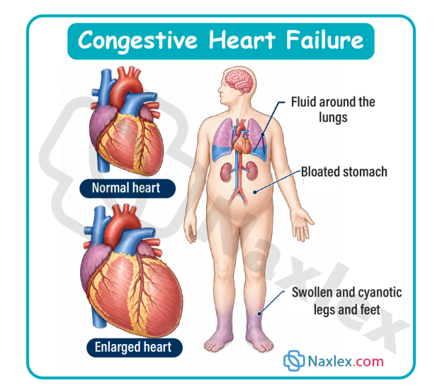
Epidemiology, Etiology, Risk Factors, And Pathophysiology
Epidemiology & Etiology
Pediatric congestive heart failure is not a primary disease entity but rather a clinical syndrome resulting from an underlying structural or functional cardiac abnormality.
- Infants (Under 1 year): The vast majority of cases (greater than 80%) are caused by structural congenital heart defects (CHDs). These are predominantly left-to-right shunting defects (e.g., large VSD, PDA, AVSD) or obstructive lesions (e.g., Coarctation of the aorta, Hypoplastic left heart syndrome).
- Children and Adolescents: Acquired cardiovascular conditions dominate this demographic. These include myocarditis (often viral, such as Coxsackievirus B or Adenovirus), dilated or hypertrophic cardiomyopathies, rheumatic heart disease (RHD) following an untreated Group A beta-hemolytic Streptococcal pharyngeal infection, or chemotherapy-induced cardiotoxicity (e.g., doxorubicin).
Risk Factors
- Prenatal Factors: Maternal diabetes, maternal lupus, intrauterine viral exposure (rubella, CMV), and advanced maternal age.
- Genetic and Chromosomal Syndromes: Down syndrome (Trisomy 21), Turner syndrome, Williams syndrome, and DiGeorge syndrome (22q11.2 deletion).
- Acquired Conditions: Severe, uncorrected chronic anemia, chronic renal failure causing fluid overload, or acute bacterial endocarditis.
Pathophysiology & Compensatory Mechanisms
When cardiac output falls below the metabolic demands of the pediatric tissues, the body initiates three primary compensatory pathways to maintain homeostasis. Although helpful in the short term, their sustained activation damages the heart muscle.
- Sympathetic Nervous System (SNS) Stimulation:
- Decreased systemic perfusion stimulates baroreceptors, triggering the release of epinephrine and norepinephrine.
- This results in tachycardia (to boost cardiac output) and increased systemic vascular resistance (vasoconstriction), shifting blood toward vital organs but raising the workload (afterload) the heart must pump against.
- Renin-Angiotensin-Aldosterone System (RAAS) Activation:
- Reduced renal perfusion prompts the kidneys to secrete renin, converting angiotensinogen to angiotensin I, which is converted to angiotensin II by ACE.
- Angiotensin II causes potent systemic vasoconstriction and stimulates the adrenal cortex to release aldosterone, causing significant sodium and water retention. This elevates blood volume (preload), leading to systemic and pulmonary edema.
- Myocardial Structural Remodeling:
- The heart muscle fibers stretch to accommodate the increased blood volume (ventricular dilation) to increase contractility via the Frank-Starling law. Over time, chronic wall stress leads to ventricular hypertrophy (thickening of the muscle walls), which increases myocardial oxygen demand and decreases ventricular compliance.
Image Title: Pediatric Heart Failure Pathophysiology
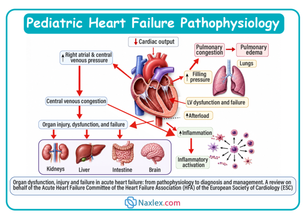
Classification
Pediatric heart failure can be classified based on the anatomical side of the heart that is primarily dysfunctional, or by the clinical presentation of systemic versus pulmonary symptoms.
Right-Sided Heart Failure
- Definition: The right ventricle is unable to pump blood effectively into the pulmonary artery.
- Mechanism: Backpressure develops in the right atrium, leading to venous congestion in the systemic circulation.
- Primary Causes: Pulmonic stenosis, uncorrected left-to-right shunts (causing secondary right ventricular strain), or pulmonary hypertension.
Left-Sided Heart Failure
- Definition: The left ventricle is unable to pump blood into the systemic circulation.
- Mechanism: Pressure increases in the left atrium, backing blood up into the pulmonary veins and capillaries.
- Primary Causes: Large VSD, PDA, Coarctation of the Aorta, Aortic Stenosis, or acute myocarditis.
High-Output Heart Failure
- Definition: The heart muscle is structurally normal and pumping at or above capacity, but cannot satisfy the body's unusually high metabolic demands.
- Primary Causes: Severe chronic anemia, systemic arteriovenous fistulas, or severe hyperthyroidism.
Clinical Manifestations
Children with heart failure present with signs related to systemic venous congestion, pulmonary congestion, and low systemic cardiac output.
Right-Sided Heart Failure (Systemic Venous Congestion)
- Hepatomegaly: Tenderness and palpable liver edge more than 2 cm below the right costal margin (caused by portal venous pooling).
- Periorbital edema: Fluid accumulation in the loose tissues around the eyes, especially visible in the morning.
- Sudden weight gain: A reliable indicator of fluid retention in infants, preceding noticeable dependent edema.
- Peripheral edema: Seen in older children as pitting edema over the shins, ankles, or sacrum.
- Distended jugular veins: Evident in older pediatric patients; rare in infants due to short neck anatomy.
Left-Sided Heart Failure (Pulmonary Congestion)
- Tachypnea: Respiratory rates exceeding 60 breaths/minute in infants, driven by fluid-congested, stiff lungs.
- Dyspnea, orthopnea, and retractions: Increased work of breathing with intercostal, subcostal, and suprasternal retractions, nasal flaring, and expiratory grunting.
- Pulmonary crackles/wheezing: Auscultated over the lung bases due to fluid accumulation in alveolar and interstitial spaces.
- Persistent dry cough: Caused by bronchial mucosal edema or compression of the left main bronchus by an enlarged left atrium.
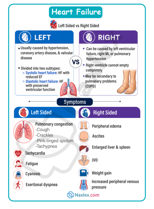
Cardiac Assessment Findings
A targeted physical assessment of the cardiovascular system reveals objective data regarding myocardial dysfunction and compensatory adaptations:
- Tachycardia: A persistent, resting heart rate elevated above normal developmental limits (e.g., sleeping infant heart rate greater than 160 bpm; child heart rate greater than 100 bpm). This is the earliest sign of heart failure.
- Abnormal Heart Sounds (Gallop Rhythm): Auscultation often reveals an S3 or S4 gallop rhythm, which represents ventricular filling delays and altered wall compliance in a volume-overloaded or stiff ventricle.
- Pathological Murmurs: Distinct structural murmurs help pinpoint the underlying cause (e.g., a loud, harsh holosystolic murmur at the lower left sternal border for a VSD, or a continuous machinery-like murmur beneath the left clavicle for a PDA).
- Precordial Activity: An active precordium, parasternal lifts, or displaced Point of Maximal Impulse (PMI) inferiorly or laterally due to ventricular dilation and hypertrophy.
- Diminished Peripheral Pulses: Systemic vasoconstriction creates weak, thready peripheral pulses (brachial, radial, pedal) compared to strong, bounding central pulses (femoral, carotid).
- Altered Tissue Perfusion: Cool, pale, or mottled extremities with a delayed capillary refill time greater than 3 seconds.
Impaired Cardiac Output & Increased Metabolic Demand
- Feeding intolerance: The infant becomes diaphoretic (sweaty), particularly on the forehead, and grows exhausted after taking only a small volume of formula or breast milk.
- Failure to thrive (FTT): Poor weight gain and linear growth retardation because the child burns excessive calories simply trying to breathe.
- Tachycardia: Resting heart rate persistently elevated above normal limits for age (e.g., greater than 160 bpm in sleeping infants) due to sympathetic drive.
- Weak peripheral pulses and delayed capillary refill: Reduced perfusion to extremities leads to pale, mottled, or cool skin and a capillary refill time greater than 2 to 3 seconds.
Chronic Signs of Hypoxemia
When heart failure is caused by cyanotic heart disease or advanced, uncorrected left-to-right shunting that has transitioned into pulmonary hypertension (Eisenmenger syndrome), chronic cellular hypoxia triggers distinct physical adaptations:
- Digital Clubbing: Proliferation of vascular connective tissue beneath the nail beds of the fingers and toes, causing the profile angle between the nail and the nail base to exceed 180 degrees. This results from chronic capillary hypoxia and the localized release of growth factors.
- Polycythemia: In response to chronic hypoxia, the kidneys increase erythropoietin production, stimulating bone marrow to produce excess red blood cells. While this raises oxygen-carrying capacity, it increases blood viscosity and escalates the risk of thromboembolic events (e.g., stroke).
- Squatting Posture (Knee-Chest Positioning): Seen in walking children during episodes of exertion or hypoxemia. Squatting mechanically increases systemic vascular resistance, reducing right-to-left shunting and forcing more blood through the lungs to improve oxygenation.
- Chronic Cyanosis: A persistent bluish discoloration of the mucous membranes, tongue, and nail beds, indicating that unoxygenated hemoglobin levels exceed 5g/dL.
Diagnostic Evaluation
Chest X-Ray (CXR)
- Cardiomegaly: Reveals an enlarged heart silhouette (cardiothoracic ratio greater than 55% in infants or greater than 50% in older children).
- Pulmonary congestion: Shows prominent pulmonary vascular markings, interstitial edema, and pleural effusions.
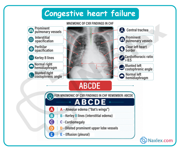
Electrocardiogram (ECG)
- Chamber hypertrophy: Shows increased voltage waves indicating right, left, or biventricular hypertrophy.
- Arrhythmias: Identifies sinus tachycardia, atrial flutter, or premature ventricular contractions resulting from myocardial stretch.
-
Echocardiogram (Echo)
- Anatomical assessment: The primary tool used to diagnose underlying congenital heart defects or confirm cardiomyopathy.
- Functional measurement: Measures ejection fraction (EF) and fractional shortening (FS) to quantify left ventricular systolic function, and tracks structural wall thickness.
-
Laboratory Biomarkers
- Brain natriuretic peptide (BNP): Released by ventricular myocytes in response to high wall stretch and volume overload; used to differentiate cardiac from primary respiratory causes of distress.
- Serum Electrolytes: Evaluated frequently because renal hypoperfusion can alter sodium and potassium balance, which is further modified by diuretic therapies.
Therapeutic Management
The main objectives of therapeutic management are to reduce volume overload (preload), decrease systemic vascular resistance (afterload), and improve myocardial contractility.
Pediatric CHF Medication Guide
|
Medication Class |
Mechanism of Action (MOA) |
Examples |
Indications |
Precautions / Contraindications |
Parameters to Monitor |
Nursing Insights & Side Effects |
|
Cardiac Glycosides |
Inhibits the sodium-potassium ATPase pump, increasing intracellular calcium concentration to boost myocardial contractility (positive inotrope) while prolonging the AV node refractory period to slow the heart rate (negative chronotrope). |
Digoxin (Lanoxin) |
Increases force of myocardial contraction; increases cardiac output while slowing heart rate. |
Contraindicated in ventricular fibrillation or heart block. Hold if apical pulse is less than 90–110 bpm in infants or less than 70 bpm in older children. |
• Apical pulse (1 full minute) • Serum potassium levels • Serum digoxin levels (0.5-2.0 ng/mL) • Renal function (BUN/creatinine) |
Narrow therapeutic index. Toxicity signs include bradycardia, anorexia, and unexplained vomiting. Hypokalemia significantly increases the risk of digoxin toxicity. |
|
Loop Diuretics |
Inhibits the Na+/K+/2Cl- cotransporter in the thick ascending loop of Henle, blocking the reabsorption of sodium, chloride, and water, which drives rapid fluid excretion. |
Furosemide (Lasix) |
Decreases intravascular volume (preload) by inducing rapid diuresis; resolves pulmonary and systemic edema. |
Contraindicated in anuria. Use with extreme caution in pre-existing severe electrolyte depletion. |
• Daily weights at identical times • Strict intake/output (I/O) volumes • Serum electrolytes (especially K+, Cl-, Na+) • Blood pressure |
Causes hypokalemia, hypochloremia, and dehydration. Ototoxicity can occur with rapid intravenous push. Monitor for muscle weakness or cramping due to low potassium. |
|
Potassium-Sparing Diuretics |
Competitively antagonizes aldosterone receptors in the distal convoluted tubule and collecting duct, blocking sodium-water reabsorption while conserving potassium ions. |
Spironolactone (Aldactone) |
Weak diuretic; used as an adjunct to loop diuretics to prevent potassium wasting while managing fluid retention. |
Contraindicated in acute renal failure, anuria, or pre-existing hyperkalemia. |
• Serum potassium and sodium • Renal function markers (BUN/creatinine) • Blood pressure |
Risk of hyperkalemia, especially if given with ACE inhibitors. Avoid potassium supplements or excessive dietary potassium intake. Assure adequate urine output before giving. |
|
ACE Inhibitors |
Blocks the Angiotensin-Converting Enzyme, preventing the conversion of Angiotensin I to the potent vasoconstrictor Angiotensin II. This promotes systemic vasodilation, reduces aldosterone secretion, and decreases afterload. |
Captopril, Enalapril |
Reduces afterload by promoting systemic vasodilation, making it easier for the weak ventricle to pump blood forward. |
Contraindicated in cases of angioedema or pregnancy. Caution with concurrent potassium-retaining medications. |
• Blood pressure (before and after) • Serum potassium • Renal function (creatinine) • White blood cell count |
Monitor blood pressure closely for profound hypotension, especially after the first dose. Watch for a persistent, dry ACE cough and hyperkalemia. |
Nutritional and Supportive Care
- High-calorie density feeding: Standard formula or breast milk (20 kcal/oz) is often concentrated to 24 to 30 kcal/oz using commercial fortifiers or glucose polymers. This delivers essential calories in a smaller fluid volume, preventing volume overload.
- Feeding schedule optimization: Limit active nipple feedings (breast or bottle) to a maximum of 20 to 30 minutes to minimize energy expenditure. Administer the remaining volume or entire feed via a gavage/nasogastric (NG) tube if the infant exhibits respiratory distress or fatigue.
- Oxygen administration: Administer supplemental humidified oxygen with caution.
Critical Safety Note: In infants with large left-to-right shunts (like VSD or PDA), oxygen acts as a potent pulmonary vasodilator. Giving too much oxygen can reduce pulmonary vascular resistance further, worsening the left-to-right shunt and flooding the lungs.
Nursing Insight: Administer pediatric CHF medications at the same times daily, ideally one hour before or two hours after feeds, using a calibrated oral syringe. Do not mix the medications directly into formula or breast milk bottles, as the infant may not finish the entire volume, resulting in an incomplete dose. Never readminister a dose if the child vomits. If a dose is missed by less than 4 hours, give it immediately; if over 4 hours, skip it. Never double doses. Contact the provider if two consecutive doses are missed.
Nursing Care Plan
Priority Nursing Diagnoses (NANDA-I Framework)
- Decreased Cardiac Output related to structural myocardial defects or altered myocardial contractility as evidenced by tachycardia, weak peripheral pulses, and delayed capillary refill.
- Excess Fluid Volume related to activation of the renin-angiotensin-aldosterone axis and compromised renal perfusion as evidenced by hepatomegaly, periorbital edema, and rapid weight gain.
- Ineffective Breathing Pattern related to pulmonary venous congestion and fluid accumulation within the alveoli as evidenced by tachypnea, grunting, nasal flaring, and intercostal retractions.
- Imbalanced Nutrition: Less Than Body Requirements related to high metabolic work of breathing and feeding intolerance as evidenced by inability to finish feeds, diaphoresis during feeding, and weight loss or failure to thrive.
Nursing Interventions & Rationales
The Strategy of Clustering Care
The organizing principle of pediatric heart failure nursing care is clustering care. This involves grouping multiple assessments, medication administrations, and nursing tasks into a single, coordinated block of time, followed by long, uninterrupted rest periods.
- Assess Patient State and Plan Layout (Pre-intervention): Review the schedule. Coordinate with laboratory, radiology, and therapy teams so all diagnostic samplings align with the planned nursing block. Never wake an infant just to check a routine parameter unless clinically unstable.
- Execute Clustered Nursing Tasks (Maximum 15–20 minutes): Gently perform essential interventions in a single session: check vital signs, auscultate heart and lung sounds, weigh the infant (if indicated), change diapers, and administer cardiac medications. Keep environmental stimuli, such as bright lights and loud noises, to a absolute minimum.
- Deliver High-Density Nutrition: Administer fortified, high-calorie nutrition. If the infant is bottle-feeding, stop at the 20-to-30-minute mark. Administer any remaining volume via the nasogastric tube while the infant rests to prevent metabolic exhaustion.
- Position for Optimal Ventilation: Place the infant in an elevated semi-Fowler or high-Fowler position (using an infant seat or elevating the head of the bed to a 30-to-45-degree angle). This lowers the diaphragm, maximizing chest expansion and decreasing venous return to ease the workload on the heart.
- Enforce Extended, Uninterrupted Rest (2 to 3 hours): Dim the lights, cluster family support at the bedside, and leave the infant completely undisturbed. This reduces the sympathetic drive, lowering the heart rate and blood pressure, which conserves energy and limits oxygen demand.
Complications
Digitalis (Digoxin) Toxicity
Because pediatric patients have immature renal clearance systems, they are at high risk for Digoxin toxicity.
- Clinical presentation: Bradycardia, poor feeding, nausea, and frequent vomiting (often mistaken for spitting up).
- Antidote: Digoxin Immune Fab (Digibind) is administered for severe, life-threatening arrhythmias or toxic serum levels.
Metabolic Exhaustion and Failure to Thrive
If caloric density cannot compensate for the high energy expenditure of breathing, the child's body breaks down muscle and fat stores. This results in severe linear growth failure, delayed cognitive development, and a weakened immune system.
Cardiopulmonary Collapse
Acute decompensation can occur when compensatory mechanisms fail. This leads to profound cardiogenic shock, severe pulmonary edema, severe metabolic acidosis, and multi-organ failure.
Prognosis
The long-term outlook for a child with heart failure depends heavily on whether the primary cause can be reversed or surgically corrected.
- Surgically correctable defects: Children with left-to-right shunts (VSD, ASD, PDA) show rapid resolution of heart failure symptoms and excellent catch-up growth once the defect is closed.
- Chronic cardiomyopathies: Structural heart muscle diseases have a more guarded prognosis. If medical management fails to halt ventricular remodeling, these patients may require long-term mechanical circulatory support (Ventricular Assist Devices) or a cardiac transplant.
Summary
- Congestive heart failure (CHF) in pediatric patients is a complex clinical syndrome characterized by the heart's inability to pump an adequate supply of oxygenated blood to meet the metabolic demands of the body.
- In infants, the condition is almost exclusively driven by structural congenital heart defects that cause a left-to-right shunt (such as ventricular septal defects or patent ductus arteriosus). These defects flood the pulmonary system with excess fluid volume.
- In older children, heart failure is more often an acquired condition resulting from viral myocarditis, cardiomyopathies, or rheumatic heart disease.
- When cardiac output drops, the pediatric body activates neurohormonal pathways, specifically the sympathetic nervous system and the renin-angiotensin-aldosterone system.
- Although these systems maintain core blood pressure via tachycardia, vasoconstriction, and fluid retention, their chronic activation overloads the heart chambers and causes systemic and pulmonary edema.
- The clinical presentation in children varies by age, with infants showing distinct signs of distress.
- Left-sided heart failure causes pulmonary congestion, which manifests as tachypnea, grunting, intercostal retractions, and rales. Right-sided heart failure causes systemic venous congestion, leading to hepatomegaly, periorbital edema, and sudden fluid weight gain.
- A key clinical feature of pediatric heart failure is feeding intolerance, where infants become diaphoretic and exhausted during feeds, resulting in failure to thrive because their caloric needs outpace their intake.
- Diagnostic evaluation relies on chest X-rays to assess for cardiomegaly, ECGs to screen for hypertrophy or arrhythmias, and echocardiograms to evaluate ventricular function and visualize structural defects.
- Therapeutic management focuses on reducing fluid volume with loop diuretics (Furosemide), decreasing afterload with ACE inhibitors, and improving contractility with cardiac glycosides (Digoxin).
- Nutritional adjustments are critical; nurses must provide high-calorie, concentrated formulas and limit active feeding times to under 30 minutes, using nasogastric gavage for any remaining volume to prevent metabolic exhaustion.
- The core nursing responsibilities include monitoring fluid balance through daily weights and precise intake/output measurements, watching for signs of Digoxin toxicity (such as bradycardia or vomiting), and supporting the child's respiratory effort.
- The long-term prognosis is excellent for children with correctable structural defects, but remains guarded for those with progressive cardiomyopathies who may ultimately require cardiac transplantation.
Naxlex
Videos
Login to View Video
Click here to loginTake Notes on Acquired Cardiovascular Disorders: Congestive Heart Failure
This filled cannot be empty
Join Naxlex Nursing for nursing questions & guides! Sign Up Now