
CONGENITAL HEART DISEASES: MIXED DEFECTS
Lessons
Notes Highlighting is available once you sign in. Login Here.
Objectives
- Classify congenital heart diseases characterized by mixed blood flow, distinguishing them from simple left-to-right shunting mechanisms.
- Describe the anatomical defects and abnormal systemic-pulmonary connections in Transposition of the Great Vessels (TGV), Total Anomalous Pulmonary Venous Connection (TAPVC), Truncus Arteriosus, and Hypoplastic Left Heart Syndrome (HLHS).
- Identify the clinical manifestations of mixed blood defects, highlighting profound cyanosis, rapidly developing congestive heart failure (CHF), and differential oxygen saturations.
- Explain the vital role of transitional fetal structures, specifically the Patent Ductus Arteriosus (PDA) and Foramen Ovale, in maintaining life before surgical intervention.
- Interpret diagnostic findings from echocardiograms, chest X-rays (such as the "egg-on-a-string" or "snowman" signs), and cardiac catheterization.
- Outline urgent medical management (including Prostaglandin E1 infusion) and complex multi-stage surgical corrections.
- Develop a comprehensive nursing care plan for pediatric patients exhibiting critical cyanosis, metabolic stress, and poor perfusion.
- Recognize severe post-operative complications, including pulmonary hypertensive crises, low cardiac output syndrome, and arrhythmias.
Introduction
- Congenital Heart Diseases (CHDs): Mixed Defects represent a highly complex, critical category of cardiac anomalies characterized by the survival-dependent mixing of oxygenated and unoxygenated blood.
- Unlike simple lesions, mixed defects involve a profound anatomical restructuring where the systemic and pulmonary circulations function either as isolated, parallel loops or drain into a single, shared cardiac chamber.
- The defining pathophysiological hallmark of these defects, which include Transposition of the Great Vessels (TGV), Total Anomalous Pulmonary Venous Return (TAPVR), and Truncus Arteriosus, is bidirectional shunting.
- Because oxygen-rich and oxygen-poor blood completely blend within the heart or great vessels, the blood ejected into the systemic circulation is permanently desaturated. This results in progressive tissue hypoxia and profound, visible cyanosis that does not resolve with supplemental oxygen.
- In the neonatal period, these defects constitute a medical emergency. Survival is entirely dependent on the patency of fetal pathways, such as a Patent Ductus Arteriosus (PDA) or Foramen Ovale, which act as the sole channels for cross-mixing.
- As these fetal pathways naturally begin to close hours or days after birth, blood mixing drops catastrophically, triggering severe metabolic acidosis, respiratory distress, and rapid cardiovascular collapse.
- Advanced nursing and medical management center on keeping these vital mixing pathways open. The immediate initiation of a continuous prostaglandin E1 (PGE1) infusion is required to maintain ductal patency.
- Paradoxically, high-flow supplemental oxygen must be tightly restricted; because oxygen is a potent pulmonary vasodilator and ductal constrictor, over-oxygenation can flood the lungs, starve systemic organs, and close the life-sustaining PDA.
- Palliative interventions like a balloon atrial septostomy provide temporary stabilization until definitive, highly complex open-heart surgical correction can be performed.
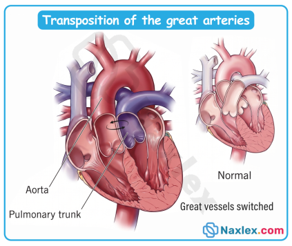
Transposition of the Great Vessels
Transposition of the Great Vessels (TGV) is a cyanotic congenital heart anomaly characterized by a complete reversal of the anatomical positions of the major outflow tracts. The aorta erroneously arises directly from the right ventricle, and the pulmonary artery arises from the left ventricle. This creates two separate, parallel circulatory loops rather than the normal series circuit, preventing oxygenated blood from reaching the systemic circulation unless a mixing shunt coexists.
Epidemiology
- TGV accounts for approximately 5% to 7% of all congenital heart defects.
- It is the most common cyanotic heart defect presenting in the immediate neonatal period.
- There is a distinct male-to-female predilection, with a ratio of approximately 3:1.
- Occurs in roughly 20 to 30 per 100,000 live births.
Etiology
- Genetic factors: Often isolated, but can be associated with specific gene mutations (such as PROSIT24 or GDF1) or chromosomal syndromes, though less frequently linked to Down syndrome than septal defects.
- Environmental factors: Strongly linked to maternal pre-gestational diabetes, maternal obesity, exposure to organic solvents, and advanced maternal age during the first trimester.
- Embryological failure: Results from the abnormal spiraling and partitioning of the truncus arteriosus by the aorticopulmonary septum during the 5th to 8th weeks of fetal development.
Pathophysiology
- Parallel Circulation: Unoxygenated systemic venous return enters the right atrium, passes to the right ventricle, and is pumped directly back out to the body via the transposed aorta. Simultaneously, oxygenated pulmonary venous return enters the left atrium, passes to the left ventricle, and is pumped back into the lungs via the transposed pulmonary artery.
- Survival Dependency: Life is incompatible with birth unless an anatomical communication exists to allow mixing of the two circuits. Mixing typically occurs via a Patent Ductus Arteriosus (PDA), Foramen Ovale/ Atrial Septal Defect (ASD), or Ventricular Septal Defect (VSD).
- Hypoxemia and Tissue Hypoxia: As the PDA begins to close postnatally, systemic oxygen saturation drops precipitously, leading to severe metabolic acidosis and tissue hypoxia.
Clinical Manifestations
Symptoms present immediately at birth or within the first hours of life, depending on the degree of mixing through fetal shunts.
- General: Profound, progressive cyanosis that does not improve with supplemental oxygen administration (unresponsive to hyperoxia challenge). Tachypnea and signs of respiratory distress (grunting, flaring, retractions) emerge as tissue hypoxia worsens.
- Growth/Feeding: Poor feeding performance and rapid fatigue during attempts to suckle.
- Cardiac Auscultation:
- S1: Normal.
- S2: Typically single and loud, because the anteriorly transposed aorta closes closer to the chest wall, obscuring the pulmonic closure sound.
- Murmur: Often completely absent if the interventricular septum is intact. If a VSD or PDA is present, a systolic murmur or continuous murmur may be heard, respectively.
Diagnostic Evaluation
- Echocardiography (ECHO): The gold standard diagnostic tool. It directly visualizes the ventriculoarterial discordance, confirming that the aorta arises from the right ventricle and the pulmonary artery from the left ventricle, while mapping out any concurrent mixing lesions (ASD, VSD, PDA).
- Electrocardiogram (ECG): Usually reveals right axis deviation and right ventricular hypertrophy (RVH) because the right ventricle continues to pump against high systemic vascular resistance.
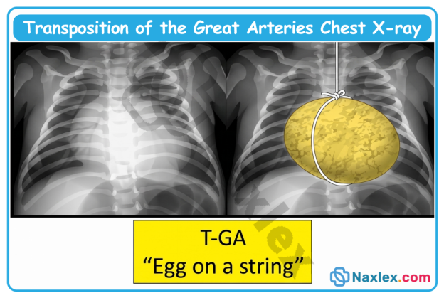
- Chest X-ray (CXR): Characteristically demonstrates cardiomegaly with a narrow mediastinal shadow, classically described as an "egg-on-a-string" appearance, along with increased pulmonary vascular markings.
Image Title: Transposition of the Great Arteries Chest X-ray
- Hyperoxia Test: Administering 100% fraction of inspired oxygen (FiO2) fails to significantly raise the partial pressure of arterial oxygen (PaO2), pointing directly to a right-to-left structural shunt.
Therapeutic Management
A. General & Medical Principles
- Prostaglandin E1 (PGE1) Infusion: Initiated immediately via a secure central or peripheral line. PGE1 prevents the physiological closure of the patent ductus arteriosus, maintaining critical mixing of systemic and pulmonary blood.
- Therapeutic Sub-ambient Oxygen/Minimal Handling: High oxygen concentrations reduce pulmonary vascular resistance and may hasten ductal closure; therefore, oxygen supplementation is kept minimal, targeting oxygen saturations between 75% and 85%.
- Emergency Balloon Atrial Septostomy (Rashkind Procedure): Performed in the cardiac catheterization lab or at the bedside under ECHO guidance if severe hypoxia persists despite PGE1. A balloon catheter is advanced into the right atrium, through the foramen ovale into the left atrium, inflated, and pulled back forcefully to tear the interatrial septum, creating a large ASD for blood mixing.
B. Surgical Management
- Indication: Definitively required for all neonates diagnosed with TGV, typically executed within the first 1 to 2 weeks of life.
- Procedure (Arterial Switch Operation / ASO): The definitive corrective surgery (Jatene procedure). The transposed aorta and pulmonary artery are transected above the valves and switched to their correct anatomical ventricles. Crucially, the coronary arteries must be meticulously excised from the native aorta and replanted into the neoaorta.
Post-Operative Nursing Interventions
Post-Op Day 0 to 1 (ICU Phase)
- Maintain continuous mechanical ventilation and closely analyze arterial blood gases (ABGs) to optimize acid-base balance and oxygenation.
- Monitor hourly chest tube output. Report drainage greater than 3 mL/kg/hr for 3 consecutive hours or greater than 5 mL/kg in any single hour, as this signals acute post-operative hemorrhage.
- Provide continuous ECG monitoring; closely watch for bradyarrhythmias, heart blocks, or ST-segment changes that indicate coronary artery compression or spasm following replantation.
- Maintain a strict fluid balance profile via an indwelling Foley catheter, ensuring a minimum urine output of 1 mL/kg/hr.
- Regularly check central and peripheral perfusion status (ensure capillary refill time remains less than 3 seconds).
Post-Op Day 2 to 3 (Transition Phase)
- Gradually wean from mechanical ventilation and supplemental oxygen as tolerated by the patient.
- Slowly advance nutrition from NPO status to clear liquids or specialized infant formula once bowel sounds return and extubation is successful.
- Implement respiratory therapy support (e.g., gentle chest physiotherapy or tactile stimulation to encourage deep crying) to prevent micro-atelectasis.
- Ensure multimodal pain management utilizing scheduled intravenous acetaminophen or ketorolac with low-dose opioid rescue choices.
Post-Op Day 4 to Discharge
- Regularly inspect the sternotomy surgical incision site for signs of localized infection (warmth, purulent drainage, erythema, or wound dehiscence).
- Assist the infant's caregivers with safe handling and holding techniques during gradual increases in ambient activity.
- Deliver comprehensive discharge and parent education, explicitly instructing caregivers never to lift the infant by their arms or under the armpits for 4 to 6 weeks to prevent structural mechanical stress on the healing sternum.
Nursing Diagnosis (Post-Op)
Post-Op Day 1
- Decreased Cardiac Output related to surgical myocardial ischemia, coronary artery translocation alterations, or arrhythmia development.
- Impaired Gas Exchange related to cardiopulmonary bypass effects, mechanical ventilation dependencies, or ventilation-perfusion mismatching.
- Risk for Fluid Volume Deficit related to chest tube blood loss, surgical hemorrhage, or aggressive post-bypass diuretic management.
Post-Op Day 2 to 3
- Acute Pain related to surgical sternotomy access, chest tube placement irritation, and frequent nursing manipulations.
- Ineffective Airway Clearance related to thick retained respiratory secretions, relative immobility, and post-extubation incisional splinting.
- Risk for Infection related to multiple indwelling lines (central venous lines, arterial lines) and the median sternotomy incision.
Post-Op Day 4+
- Deficient Knowledge (Caregiver) related to specialized infant home care routines, recognition of cardiac decompensation signs, and medication tracking.
- Activity Intolerance related to prolonged surgical recovery, decreased caloric intake during the acute illness, and generic physical weakness.
Complications
- Cardiac: Acute myocardial infarction or ischemia due to coronary artery kinking or stenosis at the surgical reimplantation sites; supraventricular arrhythmias, or neo-aortic root dilation.
- Pulmonary: Pulmonary artery stenosis at the site of the surgical anastomosis.
- Surgical: Postpericardiotomy syndrome, phrenic nerve injury causing diaphragmatic paralysis, or paradoxical systemic emboli.
Prognosis
- Without surgical intervention, the mortality rate is approximately 30% within the first week of life, and exceeds 90% by 1 year of age.
- Following a successful Arterial Switch Operation (ASO), the long-term prognosis is excellent, boasting a survival rate greater than 95% at 20 years.
- Most survivors enjoy a completely normal life span and normal exercise tolerance, requiring only lifelong periodic follow-up with a pediatric cardiologist.
