
BIOL 252 Anatomy And Physiology II Module 2 Proctored Exam
BIOL 252 Anatomy And Physiology II Module 2 Proctored Exam
Total Questions : 69
Showing 10 questions Sign up for more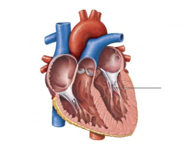
Explanation
A. Chordae tendinae: The structure pointed to by the line is the chordae tendineae. The chordae tendineae are strong, fibrous collagen cords that connect the atrioventricular valve leaflets (mitral and tricuspid valves) to the papillary muscles within the ventricles. They prevent valve prolapse during ventricular systole. When the ventricles contract, papillary muscles also contract, maintaining tension on the chordae tendineae to keep the valves closed and prevent backflow into the atria.
B. Papillary Muscles: Papillary muscles are cone-shaped projections from the ventricular walls that anchor chordae tendineae. During ventricular contraction, they prevent valve prolapse by maintaining tension on valve leaflets, ensuring unidirectional blood flow
C. Septal leaflet: The septal leaflet is part of the tricuspid valve attached to the interventricular septum. Chordae tendineae connect it to papillary muscles, stabilizing the valve during systole and preventing backflow into the right atrium.
D: Pulmonary vein: Pulmonary veins are vessels transporting oxygenated blood from the lungs to the left atrium. They are not directly connected to chordae tendineae but are adjacent to atrial structures influencing left atrial filling and valve dynamics.
Identify the letter of the heart component that is being described in the statement.
Shallow depression that is a remnant of the foramen ovale.
Explanation
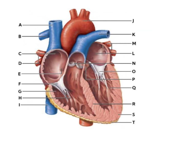
N: Pulmonary valve- The pulmonary valve is a semilunar valve between the right ventricle and pulmonary artery. It prevents backflow into the ventricle during diastole, facilitating blood flow toward the lungs for oxygenation.
E: Fossa ovalis - The fossa ovalis is a shallow, thumb-sized depression located in the interatrial septum (the wall separating the right and left atria). In a developing fetus, the foramen ovale is an open "tunnel" that allows blood to bypass the lungs by flowing directly from the right atrium to the left atrium. Once a baby takes their first breath, the pressure changes in the heart cause a flap of tissue to close over this opening. Over time, it fuses shut, leaving behind the shallow indensee fossa ovalis.
O: chordae tendinae: Chordae tendineae are fibrous cords connecting the atrioventricular valve leaflets (mitral and tricuspid) to papillary muscles. They prevent valve prolapse during ventricular contraction, ensuring unidirectional blood flow.
J: Aortic arch- The aortic arch is the curved portion of the aorta that distributes oxygenated blood from the left ventricle to systemic arteries. It contains baroreceptors and helps regulate blood pressure.
The nurse plans to use role playing as a therapeutic measure. Which individual is most likely to benefit from this type of therapeutic intervention?
Explanation
A. An adolescent who is depressed over not being accepted by peers: Role playing allows adolescents to practice social interactions, develop communication skills, and explore coping strategies in a safe environment. It can help the client gain confidence, improve problem-solving abilities, and prepare for real-life peer situations.
B. Hyperactive 4-year-old who has recently been tested for autism: While play therapy is useful for young children and those with autism spectrum disorder, structured role playing may be too abstract for a 4-year-old. Developmental level and attention span may limit the effectiveness of role playing in this age group.
C. An adult with schizophrenia who often refuses to take prescribed antipsychotic medications: Role playing is generally less effective for clients with active psychotic symptoms such as delusions or hallucinations. Medication adherence issues in schizophrenia are better addressed through psychoeducation, motivational interviewing, and structured support.
D. An older adult resident of a long-term care facility who sometimes takes other residents' belongings: While role playing could be used to teach social norms, behavioral interventions such as redirection, supervision, and environmental modifications are more practical and immediately effective for managing problematic behaviors in older adults with cognitive decline.
Identify the highlighted section using the drop down below.
What structure of the electrical conduction system passes through the highlighted area?
Explanation
Correct answer:
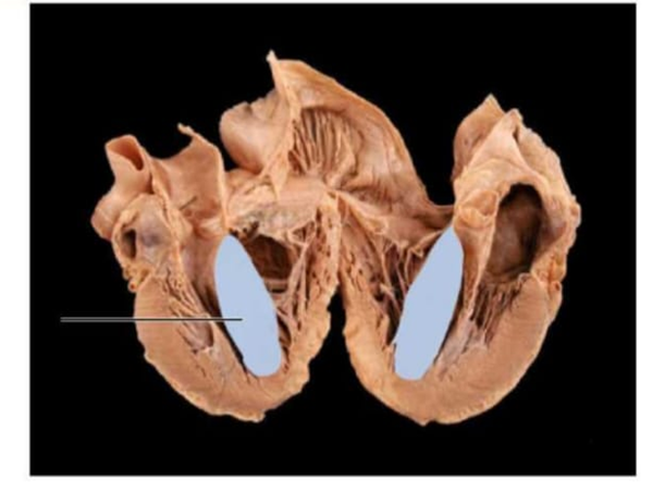
- Interventricular septum
- Atrioventricular (AV) bundle / Bundle of His
The highlighted area represents the interventricular septum, the thick muscular wall that separates the right and left ventricles. It forms the medial wall of both ventricles and extends from the atrioventricular valves superiorly to the apex inferiorly. Its primary physiologic function is to prevent mixing of oxygenated blood in the left ventricle with deoxygenated blood in the right ventricle while also contributing to ventricular contraction. The electrical conduction structure that passes through this area is the atrioventricular (AV) bundle / Bundle of His. The Bundle of His passes from the atrioventricular node into the membranous portion of the interventricular septum. It then divides into the right and left bundle branches, which travel along the septum toward the apex to distribute electrical impulses to both ventricles. This allows coordinated ventricular depolarization and synchronized contraction.
Match the structure to its description.
|
Description |
Structure |
|
A cluster of cells located in the interatrial septum that delays the electrical signal before it passes to the ventricles. |
dropdown
|
|
The pacemaker of the heart, located in the right atrium, responsible for initiating the heartbeat. |
dropdown
|
|
The valve located between the right atrium and right ventricle. |
dropdown
|
|
The layer of the pericardium that covers the heart directly, also known as the epicardium. |
dropdown
|
|
Blood vessels carrying oxygenated blood from the lungs to the left atrium. |
dropdown
|
Explanation
Correct answer:
- A cluster of cells located in the interatrial septum that delays the electrical signal before it passes to the ventricles: Atrioventricular node
- The pacemaker of the heart, located in the right atrium, responsible for initiating the heartbeat: Sinoatrial node
- The valve located between the right atrium and right ventricle: Tricuspid valve
- The layer of the pericardium that covers the heart directly, also known as the epicardium: Visceral pericardium
- Blood vessels carrying oxygenated blood from the lungs to the left atrium: Pulmonary veins
• Atrioventricular node: The AV node is located in the interatrial septum and functions to delay the electrical impulse received from the SA node. This delay allows the atria to contract and complete ventricular filling before ventricular contraction. Its position and timing role are crucial for coordinated cardiac conduction.
• Sinoatrial node: The SA node, located in the right atrium near the superior vena cava, acts as the heart’s natural pacemaker. It generates electrical impulses that initiate each heartbeat, setting the rhythm for the entire heart. Proper SA node function is essential for synchronized atrial and ventricular activity.
• Tricuspid valve: The tricuspid valve sits between the right atrium and right ventricle, preventing backflow of blood during ventricular contraction. Its three leaflets open during diastole to allow atrial emptying into the ventricle. Proper valve function maintains unidirectional blood flow and cardiac efficiency.
• Visceral pericardium: Also called the epicardium, this layer of the pericardium lies directly on the heart surface. It provides protection, reduces friction during heartbeats, and contains blood vessels supplying the myocardium. Its anatomical position distinguishes it from the parietal pericardium.
• Pulmonary veins: Pulmonary veins carry oxygen-rich blood from the lungs to the left atrium. They are unique among veins as they transport oxygenated rather than deoxygenated blood. Their flow ensures that systemic circulation receives oxygenated blood.
Blood flows from the right coronary artery into the
Explanation
A. Pulmonary artery: The pulmonary artery originates from the right ventricle and is positioned anterior to the ascending aorta as it exits the heart. Its physiologic role is to transport deoxygenated blood to the lungs for oxygenation. It is part of the pulmonary circulation and does not arise from or receive blood flow from the right coronary artery, which supplies myocardium.
B. Left anterior descending artery: The left anterior descending artery, also known as the anterior interventricular artery, branches from the left coronary artery and runs within the anterior interventricular sulcus toward the apex. It supplies the anterior wall of the left ventricle and the anterior two-thirds of the interventricular septum.
C. Circumflex artery: The circumflex artery arises from the left coronary artery and courses in the left atrioventricular (coronary) sulcus. It supplies the lateral and posterior portions of the left ventricle and may contribute to left atrial perfusion. Its anatomical origin from the left coronary artery excludes it from being a branch of the right coronary artery.
D. Anterior interventricular artery: The anterior interventricular artery lies in the anterior interventricular groove between the right and left ventricles. It provides blood supply to the interventricular septum and the anterior surfaces of both ventricles. As a branch of the left coronary artery, it does not represent a continuation of blood flow from the right coronary artery.
E. Right marginal artery: The right marginal artery is a direct branch of the right coronary artery and travels along the acute margin of the heart toward the apex. It supplies the right ventricular free wall and contributes to perfusion of the right myocardium. Its anatomical course and origin confirm that blood flows from the right coronary artery into the right marginal artery.
The pulmonary veins carry deoxygenated blood from the lungs to the left atrium.
Explanation
Correct answer: False
The pulmonary veins are paired vessels located within the thoracic cavity that extend from each lung to the posterior aspect of the left atrium. After blood passes through the pulmonary capillaries surrounding the alveoli, carbon dioxide diffuses out and oxygen diffuses into the bloodstream. The pulmonary veins then transport this oxygenated blood back to the heart. Their physiological role is to ensure that oxygen-rich blood enters the left atrium, flows into the left ventricle, and is subsequently pumped through the aorta to supply systemic circulation. This unique function distinguishes pulmonary veins from systemic veins, which typically carry deoxygenated blood.
Explanation
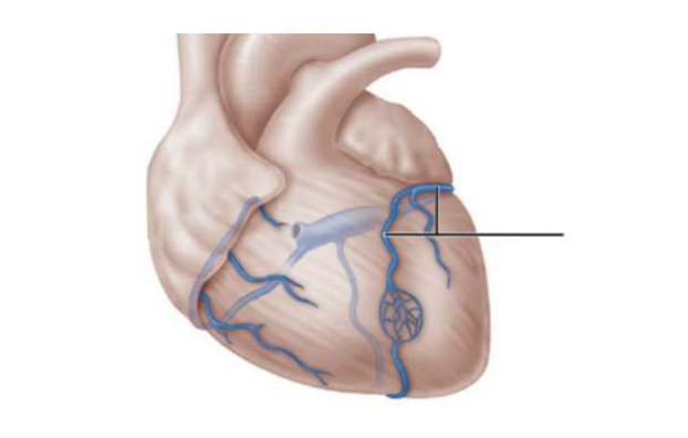
A. Small cardiac vein: The small cardiac vein runs along the right margin of the heart and drains blood from the right atrium and ventricle. It empties into the coronary sinus, facilitating venous return to the right atrium.
B. Coronary sinus: The coronary sinus is a large venous channel on the posterior aspect of the heart. It collects most cardiac venous blood and drains directly into the right atrium, completing coronary circulation.
C. Anterior cardiac vein: The anterior cardiac veins run along the anterior surface of the right ventricle. They bypass the coronary sinus, draining directly into the right atrium, and contribute to the venous return from the right ventricular myocardium.
D. Superior vena cava: The superior vena cava is a major systemic vein that returns deoxygenated blood from the upper body, including the head, neck, and upper limbs, directly into the right atrium of the heart.
E. Middle cardiac vein; The middle cardiac vein runs in the posterior interventricular sulcus, draining the posterior portion of both ventricles. It empties into the coronary sinus, ensuring efficient venous return from the heart’s posterior myocardium.
F. Great cardiac vein: The blood vessel highlighted in the image is the great cardiac vein located on the anterior surface of the heart. The great cardiac vein begins at the apex of the heart and eventually curves around the left side of the heart (within the coronary sulcus) to empty into the coronary sinus on the posterior side. It is the principal vein of the anterior heart.
A patient with a defective tricuspid valve undergoes valve replacement surgery. After surgery, the patient should be monitored for proper opening of the valve during which part of the cardiac cycle?
Explanation
A. During pulmonary circulation: Pulmonary circulation refers to the movement of blood from the right ventricle through the pulmonary trunk to the lungs and back to the left atrium. The tricuspid valve is between the right atrium and right ventricle and does not open or close in response to pulmonary vascular events. Its function is confined to regulating flow between the right atrium and right ventricle.
B. During right ventricular systole: Right ventricular systole occurs when the right ventricle contracts to eject blood through the pulmonary valve into the pulmonary trunk. During this phase, the tricuspid valve should be closed to prevent regurgitation of blood back into the right atrium. Proper valve competence at this time prevents right atrial volume overload.
C. During left ventricular systole: Left ventricular systole involves contraction of the left ventricle and ejection of blood through the aortic valve into systemic circulation. This phase is functionally and anatomically related to the mitral and aortic valves on the left side of the heart. The tricuspid valve on the right side is not involved in left ventricular contraction dynamics.
D. During right atrial systole: Right atrial systole occurs at the end of ventricular diastole when the right atrium contracts to push blood through the tricuspid valve into the right ventricle. The tricuspid valve must be open during this phase to allow forward flow into the right ventricular chamber. Its proper opening ensures adequate right ventricular filling prior to ventricular systole.
E. During atrial diastole: Atrial diastole refers to the relaxation phase of the atria when they are filling with venous blood from the venae cavae. Although the tricuspid valve may be open during part of ventricular diastole due to pressure gradients, atrial contraction is the specific phase that actively requires valve opening for optimal ventricular filling.
Which letter represents ventricular depolarization?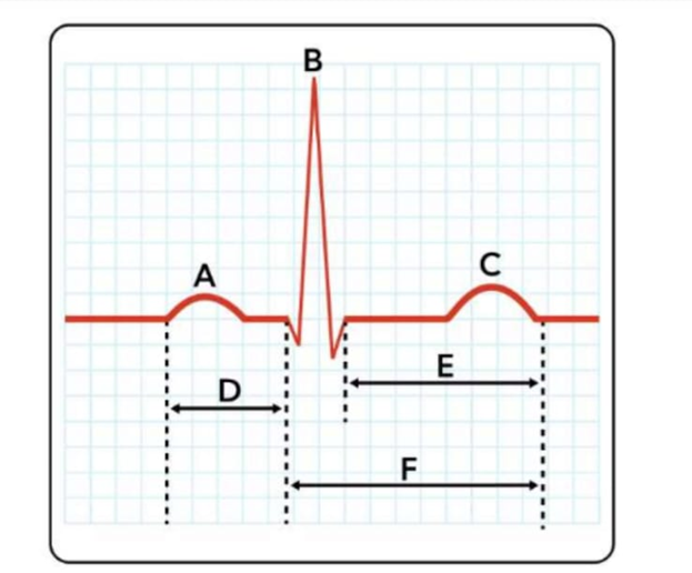
Explanation
Correct answer: B.
B: Ventricular depolarization is the electrical activation of the ventricles that triggers ventricular contraction. It occurs after the impulse travels from the sinoatrial . On an electrocardiogram (ECG), ventricular depolarization is represented by the QRS complex, labelled as B in the diagram.
D: artrial depolarization - Atrial depolarization initiates atrial contraction, driven by impulses from the SA node. This electrical activity spreads across atrial myocardium, generating the P wave on the ECG and enabling ventricular filling.
E: ventricular repolarization - Ventricular repolarization restores the ventricles to their resting state after contraction. Represented by the T wave, it prepares the heart for the next depolarization, ensuring rhythmic cardiac cycles and effective pumping.
You just viewed 10 questions out of the 69 questions on the BIOL 252 Anatomy And Physiology II Module 2 Proctored Exam Exam. Subscribe to our Premium Package to obtain access on all the questions and have unlimited access on all Exams. Subscribe Now

