





 Video Library


Please set your exam date
Acquired Cardiovascular Disorders: Kawasaki Disease
Study Questions
Practice Exercise 1
A nurse is reviewing the medical records of a group of pediatric patients. Which of the following children is at the highest statistical risk for developing Kawasaki disease?
Explanation
Kawasaki diseaseis an acute, self-limiting systemic vasculitisthat primarily affects medium-sized arteries, with a strong predilection for the coronary arteries.The etiology remains unknown, but it is widely understood to result from an abnormal immunological response to an infectious trigger in genetically susceptible individuals. Epidemiological studies consistently show that the disease has a distinct distribution based on age, sex, and ethnicity,which serves as the primary basis for identifying high-risk populations.
Rationale for correct answer:
2.A 14-month-old male of Japanese descentrepresents the peak demographic risk group for Kawasaki disease. Approximately 75% to 80% of cases occur in children under the age of 5 years, with a peak incidence between 12 and 24 monthsof age. Additionally, the disease is 1.5 times more common in males than females, and children of East Asian heritage,particularly Japanese or Korean descent, exhibit the highest incidence rates globally.
Rationale for incorrect answers:
1.A 9-year-old female of Caucasian descentis at a significantly lower risk for Kawasaki disease. Children older than 5 years represent a small minority of cases, and the disease is relatively rare in older school-aged children.Furthermore, females and individuals of Caucasian ethnicityhave a statistically lower incidence compared to males of East Asian descent.
3.A 4-month-old female of African American descenthas multiple demographic factors that place her at lower risk. Although infants under 6 months can develop the disease and are at high risk for coronary aneurysms, the overall incidence is low in this very young age bracket.Additionally, females of African American heritagedo not represent the highest statistical risk cohort.
4.An 8-year-old male of Hispanic descentis not in the highest risk category. While being male is a risk factor, his advanced age of 8 years is well outside the peak toddler age groupof 1 to 2 years. Children of Hispanic descentalso have a lower incidence of Kawasaki disease compared to those of East Asian descent.
Test-taking strategy:
- Analyze the scenario/question:The clinical stem asks the nurse to identify the pediatric patient at the highest statistical risk for developing Kawasaki disease based on epidemiological data.
- Evaluate demographic risk factors:Apply knowledge of the age, sex, and ethnic distribution of Kawasaki disease to triage the choices.
- Assess age distribution:Focus on the peak range, which is under 5 years of age, specifically between 12 and 24 months.This immediately prioritizes Choice 2and Choice 3over Choice 1 andChoice 4.
- Assess sex distribution:Recall that males are affected significantly more often than females, which favors Choice 2over Choice 3.
- Assess ethnic distribution:Identify that East Asian descent(specifically Japanese or Korean) carries the highest relative risk worldwide.
- Synthesize the findings:Combining these three factors makes the 14-month-old Japanese male the highest-risk candidate.
Take home points
- Kawasaki disease is most commonly diagnosed in children under the age of 5 years, with the peak incidence occurring in toddlers aged 12 to 24 months.
- Epidemiological data shows a clear male-to-female predominance, with males being approximately 1.5 times more likely to acquire the condition.
- The highest incidence rates of Kawasaki disease are documented globally among children of East Asian descent, particularly those of Japanese and Korean heritage.
- Understanding these demographic risk factors helps clinicians maintain a high index of suspicion to prevent critical coronary artery complications.
A nurse is conducting a staff education program on Kawasaki disease (KD). During which phase of KD is a pediatric client at the highest risk for developing coronary artery aneurysms and associated thromboses?
Explanation
Kawasaki diseaseis a self-limiting systemic vasculitis that evolves through distinct clinical phases, each characterized by specific pathophysiological changes. The progression from acute inflammation to vascular remodeling dictates the timing of life-threatening complications. Understanding the precise timing of coronary artery pathologyis essential for scheduled echocardiographic monitoring and the titration of antiplatelet therapies to prevent myocardial infarction.
Rationale for correct answer:
4.The subacute phaserepresents the period of highest risk for the development of coronary artery aneurysms and thrombosis. This phase begins around day 11 to 25 of the illness,as the acute fever resolves, and is marked by coronary artery vasculitispaired with a rapid elevation in platelet counts(thrombocytosis). The combination of stretching, damaged arterial walls, and hypercoagulable blood dramatically raises the risk of aneurysm formation and acute coronary occlusion.
Rationale for incorrect answers:
1.The convalescent phase,occurring from approximately week 6 to 8 after onset, is a period of clinical recovery. During this phase, all clinical signs of illness have resolved, and laboratory markers like the erythrocyte sedimentation rate and platelet count return to baseline. While existing aneurysms may continue to be monitored, the acute risk of developing new aneurysmsor experiencing sudden thromboses is extremely lowcompared to the subacute stage.
2.The chronic remodeling phaseoccurs months to years after the initial illness has resolved. During this stage, damaged coronary vessels undergo fibrous scar tissue formation, calcification, or gradual stenosis rather than acute aneurysm formation.The risk of acute vessel ruptureor sudden platelet-driven thrombotic occlusion is characteristic of the much earlier, active inflammatory phases of the disease.
3.The acute phase,spanning the first 10 days of illness,is characterized by high, persistent fever, conjunctivitis, strawberry tongue, and intense systemic inflammation. Although microscopic vascular inflammationbegins during this phase, structural arterial wall dilation has not typically progressed to the point of clinically detectable aneurysms. The thrombocytosisthat drives clot formation in dilated vessels has also not yet reached its peak.
Test-taking strategy:
- Analyze the scenario/question:The question asks the nurse to identify the specific clinical phase of Kawasaki disease when a pediatric client is at the highest risk for developing coronary artery aneurysms and associated thromboses.
- Evaluate pathophysiological phases:
- Recall that Kawasaki disease has three primary clinical phases: acute, subacute, and convalescent.
- Analyze the characteristics of the subacute phase:It is defined by the resolution of fever, desquamation of the skin, and a massive rise in platelet counts (often exceeding 1,000,000/mm³).
- Synthesize the vascular changes: The combination of active arterial wall necrosis and extreme thrombocytosis during the subacute phase(Choice 4)creates the perfect storm for arterial dilation (aneurysm) and thrombus formation.
- Rule out Choice 3(acute phase) because structural arterial changes are just beginning and platelets are often normal.
- Rule out Choice 1(convalescent) and Choice 2(chronic remodeling) because active inflammation has settled and healing or scarring has begun.
Take home points
- The subacute phase of Kawasaki disease carries the highest risk for coronary artery aneurysm development and acute thrombotic occlusion.
- This high-risk period corresponds with the onset of profound, reactive thrombocytosis and the resolution of the initial high fever.
- The acute phase is characterized by intense, generalized systemic inflammation and classic diagnostic features but precedes clinical aneurysm formation.
- The convalescent phase marks the normalization of laboratory values and clinical healing, shifting focus from acute safety to long-term cardiac surveillance.
A nurse is conducting a staff education program on Kawasaki disease (KD). The pathophysiology of KD is characterized by which of the following processes?
Explanation
Kawasaki diseaseis an acute, self-limiting systemic vasculitisthat primarily targets medium-sized blood vessels throughout the body. While the inflammatory process is widespread, its predilection for the coronary arteriesis the primary driver of morbidity and mortality in pediatric patients. The underlying pathology involves a progressive, immune-mediated infiltration of inflammatory cells into the vessel walls, leading to structural weakness, luminal dilatation, and a high risk of cardiovascular complications.
Rationale for correct answer:
3.The hallmark pathophysiology of Kawasaki disease is widespread inflammatory damage to medium-sized muscular arteries.The inflammatory cascade begins in the adventitia and progresses to the media and intima, leading to the destruction of elastin and collagen fibers.This structural degradation weakens the arterial walls, predisposing the patient to coronary artery aneurysmsand localized thrombosis.
Rationale for incorrect answers:
1.Kawasaki disease does not involve autoimmune destruction of the cardiac valves leading to valvular stenosis.Valvular damage and subsequent stenosis are classic features of rheumatic heart disease,which is triggered by an untreated group A streptococcal infection. While Kawasaki disease can cause mild, transient valvulitis,it does not characteristically lead to chronic valvular stenosis.
2.The deposition of immune complexes in the renal glomerulidescribes the pathophysiology of post-streptococcal glomerulonephritisor lupus nephritis. Kawasaki disease is not characterized by immune-complex mediated glomerulonephritis. Although transient sterile pyuriacan occur due to localized urethritis, renal parenchymal injury and glomerular deposition are not features of this vasculitis.
4.Obstructive hypertrophy of the left ventricular outflow tractis the defining feature of hypertrophic cardiomyopathy.This is a genetic cardiovascular disorder characterized by asymmetric septal hypertrophy, which is entirely unrelated to the inflammatory vasculitisseen in Kawasaki disease. Kawasaki disease may cause myocarditis or pericarditis, but it does not cause structural muscular hypertrophy.
Test-taking strategy:
- Analyze the scenario/question:The question asks the nurse to identify the primary pathophysiological process that characterizes Kawasaki disease.
- Evaluate pathophysiological mechanisms:
- Recall the classification of Kawasaki disease: It is widely classified as an acute, febrile, systemic vasculitis of childhood.
- Identify the target vessels: The disease predominantly affects medium-sized muscular arteries, with a dangerous affinity for the coronary arteries(Choice 3).
- Rule out Choice 1:Valvular stenosis is associated with rheumatic fever,not the vasculitis of Kawasaki disease.
- Rule out Choice 2:Glomerular immune-complex deposition is typical of renal autoimmune disordersor post-infectious sequelae.
- Rule out Choice 4:Left ventricular outflow tract obstruction is a structural anomaly of hypertrophic cardiomyopathy,not an inflammatory vascular disease.
Take home points
- Kawasaki disease is characterized by a multi-systemic, inflammatory vasculitis that mainly targets medium-sized muscular arteries.
- The primary site of clinical concern is the coronary arterial bed, where intense inflammation can cause structural wall failure and aneurysms.
- The vascular damage is driven by an immune-mediated infiltration of neutrophils, T-lymphocytes, and macrophages into the vessel walls.
- Unlike rheumatic heart disease, Kawasaki disease does not primarily target or cause chronic stenotic destruction of the cardiac valves.
A nurse understands that the peak incidence of Kawasaki Disease occurs within which age range?
Explanation
Kawasaki diseaseis an acute, febrile systemic vasculitis of childhood. The epidemiological profile of this disease shows a distinct age-dependent distribution, with the vast majority of cases clustered in early childhood. Recognizing the peak age of onset is clinically vital, as younger infants often present with atypical clinical featuresthat can delay diagnosis and increase the risk of coronary artery sequelae.
Rationale for correct answer:
2.The peak incidence of Kawasaki disease occurs in infants and toddlers aged 6 months to 2 years.Approximately 75% to 80% of all diagnosed cases occur in children under the age of 5 years. Within this cohort, the incidence is heavily concentrated in children under 2 years of age,making this the highest risk period for presentation.
Rationale for incorrect answers:
1.Kawasaki disease is extremely rare in neonates from birth to 28 days.This low incidence is widely attributed to the protective effect of maternal antibodiestransferred transplacentally during gestation. If a neonate does develop the condition, they are at exceptionally high risk for incomplete atypical presentationsand rapid giant coronary aneurysm development.
3.School-aged children aged 6 to 10 yearsrepresent a small minority of cases. While the disease can occasionally occur in older children, the statistical incidence declines sharply after the age of 5 years.Older children also tend to experience longer delays in diagnosis due to a lower index of suspicionby clinicians.
4.Adolescents aged 12 to 18 yearsare rarely diagnosed with Kawasaki disease. The condition is almost exclusively a disease of early childhood.When adolescents do present with systemic vasculitis, clinicians must first rule out other pediatric rheumatological conditionssuch as polyarteritis nodosa or systemic lupus erythematosus.
Test-taking strategy:
- Analyze the scenario/question:The clinical question asks the nurse to identify the specific age range associated with the peak incidence of Kawasaki disease.
- Evaluate epidemiological age data:
- Recall that Kawasaki disease is predominantly a disease of young childhood, with approximately 80% of cases presenting under 5 years of age.
- Identify the peak narrow window: The highest density of cases is seen between 12 and 24 monthsof age.
- Rule in Choice 2:This range (6 months to 2 years) perfectly aligns with the peak toddler demographic.
- Rule out Choice 1:Neonates are protected by passive maternal immunity.
- Rule out Choice 3and Choice 4:Both represent older cohorts where the disease is statistically rare.
Take home points
- The highest statistical concentration of Kawasaki disease cases is found in infants and toddlers aged 6 months to 2 years.
- Passive maternal immunity is thought to protect neonates and very young infants under 3 months from developing the condition.
- Diagnosis in children older than 5 years or adolescents is uncommon and often characterized by atypical symptoms and delayed treatment.
- Early age-appropriate recognition is crucial because infants under 1 year have the highest relative rate of coronary artery aneurysm formation.
A nurse is explaining the etiology of Kawasaki disease to a parent. Which statement by the nurse is most accurate?
Explanation
Kawasaki diseaseis an acute, self-limiting systemic vasculitis whose exact etiology remains one of the most investigated mysteries in pediatric medicine. Decades of epidemiological and immunological research point toward a complex multifactorial disease model.The condition is widely understood to result from an exaggerated, abnormal inflammatory cascade triggered by an environmental or infectious agent, which selectively targets individuals with a genetic predisposition.
Rationale for correct answer:
3.The most accurate etiology of Kawasaki disease is an abnormal immunological response to an environmental or infectious triggerin a genetically susceptible child. While the specific causative pathogen has not been isolated, the clinical presentation, seasonal peaks, and epidemic waves point toward an infectious trigger.This trigger initiates a massive inflammatory cascadeonly in children with predisposing genetic variations.
Rationale for incorrect answers:
1.Kawasaki disease is not a genetic condition inherited in an autosomal dominant pattern.While genetic susceptibility is strongly supported by high familial concordance rates and a elevated incidence in East Asian populations, it does not follow Mendelian inheritance.There is no single gene mutation responsible for direct autosomal transmission.
2.Kawasaki disease is not a direct bacterial infection of the heart tissue and does not require long-term intravenous antibiotics.It is an inflammatory vasculitisrather than an infectious endocarditis or myocarditis. Treatment relies on immunomodulatory therapiessuch as intravenous immunoglobulin and aspirin, rather than bactericidal or bacteriostatic agents.
4.Kawasaki disease is not caused by a nutritional deficiency in essential fatty acidsduring early infancy. There is no scientific evidence linking lipid profiles, dietary fatty acid intake, or any other nutritional deprivationto the onset of this vasculitis. The disease is characterized by an acute inflammatory responserather than a metabolic or nutritional defect.
Test-taking strategy:
- Analyze the scenario/question:The nurse is explaining the etiology of Kawasaki disease to a parent. The nurse must identify the statement that most accurately reflects current scientific consensus on what causes the disease.
- Evaluate etiology concepts:
- Recall that the precise cause of Kawasaki disease is officially classified as unknown but multifactorial.
- Evaluate Choice 3:This statement correctly integrates the dual-pathway model of an infectious trigger acting on a genetically susceptible host.
- Rule out Choice 1:Although genetic factors influence susceptibility, there is no autosomal dominant inheritance pattern.
- Rule out Choice 2:Antibiotics are ineffective, proving it is not a direct active bacterial infectionof cardiac structures.
- Rule out Choice 4:The pathology is immunologic and inflammatory, entirely ruling out a nutritional etiology.
Take home points
- The etiology of Kawasaki disease is widely accepted as an abnormal, hyper-inflammatory immune response to an unidentified environmental or infectious trigger.
- Genetic susceptibility plays a significant role in pathogenesis, though the condition does not follow classic Mendelian inheritance patterns.
- Because the condition is not a direct bacterial infection, standard antibiotic therapy has no therapeutic role in its clinical management.
- The primary therapeutic goal is to suppress the exaggerated immune response using immunomodulators to prevent coronary artery complications.
A nurse is assessing an infant with suspected Kawasaki disease. Why is the disease relatively rare in infants under 3 months of age?
Explanation
Kawasaki diseaseis an acute, systemic vasculitis of childhood with a distinct, age-dependent epidemiological profile. While the disease primarily affects children under the age of 5 years, it is statistically rare in the first 3 months of life. This transient resistance is attributed topassive immunity,which shields the neonate during early development. Understanding this protective mechanism is clinically valuable, as infants who do break through this barrier often present with atypical clinical manifestationsthat delay diagnosis and increase the risk of coronary artery aneurysms.
Rationale for correct answer:
2.The presence of protective maternal antibodies circulating in the infant's bloodstreamprovides transient passive immunityagainst the unknown environmental or infectious triggers of Kawasaki disease. During the third trimester of pregnancy, maternal immunoglobulin G (IgG) is transplacentally transferredto the fetus. These antibodies persist in the infant's circulation for the first few months of life, preventing the abnormal immunological cascadethat characterizes the condition.
Rationale for incorrect answers:
1.An immature immune system does not prevent an inflammatory responsein infants under 3 months of age. Although their adaptive immunity is developing, young infants possess a fully functional innate immune systemcapable of producing massive inflammatory responses. When exposed to other pathogens, they can develop severe, systemic inflammation, meaning an incapability to mount inflammationis not the reason for the rarity of Kawasaki disease.
3.High levels of fetal hemoglobindo not prevent arterial wall inflammation. Fetal hemoglobin (HbF) has a high affinity for oxygen to facilitate oxygen transport across the placenta, but it has no immunomodulatory or anti-inflammatory properties.It does not interact with the vascular endothelium or the cytokines that drive systemic vasculitis.
4.Infants in this age range do not have a different coronary artery anatomythat protects them from vasculitis. While the physical size of the coronary arteries is smaller in young infants, the histological structureof the adventitia, media, and intima is identical to that of older infants. The structural vulnerability to elastin and collagen degradationremains the same once the inflammatory cascade is triggered.
Test-taking strategy:
- Analyze the scenario/question:The question asks why Kawasaki disease is relatively rare in infants under 3 months of age.
- Evaluate immunological concepts:
- Recall that the lowest incidence of Kawasaki disease is in the neonatal and early infantile periods.
- Evaluate Choice 2:This statement correctly identifies transplacentally acquired maternal antibodiesas the biological shield providing passive protection during early infancy.
- Rule out Choice 1:Infants are highly capable of mounting severe inflammatory responses, so an immature immune systemis not protective.
- Rule out Choice 3:Fetal hemoglobin is a physiological oxygen carrier and has no protective vascular roleagainst vasculitis.
- Rule out Choice 4:Coronary anatomy is structurally consistent across infancy, ruling out an anatomical defense mechanism.
Take home points
- Kawasaki disease is rare in infants under 3 months of age due to the protective effect of transplacentally acquired maternal IgG antibodies.
- Young infants who do develop the disease are at a high risk for incomplete or atypical presentations, which can delay critical treatment.
- The transient protection provided by passive maternal immunity gradually wanes as maternal antibody levels decline over the first 6 months of life.
- An immature adaptive immune system does not prevent infants from mounting severe, systemic inflammatory responses to infectious triggers.
A nurse is conducting a staff education program on Kawasaki disease (KD). Which of the following hematological alterations typically peaks during the subacute phase of KD, escalating the risk of coronary artery thrombosis?
Explanation
Kawasaki diseaseis an acute, systemic vasculitisof childhood. The progression of this inflammatory disorder is accompanied by significant, predictable hematological shifts that alter the hemorheological profile of the blood. During the transition from the acute to the subacute phase, the bone marrow exhibits a reactive myeloproliferative response, resulting in a dramatic change in cell counts. Recognizing this peak hematological alteration is critical for timing antiplatelet therapies to prevent ischemic myocardial injury.
Rationale for correct answer:
2.Marked thrombocytosis,characterized by a reactive elevation in platelet count often exceeding 1,000,000/mm³, typically peaks during the subacute phase of Kawasaki disease (weeks 2 to 3). This profound increase in circulating platelets, combined with severe coronary artery dilationand endothelial damage, dramatically escalates the risk of coronary artery thrombosis.
Rationale for incorrect answers:
1.Severe neutropeniais not a characteristic finding of Kawasaki disease. The acute phase of this vasculitis is instead marked by a pronounced leukocytosis with a neutrophilic predominance.This reflects the intense, active systemic inflammatory response. Neutropenia would suggest an alternative myelosuppressive pathology or a severe atypical viral infection rather than Kawasaki disease.
3.Microcytic anemiacan occur as a chronic manifestation of systemic inflammation (anemia of chronic disease), but it is not the primary driver of thrombosis. Although a normocytic, normochromic anemia is common during the acute inflammatory phase due to erythrocyte destructionand active inflammation, a microcytic, hypochromic profilesuggests iron deficiency and does not peak to cause acute coronary occlusion.
4.A prolonged prothrombin timeis not a typical hematological alteration in Kawasaki disease. The coagulation cascade remains largely balanced in uncomplicated cases, and the risk of clotting is driven by hyperreactive plateletsand endothelial injury rather than a clotting factor deficiency.A prolonged prothrombin time would instead suggest hepatic dysfunction or disseminated intravascular coagulation.
Test-taking strategy:
- Analyze the scenario/question:The question asks the nurse to identify the specific hematological alteration that typically peaks during the subacute phase of Kawasaki disease, directly increasing the risk of coronary artery thrombosis.
- Evaluate pathophysiological phases and hematology:
- Recall that the subacute phase (days 11 to 25) is marked by the resolution of fever, skin peeling, and a massive surge in platelet count.
- Evaluate Choice 2:Thrombocytosis refers to an elevated platelet count, which directly promotes clot formation (thrombosis) in dilated coronary arteries. This represents the correct physiological hazard.
- Rule out Choice 1:The disease causes a high white blood cell count (leukocytosis), making neutropeniaan incorrect clinical description.
- Rule out Choice 3:While mild anemia occurs due to inflammation, microcytic anemiais not the primary subacute driver of vascular thrombosis.
- Rule out Choice 4:Coagulation factors are not typically depleted, meaning a prolonged prothrombin timeis not a standard feature of this phase.
Take home points
- Marked thrombocytosis is a hallmark hematological finding of the subacute phase of Kawasaki disease, peaking between 2 and 3 weeks after fever onset.
- The combination of slow blood flow in dilated coronary aneurysms and an extremely high platelet count creates a massive risk for acute coronary thrombosis.
- Low-dose aspirin therapy is initiated primarily during this subacute phase to provide essential antiplatelet effects until platelet counts return to baseline.
- Leukocytosis with a neutrophilic shift is typical of the acute phase, whereas the platelet count remains normal initially and rises later.
A nurse is caring for a child in the acute phase of Kawasaki disease. The nurse should anticipate that active myocarditis during this phase may manifest as:
Explanation
Kawasaki disease is an acute systemic vasculitisof childhood. Active myocarditis during the acute phase involves intense myocardial inflammation,which can cause left ventricular dysfunction,mitral valve regurgitation,and heart failure.
Rationale for correct answer:
2.Active myocarditis causes transient myocardial hypokinesis,impairing left ventricular contractility. This systolic impairment leads to diminished cardiac output and ventricular dilation. Dilation of the mitral valve annulus causes mitral regurgitation.This valvular incompetence manifests clinically as a high-pitched apical systolic murmur.
Rationale for incorrect answers:
1.Active pediatric myocarditis typically causes compensatory tachycardia,not bradycardia, to maintain perfusion. Reduced cardiac output yields hypotensionrather than hypertension. The fast heart rate remains characteristically out of proportion to fever. Therefore, bradycardia and high blood pressure do not indicate myocarditis.
3.Cyanosisis not a primary manifestation of Kawasaki disease myocarditis. Right-to-left shunting belongs to cyanotic defects like tetralogy of Fallot,not acute inflammation. Myocarditis leads to congestive heart failure with pulmonary congestion. This pathological process does not involve any intracardiac shunting.
4.Inflamed myocardium decreases stroke volume, which produces weak and thready peripheral pulses.Compensatory mechanisms increase systemic vascular resistance to maintain blood pressure. Bounding pulses represent states like distributive shockor fluid overload. Therefore, myocarditis manifests with diminished perfusion and systemic vasoconstriction.
Test-taking strategy:
- Analyze the scenario/question:The pediatric patient is in the acute phase of Kawasaki disease, and the nurse must identify the clinical manifestation of active myocarditis, which impacts myocardial contractility.
- Correlate pathology with clinical signs:
- Rule in Choice 2:Active myocarditis decreases ventricular contractility, resulting in left ventricular dysfunction,while valvular inflammation causes mitral regurgitation, manifesting as an apical systolic murmur.
- Rule out Choice 1:Myocardial compromise reduces cardiac output, which triggers compensatory tachycardia and hypotension, making bradycardiaand hypertension incorrect.
- Rule out Choice 3:Right-to-left shunting causes severe cyanosis in structural heart defects, whereas myocarditis causes congestive failure leading to pulmonary congestion without intracardiac shunts.
- Rule out Choice 4:Poor cardiac output reduces pulse volume, leading to weak pulses and increased systemic vascular resistanceto maintain blood pressure, rather than decreased resistance and bounding pulses.
Take home points
- Active myocarditis is a common acute complication of Kawasaki disease that leads to left ventricular systolic dysfunction and transient mitral regurgitation.
- Key clinical indicators of myocarditis in pediatric patients include tachycardia that is disproportionate to fever, a gallop rhythm, and an apical systolic murmur.
- Unlike structural cyanotic heart defects, acute myocarditis does not present with right-to-left intracardiac shunting or severe arterial desaturation.
- Reduced myocardial contractility results in low cardiac output, manifesting as hypotension and weak, thready peripheral pulses rather than bounding pulses.
A nurse explains to a student nurse that the term "vasculitis" in Kawasaki disease refers to:
Explanation
Kawasaki disease causes an acute systemic vasculitisfeaturing extensive coronary arteryinflammation. This inflammatory cascade disrupts the vessel layers, leading to aneurysmal dilationand potential myocardial infarctionin pediatric patients.
Rationale for correct answer:
2.Kawasaki disease causes a generalized transmural inflammation of medium-sized arteries.This pathological process destroys the internal elasticlamina of the vessel walls. Consequently, structural integrity is lost across all 3 vascular tunics. This diffuse inflammation constitutes the classic definition of vasculitis.
Rationale for incorrect answers:
1.Severe vasospasmscharacterize functional vascular disorders such as Raynaud phenomenon.Kawasaki disease involves chronic cellular infiltration rather than temporary constriction. The pathology focuses on structural damage within the vessel walls. Thus, simple peripheral venous spasms do not define this inflammatory vasculopathy.
3.Bacterial colonization of venous valvesoccurs in infective endocarditis or septic thrombophlebitis. Kawasaki disease is an aseptic, non-infectious inflammatory process. No viable pathogens reside inside the vascular system. Therefore, bacterial pathogens do not cause this pediatric coronary vasculitis.
4.Congenital narrowing of the aortais a structural defect known as coarctation of the aorta.This condition represents a localized mechanical obstruction present at birth. Kawasaki disease is an acquired systemic inflammatory illness. Therefore, congenital aortic lesions are unrelated to this acquired vasculitis.
Test-taking strategy:
- Analyze the scenario/question:The question asks for the definition of "vasculitis" specifically within the context of Kawasaki disease pathology.
- Apply pathophysiological principles:
- Rule in Choice 2:Recall that the suffix "-itis" indicates inflammation, and "vascul-" refers to blood vessels, making transmural inflammation of all three vascular layers the correct definition of panvasculitis.
- Rule out Choice 1:Spasms of veins represent functional vasomotor changes, not the structural inflammatory damage of vessel walls.
- Rule out Choice 3:Microbial colonization of valves describes bacterial endocarditis, whereas Kawasaki disease is an asepticsystemic process.
- Rule out Choice 4:Congenital narrowing describes a localized anatomical stenosis, not an acquired inflammatory vascular pathology.
Take home points
- Vasculitis in Kawasaki disease involves transmural inflammation affecting the intima, media, and adventitia of medium-sized arteries.
- The inflammatory process damages the internal elastic lamina, predisposing pediatric patients to coronary artery aneurysms.
- Unlike infective cardiovascular conditions, Kawasaki disease is an aseptic inflammatory syndrome that does not involve bacterial colonization.
- Clinical management targets systemic vascular inflammation using high-dose intravenous immunoglobulin and aspirin to prevent long-term cardiac sequelae.
A nurse is caring for a toddler with Kawasaki disease (KD). Which clinical finding should the nurse expect during the acute phase of KD as a result of active systemic inflammation?
Explanation
Kawasaki diseasecauses an acute systemic vasculitisthat manifests with prominent inflammatory signs during its initial stage. The acute phase is characterized by a high, remittent feverand classic mucocutaneous lesions, reflecting widespread immune system activation and cervical adenopathyin pediatric patients.
Rationale for correct answer:
3.The acute phase of Kawasaki disease is defined by a high fever lasting more than 5 days that is unresponsive to antibiotics. Active systemic inflammation stimulates proinflammatory cytokines,which cause high fevers and localized tissue reactions. This intense immune response typically produces unilateral or bilateral cervical lymphadenopathywith a node diameter > 1.5 centimeters.
Rationale for incorrect answers:
1.High fever,not a subnormal body temperature, is the hallmark clinical finding of the acute phase. The elevated core temperature is driven by an interleukin-mediatedpyrogenic response within the hypothalamus. Subnormal temperatures represent systemic hypothermia or cold stress, which are completely inconsistent with active vasculitis.
2.Although Kawasaki disease is a systemic inflammatory condition, it does not typically cause organomegaly of the spleen or kidneys.Clinical findings focus instead on the mucocutaneous tissuesand the cardiovascular system. Visceral organ enlargement of the kidneys and spleen points toward other conditions like lymphoproliferative disorders.
4.Pediatric patients with Kawasaki disease do not exhibit flaccid paralysis.Extreme irritability is the expected central nervous system finding, which is secondary to aseptic meneal inflammationand severe systemic discomfort. Flaccid paralysis is a hallmark of lower motor neuron lesions or acute neuromuscular diseases.
Test-taking strategy:
- Analyze the scenario/question:The pediatric patient is a toddler in the acute phase of Kawasaki disease, and the nurse must identify the expected clinical finding resulting from active systemic inflammation.
- Apply clinical knowledge:
- Rule in Choice 3:Recall that a high, persistent fever and cervical lymphadenopathyrepresent two of the classic diagnostic criteria for the acute phase of Kawasaki disease.
- Rule out Choice 1:A subnormal temperature is incorrect because active systemic inflammation triggers a high febrile response.
- Rule out Choice 2:Systemic vasculitis targets blood vessels and mucocutaneous tissue, not the parenchymal tissue of the kidneys or splenic tissue.
- Rule out Choice 4:Kawasaki disease causes extreme irritability due to aseptic meningitis, whereas flaccid paralysis is a finding associated with neuromuscular conditions.
Take home points
- The acute phase of Kawasaki disease is characterized by a high, spike-like fever that persists for several days and resists antipyretic therapy.
- Cervical lymphadenopathy, typically unilateral and measuring greater than 1.5 centimeters, is the least common but most specific diagnostic criterion.
- Other diagnostic criteria include bilateral non-purulent conjunctivitis, strawberry tongue, polymorphous rash, and erythema of the hands and feet.
- Irritability is a highly characteristic clinical feature of the acute phase, often caused by transient, mild aseptic meningitis.
Practice Excercise 2
A 2-year-old child is brought to the pediatric clinic. The parent reports a fever of 103°F (39.4°C) for 6 days. On assessment, the nurse notes bilateral, non-purulent bulbar conjunctival injection, dry, cracked lips, a strawberry tongue, and a polymorphous rash on the trunk. Which of the following conditions should the nurse suspect?
Explanation
Kawasaki diseaseis an acute systemic vasculitisof childhood. Widespread microvascular inflammation drives a prolonged high remittent feverand classic mucocutaneous manifestations, including bilateral bulbar conjunctivitis,oral mucosal changes, and a polymorphous exanthematous rash.
Rationale for correct answer:
2.Kawasaki diseasediagnostic criteria require a persistent fever lasting ≥ 5 days along with at least 4 mucocutaneous findings. This child exhibits classic signs: non-purulent conjunctivitis,strawberry tongue, cracked lips, and a truncal polymorphous rash. Prompt recognition is vital to prevent coronary artery aneurysms.
Rationale for incorrect answers:
1.Scarlet feveris a bacterial infection caused by group A Streptococcus. While it causes a strawberry tongue, it presents with exudative tonsillopharyngitisand a sandpaper-like rash. It lacks the characteristic non-purulent bulbar conjunctivitis and is successfully treated with antibiotic therapy.
3.Measlespresents with a prodromal phase of cough, coryza, and conjunctivitis. However, measles conjunctivitis is typically purulent and accompanied by photophobia and Koplik spotson the buccal mucosa. The maculopapular rash also spreads characteristically from the cephalic regiondownward.
4.Roseola infantumis caused by human herpesvirus 6. It features a high fever that abruptly subsides before the rash appears. The rash is typically rose-pink macules that start on the trunk and is not associated with conjunctival injectionor severe oral mucosalchanges.
Test-taking strategy:
- Analyze the scenario/question:A 2-year-old child presents with a fever of 103°F (39.4°C) lasting for 6 days, bilateral non-purulent conjunctivitis, strawberry tongue, dry cracked lips, and a polymorphous rash. The nurse must identify the primary medical condition.
- Correlate clinical criteria:
- Rule in Choice 2:The combination of a fever lasting > 5 days with at least 4 classic mucocutaneous signs meets the strict diagnostic criteria for Kawasaki disease.
- Rule out Choice 1:Scarlet fever involves a sandpaper-textured rash and acute pharyngitis, which are absent here, and lacks bulbar conjunctivitis.
- Rule out Choice 3: Measles involves a prominent respiratory prodrome (cough, coryza) and Koplik spots rather than dry, cracked lips and non-exudative conjunctivitis.
- Rule out Choice 4:Roseola features a rash that emerges only after the fever resolves, whereas this patient has an active fever alongside a polymorphous rash.
Take home points
- Kawasaki disease is diagnosed clinically based on a fever lasting 5 or more days combined with at least 4 of 5 classic mucocutaneous criteria.
- The classic criteria include bulbar conjunctival injection, oral mucosal changes, polymorphous rash, extremity changes, and cervical lymphadenopathy.
- Distinguishing Kawasaki disease from infectious exanthems is critical because delay in IVIG administration increases the risk of coronary aneurysms.
- Unlike measles or adenovirus, the bilateral conjunctival injection of Kawasaki disease is characteristically non-purulent with limbal sparing.
A nurse is conducting a staff education program on Kawasaki disease (KD). Which of the following clinical findings constitutes a "cardinal sign" required for the diagnosis of classic KD, in addition to a prolonged fever?
Explanation
Kawasaki diseaseis an acute systemic vasculitisaffecting small and medium-sized arteries in children. Widespread microvascular inflammationleads to a prolonged, high fever and characteristic mucocutaneous findings, including extremity changessuch as acute erythema and induration of the hands and feet.
Rationale for correct answer:
2.Changes in the extremities are a cardinal diagnostic sign of classic Kawasaki disease. During the acute phase, intense local vascular inflammation causes painful palmar erythemaand firm, non-pitting edema of the hands and feet.During the subacute phase, this progresses to characteristic periungual desquamationstarting beneath the fingernails and toenails.
Rationale for incorrect answers:
1.Kawasaki disease does not cause generalized lymphadenopathy. The lymphatic involvement in this condition is characteristically localized and presents as cervical lymphadenopathythat is typically unilateral, nonsuppurative, and exceeds 1.5 centimeters in diameter. Broadly distributed generalized adenopathypoints toward malignant or infectious processes.
3.The ocular manifestation of Kawasaki disease is characteristically a non-purulent bulbar conjunctivitis. This inflammation is bilateral, spares the limbus, and does not produce ocular discharge.The presence of purulent drainage or exudate suggests viral or bacterial infectious conjunctivitisrather than systemic vasculitis.
4.The rash of Kawasaki disease is polymorphous but never vesicular, bullous, or pustular. It typically presents as a diffuse, macular, or plaque-like erythematous exanthemon the trunk. The presence of fluid-filled vesicles suggests alternative pediatric infections such as varicella zosteror herpes simplex.
Test-taking strategy:
- Analyze the scenario/question:The nurse is identifying a cardinal diagnostic sign of classic Kawasaki disease that must accompany a prolonged fever.
- Correlate pathology with clinical criteria:
- Rule in Choice 2:Recall that extremity changes, including acute swelling and erythema followed by subacute periungual desquamation,constitute one of the five classic diagnostic criteria.
- Rule out Choice 1:Lymphadenopathy in Kawasaki disease is strictly localized to the cervical chain, not generalized lymphadenopathy.
- Rule out Choice 3:The conjunctivitis of Kawasaki disease is characteristically dry and non-purulent; exudative drainage indicates a bacterial infection.
- Rule out Choice 4:The cutaneous eruption is polymorphous but never displays fluid-filled vesicles, which are characteristic of varicella infections.
Take home points
- Extremity changes in Kawasaki disease evolve from acute erythema and edema of the palms and soles to subacute periungual peeling.
- Diagnostic criteria for classic Kawasaki disease require a fever of five or more days plus at least four cardinal mucocutaneous signs.
- The diagnostic conjunctival injection is characteristically bilateral, non-purulent, and displays classic sparing of the limbal region.
- Skin lesions in Kawasaki disease are highly polymorphous and exanthematous but absolutely exclude vesicular, pustular, or bullous lesions.
A nurse is assessing the eyes of a child with suspected Kawasaki disease. Which of the following descriptions best matches the classic conjunctivitis associated with this disorder?
Explanation
Kawasaki diseaseis an acute systemic vasculitisthat primarily targets medium-sized blood vessels in pediatric patients. The characteristic ocular inflammation manifests as a sterile, non-purulent bulbar conjunctivitisdriven by microvascular congestion, classically presenting with limbal sparingand a complete absence of exudative drainage.
Rationale for correct answer:
2.The conjunctivitisof Kawasaki disease is characteristically bilateral, non-exudative, and painless.It selectively affects the bulbar conjunctiva rather than the palpebral conjunctiva, creating a distinct ring of clearing around the cornea known as limbal sparing.Because it is an aseptic inflammatory process, there is no drainageor crusting.
Rationale for incorrect answers:
1.Unilateral redness accompanied by thick, yellow, or green purulent dischargeis highly indicative of bacterial conjunctivitis.Kawasaki disease causes a symmetric, bilateral ocular reaction that is entirely non-purulent. Bacterial infections require antibiotic therapy,whereas Kawasaki conjunctivitis resolves with systemic anti-inflammatory treatment.
3.A cobblestone appearance of the palpebral conjunctiva,intense pruritus,and watery dischargeare classic features of allergic conjunctivitis.These signs are mediated by type I hypersensitivity and mast cell degranulation. Kawasaki disease does not cause cobblestone papillaeor severe, persistent ocular itching.
4.Pale conjunctivaindicates systemic anemia,while dark infraorbital circles, or allergic shiners, are classic signs of allergic rhinitis.These chronic manifestations are associated with mucosal congestion and atopic disease. They do not represent the acute, hyperemic vascular inflammation seen in systemic vasculitis.
Test-taking strategy:
- Analyze the scenario/question:The nurse is assessing the eyes of a child with suspected Kawasaki disease to identify the classic description of conjunctivitis associated with this inflammatory disorder.
- Evaluate pathological characteristics:
- Rule in Choice 2:Recall that the conjunctival injection of Kawasaki disease is characteristically bilateral, dry, and spares the limbal boundarysurrounding the iris.
- Rule out Choice 1:Thick, yellow purulent drainage and unilateral presentation indicate a bacterial pathogenrather than sterile systemic vasculitis.
- Rule out Choice 3:Cobblestone palpebral changes and intense itching are hallmark symptoms of allergic reactionsrather than systemic arterial inflammation.
- Rule out Choice 4:Pale conjunctiva and infraorbital shadow congestion are associated with anemia and atopic allergysyndromes.
Take home points
- The conjunctival injection in Kawasaki disease is classically bilateral, non-purulent, and completely dry.
- Sparing of the limbus, the avascular zone surrounding the cornea, is a highly specific clinical sign of Kawasaki disease conjunctivitis.
- The absence of ocular discharge or palpebral follicles helps differentiate this vasculitis from bacterial or viral conjunctivitis.
- Ocular symptoms appear during the acute phase of the disease and resolve without leaving permanent ophthalmic damage.
An 8-month-old infant is admitted with an unexplained fever of 104°F (40°C) for 8 days, accompanied by irritability, but does not meet the full 4 out of 5 "CRASH" criteria. What is the priority nursing and medical response?
Explanation
Incomplete Kawasaki diseaseis a critical diagnostic challenge in infants under 12 months of age. Widespread microvascular inflammationcan cause severe coronary arteryinjury without presenting with the classic clinical criteria. Prompt evaluation with inflammatory markersand echocardiographyis vital to prevent aneurysms.
Rationale for correct answer:
2.Infants under 1 year of age are at the highest risk for developing coronary artery aneurysms and frequently present with incomplete Kawasaki disease.When an infant has an unexplained fever lasting ≥ 5 days with fewer than 4 classic criteria,guidelines dictate evaluating laboratory markerssuch as C-reactive protein and erythrocyte sedimentation rate. A diagnostic screening echocardiogrammust be performed immediately to evaluate for coronary artery abnormalities.
Rationale for incorrect answers:
1.Discharging the infant home with reassuranceis extremely dangerous and constitutes medical neglect.Delays in diagnosing Kawasaki disease in young infants directly correlate with a higher incidence of permanent coronary damage. Active monitoring and diagnostic testing are urgently required rather than outpatient discharge.
3.Administering empiric broad-spectrum antibioticswithout diagnostic investigation is incorrect because Kawasaki disease is a non-bacterial, sterilesystemic vasculitis.Antibiotic therapy will not reduce the vascular inflammation or prevent aneurysmal progression.Delaying targeted therapy like intravenous immunoglobulin while waiting 48 hours increases cardiovascular risk.
4.Restricting fluid intakebased on an unconfirmed viral diagnosis is contraindicated. Infants with prolonged fevers are at high risk forsystemic dehydrationdue to increased insensible fluid losses. Restricting fluids can compromise renal perfusion, while failing to investigate the fever allows arterial inflammationto progress unchecked.
Test-taking strategy:
- Analyze the scenario/question:An 8-month-old infant has an unexplained fever of 104°F (40°C) for 8 days and irritability but does not meet the full 4 out of 5 "CRASH" diagnostic criteria. The nurse must identify the priority response.
- Correlate age and diagnostic risk:
- Rule in Choice 2:Recall that infants under 12 months of age are highly susceptible to incomplete Kawasaki diseaseand coronary complications, necessitating immediate evaluation of inflammatory markers and an echocardiogram.
- Rule out Choice 1:Discharging a highly febrile, irritable infant with suspected incomplete vasculitis violates safety standards due to the risk of coronary abnormalities.
- Rule out Choice 3:Broad-spectrum antibiotics are ineffective against sterile vasculitis and unnecessarily delay appropriate therapywith intravenous immunoglobulin.
- Rule out Choice 4:Restricting fluids in a febrile infant is dangerous because it exacerbates dehydration and does not address the underlying vascular inflammation.
Take home points
- Infants under one year of age frequently present with incomplete Kawasaki disease, making prolonged unexplained fever a critical red flag.
- Incomplete Kawasaki disease is suspected when a pediatric patient has a fever lasting five or more days with two or three classic clinical criteria.
- Laboratory evaluation showing elevated C-reactive protein and erythrocyte sedimentation rate supports the diagnosis of incomplete vasculitis.
- An echocardiogram must be obtained promptly to assess for coronary artery dilation, ectasia, or active myocarditis.
A nurse is reviewing lab results for a child in the 3rd week of Kawasaki disease. Which of the following results is highly characteristic of the subacute phase of this disease?
Explanation
The subacute phase of Kawasaki disease,occurring in the 2nd to 3rd week, features a dramatic rise in platelet production. This reactive systemic thrombocytosisis driven by ongoing vascular and megakaryocytic stimulation,which elevates the platelet count significantly, increasing the risk of thrombus formationwithin inflamed coronary arteries.
Rationale for correct answer:
1.Extreme thrombocytosisis a hallmark feature of the subacute phase of Kawasaki disease. Platelet counts begin to climb in the 2nd week and peak during the 3rd week, often exceeding 800,000/µLor higher. This marked rise represents a reactivemyeloid responseto systemic cytokine release, returning to normal baseline levels only by the 6th to 8th week.
Rationale for incorrect answers:
2.A white blood cell count of 3,000/µLindicates leukopenia,which is not characteristic of Kawasaki disease. The acute and subacute phases are marked by systemic inflammation, typically presenting with leukocytosiswith a neutrophilic dominance. White blood cell counts are normally elevated, often ranging from 15,000/µLto 30,000/µL.
3.An erythrocyte sedimentation rate of 5 mm/hrrepresents a completely normal value. During the active subacute phase of this vasculitis, systemic inflammatory markers such as the ESR and C-reactive proteinremain significantly elevated. A normal ESR is expected only in the late convalescent phasewhen complete clinical resolution has occurred.
4.A serum albumin level of 5.5 g/dLrepresents hyperalbuminemia,which is not associated with this pathology. Active vascular inflammation causes capillary leakage, leading to hypoalbuminemia(typically < 3.0 g/dL). Normal or slightly elevated albumin values occur only after the inflammatory vasculitishas fully resolved.
Test-taking strategy:
- Analyze the scenario/question:The pediatric patient is in the 3rd week (subacute phase) of Kawasaki disease, and the nurse is identifying the highly characteristic laboratory finding.
- Correlate hematological shifts:
- Rule in Choice 1:Recall that the subacute phase is characterized by a massive, reactive elevation in platelets, making a platelet countof 850,000/µL a classic hematological finding.
- Rule out Choice 2:The inflammatory response in Kawasaki disease triggers prominent leukocytosis, not leukopeniawith low white cell counts.
- Rule out Choice 3:Inflammatory markers like the ESR remain markedly elevated in the subacute phase, making a normal ESRof 5 mm/hr incorrect.
- Rule out Choice 4:Widespread vascular permeability causes hypoalbuminemia, not hyperalbuminemia,making a high albumin level incorrect.
Take home points
- Reactive thrombocytosis is the most characteristic laboratory finding of the subacute phase of Kawasaki disease, peaking in the 3rd week.
- The massive rise in platelet counts increases blood viscosity, raising the risk of coronary artery thrombosis in damaged vessels.
- Inflammatory markers such as ESR and C-reactive protein remain elevated during this phase, gradually normalizing in the convalescent stage.
- Capillary leak syndrome during active vasculitis causes hypoalbuminemia, which serves as an indicator of severe disease progression.
A nurse is caring for a toddler with suspected Kawasaki disease. Which diagnostic test must be performed immediately to establish a baseline for coronary artery anatomy?
Explanation
Kawasaki diseaseis an acute systemic vasculitisthat primarily damages the coronary arteries. Intense vascular inflammation can lead to aneurysmal dilation,thrombosis, or stenosis, making transthoracic echocardiographythe essential imaging modality to assess cardiovascular structures and establish baseline dimensions.
Rationale for correct answer:
2.An echocardiogrammust be performed immediately upon suspicion of Kawasaki disease. This non-invasive imaging tool allows direct visualization of the coronary arteryluminal diameters to detect early ectasia or aneurysm formation. It also evaluates myocardial function, detecting left ventriculardysfunction or valvular regurgitation caused by active myocarditis.
Rationale for incorrect answers:
1.An electroencephalogrammeasures electrical activity in the brain. While children with Kawasaki disease may display extreme irritability due to mild aseptic meningitis, they do not experience cortical dysfunctionor primary seizure activity. Therefore, an EEG is not indicated to evaluate cardiovascular sequelae.
3.A computed tomography scan of the brainevaluates intracranial structures for hemorrhage, tumors, or acute trauma. Kawasaki disease is a cardiovascular-focused vasculitis that does not cause structural brainlesions or neurological deficits. Thus, a head CT scan is completely irrelevant for establishing coronary anatomy.
4.Magnetic resonance imagingof the spineis utilized to diagnose spinal cord compression, demyelinating diseases, or spinal anomalies. Kawasaki disease does not involve the spinal cord or the vertebral column. Consequently, a spinal MRI provides no useful diagnostic information regarding coronary vesselpathology.
Test-taking strategy:
- Analyze the scenario/question:The pediatric patient has suspected Kawasaki disease, and the nurse must identify the diagnostic test performed immediately to establish a baseline for coronary artery anatomy.
- Select targeted cardiac imaging:
- Rule in Choice 2:Recall that echocardiographyis the gold standard, non-invasive imaging modality used to visualize the proximal coronary arteries and assess for aneurysmal changes.
- Rule out Choice 1:An EEG evaluates electrical brain activity and is completely unrelated to tracking arterial inflammation.
- Rule out Choice 3:A brain CT scan assesses neurological tissue and does not provide any structural imaging of the coronary arteries.
- Rule out Choice 4:A spinal MRI evaluates musculoskeletal and neurological pathways, providing no clinical value for systemic vasculitismonitoring.
Take home points
- Echocardiography is the primary imaging modality used to diagnose, risk-stratify, and monitor coronary artery abnormalities in Kawasaki disease.
- Initial echocardiography should be performed as soon as Kawasaki disease is suspected to establish baseline coronary dimensions and detect early myocarditis.
- Follow-up echocardiograms are typically scheduled at two weeks and six to eight weeks after onset to monitor for delayed aneurysm development.
- In addition to coronary diameter, echocardiography assesses global ventricular function, pericardial effusions, and valvular competence.
A nurse notes "sterile pyuria" on a urinalysis report for a child with suspected Kawasaki Disease. What does this finding indicate?
Explanation
Kawasaki diseasecauses an acute systemic vasculitisthat can affect multiple organ systems, including the genitourinary tract. A sterile pyuria represents a localized urethral inflammationor aseptic interstitial nephritisthat results in white blood cells entering the urine without any detectable microbial growth.
Rationale for correct answer:
2.Sterile pyuria is a classic, transient genitourinary finding in children with Kawasaki disease. The systemic vasculitic process leads to localizedurethral inflammationand mild, self-limiting aseptic nephritis. This inflammatory response sheds leukocytes into the urine, but because the underlying pathology is non-infectious, standard urine cultures show no bacteria.
Rationale for incorrect answers:
1.A severe bacterial urinary tract infectionis characterized by pyuria accompanied by significant bacteriuria, typically defined as ≥ 100,000 colony-forming units per milliliter. Kawasaki disease does not involve bacterial pathogens,and antibiotics are completely ineffective. Therefore, sterile pyuria does not represent an active bacterial infection.
3.Glomerulonephritisis an inflammatory disease of the renal glomeruli that typically presents with hematuria, red blood cell casts, and proteinuria. While Kawasaki disease causes mild renal vasculitis,it does not characteristically present as acute glomerulonephritis with glomerular leakageor gross hematuria. Sterile pyuria is isolated to white blood cells.
4.Specimen contamination during collectionusually introduces epithelial cells and a mix of skin flora into the urine. Contamination does not selectively produce sterile pyuria, which features isolated leukocytesin the absence of bacteria. True sterile pyuria reflects an inflammatory processrather than a poor collection technique.
Test-taking strategy:
- Analyze the scenario/question:The nurse notes sterile pyuria on a urinalysis report for a child with suspected Kawasaki disease and must determine what this specific clinical finding indicates.
- Correlate Pathophysiology with Urinary Markers:
- Rule in Choice 2: Recall that the term "sterile pyuria" by definition means white blood cells (pyuria) are present in the urine, but the culture remains sterile (no bacteria), indicating aseptic urethral inflammationor nephritis.
- Rule out Choice 1: A bacterial urinary tract infection is ruled out because "sterile" indicates the complete absence of any infectious bacteria.
- Rule out Choice 3: Glomerulonephritis is incorrect because it is characterized by hematuria and proteinuria rather than an isolated finding of sterile leukocytes.
- Rule out Choice 4: Specimen contamination is ruled out because contamination introduces squamous epithelial cells and multi-organism bacterial growth rather than aseptic pyuria.
Take home points
- Sterile pyuria in Kawasaki disease is an aseptic inflammatory finding, not an indicator of a bacterial urinary tract infection.
- The white blood cells in the urine typically originate from urethral inflammation or mild, transient interstitial nephritis.
- Urine cultures in these patients will show no bacterial growth, rendering antimicrobial therapy unnecessary for this finding.
- To prevent contamination and misinterpretation, a clean-catch or catheterized specimen is preferred when assessing pediatric urinalysis.
A 3-year-old child with Kawasaki Disease has dry, cracked, and bleeding lips. Which of the following nursing interventions is most appropriate?
Explanation
Kawasaki diseasecauses an acute systemic vasculitisthat frequently manifests with severe mucocutaneous inflammation.Intense oral mucosal involvement leads to dry, erythematous, and fissured lips,which require proactive, gentle nursing care to maintain skin integrity, prevent bleeding, and minimize pediatric discomfort.
Rationale for correct answer:
1.Applying a petroleum-based ointment or lip balmis the optimal intervention to protect the dry, cracked lips. This non-irritating barrier therapy locks in moisture, promotes mucosal healing, and prevents further cracking or painful bleeding. Frequent, gentle application provides significant comfort and maintains themucosal barrier,reducing the risk of secondary infection.
Rationale for incorrect answers:
2.Scrubbing the inflamed lips with sterile gauze and salineis strictly contraindicated because it causes mechanical trauma. The fragile, inflamed tissues are highly prone to bleeding due to active vasculitisand capillary fragility. Debridement of crusts will exacerbate mucosal injury and cause extreme painto the young child.
3.Swabbing the lips with lemon-glycerin swabsmust be avoided because they cause severe irritation and dryness.The high citric acid content causes intense burning on open fissures, while glycerin acts as a humectant that eventually increases tissue dehydration.These swabs compromise, rather than support, mucosal integrity.
4.Keeping the child completely NPOis unnecessary and potentially harmful. Children with Kawasaki disease require adequate hydration to combat high fevers, and fluid restriction can precipitatesevere dehydration.Encouraging clear, cool, non-acidic fluids is essential, while complete NPO status is reserved only for surgical interventions.
Test-taking strategy:
- Analyze the scenario/question:A 3-year-old child with Kawasaki disease has dry, cracked, and bleeding lips. The nurse must identify the most appropriate, supportive nursing intervention to manage this mucocutaneous symptom.
- Correlate clinical interventions:
- Rule in Choice 1:Emollients like petroleum-based ointment are the standard, non-invasive therapy to soothe inflamed tissues and restore the lipid barrier.
- Rule out Choice 2:Scrubbing fragile, inflamed mucosal surfaces causes mechanical trauma, bleeding, and severe pain during the acute phase.
- Rule out Choice 3:Lemon-glycerin swabs are chemically irritating and dry out mucosal tissues, making them contraindicated for cracked lips.
- Rule out Choice 4:Restricting oral intake entirely is unnecessary and increases the risk of dehydration during a high, febrile state.
Take home points
- Mucosal changes, including dry, cracked, and bleeding lips, are classic diagnostic features of the acute phase of Kawasaki disease.
- Appropriate nursing care focuses on the frequent application of bland, petroleum-based ointments to maintain moisture and prevent bleeding.
- Irritating agents, such as lemon-glycerin swabs, and mechanical trauma, like scrubbing, must be strictly avoided to prevent tissue damage.
- Adequate hydration should be maintained by offering cool, non-acidic fluids, which are soothing to the painful oral cavity.
A nurse is conducting a staff education program on Kawasaki disease (KD). What size must a cervical lymph node be to meet the diagnostic criteria for KD, and what is its typical presentation?
Explanation
Kawasaki diseaseis an acute systemic vasculitisthat primarily affects infants and young children. Lymphatic involvement during the acute phase represents localized immune activation,classically presenting as a prominent, non-suppurative cervical lymphadenopathythat is typically unilateral and measures greater than 1.5 cmin diameter.
Rationale for correct answer:
2.Cervical lymphadenopathyis the least common of the cardinal diagnostic criteria but is highly specific when present. The diagnostic guidelines require a node diameter greater than 1.5 cmthat is typically unilateral, firm, and non-fluctuant.This localized, non-purulent swelling reflects intense lymphatic follicular hyperplasia secondary to systemic vascular inflammation.
Rationale for incorrect answers:
1.A lymph node size of less than 0.5 cmrepresents a normal or minimally enlarged node. Furthermore, a highly fluctuant node indicates abscess formation or suppurative bacterial lymphadenitis. Kawasaki disease lymphadenopathy is characteristically large and firm,and it is never expected to be highly fluctuant.
3.While nodes can occasionally exceed 3.0 cm, a generalized lymphadenopathypattern involving multiple non-cervical chains is not a feature of Kawasaki disease. Generalized enlargement points toward hematologic malignanciesor chronic viral infectionslike mononucleosis. The lymphadenopathy of this disease is strictly cervical and localized.
4.A node measuring exactly 1.0 cmdoes not meet the diagnostic size threshold. Additionally, a purulent presentation indicates bacterial infection, such as Staphylococcus aureus adenitis, which requires antibiotic treatment.The classic lymphadenitis of Kawasaki disease is aseptic, non-infectious, and never purulent.
Test-taking strategy:
- Analyze the scenario/question:The nurse is identifying the specific node size and typical presentation required to satisfy the cervical lymphadenopathy diagnostic criterion for Kawasaki disease.
- Correlate diagnostic criteria:
- Rule in Choice 2:Recall that the diagnostic threshold for the lymph node criterion is a diameter of greater than 1.5 cmand is typically unilateral, firm, and non-fluctuant.
- Rule out Choice 1:Small node size (< 0.5 cm) and fluctuance indicate normal anatomy or localized suppurative bacterial abscesses.
- Rule out Choice 3:Generalized lymphadenopathy is a systemic finding indicative of lymphomas or viral syndromes,not localized cervical vasculitis.
- Rule out Choice 4:A 1.0 cm node is below the diagnostic threshold, and a purulent discharge indicates a pyogenic bacterial infection.
Take home points
- Cervical lymphadenopathy is the least frequently observed of the five classic diagnostic criteria for Kawasaki disease.
- To meet the diagnostic criteria, at least one cervical lymph node must measure greater than 1.5 cm in diameter.
- The typical presentation is unilateral, firm, non-fluctuant, and non-suppurative, involving the anterior cervical chain.
- The presence of fluctuance or purulent drainage points toward a bacterial infection rather than aseptic systemic vasculitis.
A nurse is caring for a child with Kawasaki disease (KD). When assessing a child's hands and feet during the acute phase of KD, what should the nurse expect to find?
Explanation
Kawasaki diseaseis an acute pediatric vasculitistriggering endothelial activation. The acute febrile phase drives intense capillary hyperpermeability,presenting as localized cutaneous erythemaand severe subcutaneous indurationof the hands and feet.
Rationale for correct answer:
3.The acute phase of Kawasaki disease involves intense microvascular congestion and localized perivascular inflammatory infiltration. This severe vascular inflammation causes marked erythemadue to localized capillary engorgement. Concomitant capillary leakage into the surrounding dermal tissues produces extremely firm, painful peripheral induration.Consequently, the toddler experiences significant pain, swelling, and decreased physical mobility of the extremities.
Rationale for incorrect answers:
1.Kawasaki disease does not affect the lower motor neurons or peripheral sensory pathways. Flaccid weakness and sensory lossindicate a neuropathic or upper motor neuron lesion such as Guillain-Barré syndrome.This pediatric vasculitis spares myelin sheaths and motor endplates entirely. Therefore, neurological sensory deficits do not characterize acute extremity changes.
2.Deep bruising and splinter hemorrhages or petechiae under the fingernailsare not clinical features of this vasculitis. These findings typically point to microemboli from infective endocarditisor localized physical trauma.Kawasaki disease inflammation causes diffuse cutaneous changes rather than localized subungual petechiae. Thus, nail bed bruising does not indicate active systemic vasculitis.
4.Active Kawasaki disease is characterized by high, unremitting fever and dry, warm skin rather than diaphoresis. Cold, clammy skin and profuse sweatingindicate severe autonomic dysfunction or decompensated circulatory shock. This inflammatory pathology presents with warm, hyperemic extremities due to profound vasodilation. Therefore, cool and clammy skin is inconsistent with acute phase pathophysiology.
Test-taking strategy:
- Analyze the scenario/question:The nurse is assessing the hands and feet of a pediatric patient during the acute phase of Kawasaki disease to identify expected inflammatory clinical manifestations.
- Correlate pathophysiology with acute physical findings to recognize diagnostic criteria:
- Rule in Choice 3:Widespread vascular inflammation and capillary leaking cause intense erythemaand firm, painful indurationof the hands and feet.
- Rule out Choice 1:Flaccid weakness and sensory deficits indicate a neuromuscular disorderrather than systemic vascular inflammation.
- Rule out Choice 2:Deep bruising and subungual petechiae are signs of trauma or infectious microemboli, not acute cutaneous changes.
- Rule out Choice 4:Cold, clammy skin and diaphoresis are signs of circulatory collapse, whereas Kawasaki disease presents with dry, warm skin and high fevers.
- Correlate pathophysiology with acute physical findings to recognize diagnostic criteria:
Take home points
- Acute phase extremity manifestations of Kawasaki disease include intense, symmetric erythema of the palms and soles accompanied by firm, painful edema.
- These inflammatory extremity changes are transient and serve as one of the five cardinal diagnostic criteria for classic Kawasaki disease.
- Neurological deficits, localized petechiae, and clammy diaphoresis are not typical findings of the acute vasculitic phase of this illness.
- The painful swelling and redness of the hands and feet resolve as the disease transitions into the subacute peeling stage.
Practice Excercise 3
A nurse is preparing to administer Intravenous Immunoglobulin (IVIG) to a child with Kawasaki disease. Which of the following administration protocols is correct?
Explanation
Intravenous immunoglobulin (IVIG)is a sterile solution of concentrated antibodies derived from pooled human plasma. Widespread microvascular inflammationis rapidly suppressed by IVIG, but because it is a human blood product, administration carries risks of anaphylactoid reactionsand volume overload,requiring strict adherence to infusion protocols.
Rationale for correct answer:
2.Intravenous immunoglobulin is prepared from pooled human plasmaand must be handled with the same safety precautions as blood products. The infusion must start at a slow, conservative rate to monitor forsystemic hypersensitivity.Frequent assessment of vital signs, typically every 15 minutes initially,is mandatory to detect early signs of hemodynamic instability or transfusion-related reactions.
Rationale for incorrect answers:
1.Administering a full dose of IVIG as a rapid intravenous pushis strictly contraindicated. Rapid administration of highly concentrated proteins can precipitate severe anaphylactic shockor acute renal failure.Additionally, the sudden intravascular volume expansion will cause acutecardiovascular collapsein a child with active, inflamed myocardium.
3.Diluting IVIG with normal salineis incorrect because it can cause the product to precipitate out of solution. Most commercial IVIG preparations are incompatible with saline and must be infused undiluted or mixed only with 5% dextrose. Running the infusion over 24 hours unnecessarily delays therapeutic effect, as the standard infusion duration is 10 to 12 hours.
4.Intravenous immunoglobulin cannot be administered intramuscularly.The large volume required for the standard therapeutic dose of 2 g/kg would cause severe tissue necrosisand compartment syndromeif injected into skeletal muscle. Furthermore, intramuscular absorption is too slow to achieve the rapid anti-inflammatory serum levels required to prevent coronary arterydamage.
Test-taking strategy:
- Analyze the scenario/question:The nurse is preparing to administer Intravenous Immunoglobulin (IVIG) to a child with Kawasaki disease. The nurse must select the correct administration protocol.
- Correlate pharmacology with nursing action:
- Rule in Choice 2:Recall that IVIG is a pooled human plasma derivative, making it a blood productthat requires slow initial infusion and close vital sign monitoring.
- Rule out Choice 1:Rapid push administration of concentrated proteins is never performed due to the risk of anaphylactoid reactionsand circulatory overload.
- Rule out Choice 3:IVIG is incompatible with normal saline and is typically administered over 10 to 12 hours rather than a 24-hour period.
- Rule out Choice 4:Large-volume immunoglobulins are strictly contraindicated for intramuscular injectionbecause of tissue trauma and slow absorption kinetics.
Take home points
- Intravenous immunoglobulin must be treated with the same safety and monitoring protocols as any human blood product transfusion.
- Infusions must begin slowly, with vital signs monitored frequently to catch early signs of hypersensitivity, anaphylaxis, or fluid overload.
- IVIG is chemically incompatible with normal saline and should only be diluted with 5% dextrose in water if dilution is clinically indicated.
- The standard therapeutic dose of 2 g/kg is administered as a continuous intravenous infusion over a period of 10 to 12 hours.
A child with Kawasaki disease is transitioning from high-dose aspirin to low-dose aspirin. The nurse explains that low-dose aspirin (3 to 5 mg/kg/day) is primarily given for its:
Explanation
Kawasaki diseaseis an acute systemic vasculitisthat damages the structural integrity of the coronary arteries. The subacute phase features a marked reactive thrombocytosis, making the administration of low-dose aspirinessential for its anti-plateletproperties to prevent thrombus formationwithin inflamed or aneurysmal vessels.
Rationale for correct answer:
2.Low-dose aspirin(3 to 5 mg/kg/day) selectively inhibits platelet cyclooxygenase-1,reducing thromboxane A2synthesis. This biochemical blockade prevents platelet aggregation and subsequent clot formation. During the subacute and convalescent phases, this therapeutic effect is critical to prevent coronary occlusion in patients with vascular damage.
Rationale for incorrect answers:
1.Antipyretic effectsrequire much higher doses of aspirin(80 to 100 mg/kg/day) to inhibit hypothalamic prostaglandin synthesis. Low-dose aspirin has no clinically significant antipyretic activity and will not lower a fever. Defervescence is achieved during the acute phase via high-dose aspirin and immunoglobulin therapy.
3.Analgesia for joint pain and musculoskeletal achesis achieved using intermediate to high doses of aspirin.The low-dose regimen of 3 to 5 mg/kg/day is pharmacologically insufficient to achieve systemic analgesia. Alternative non-steroidal anti-inflammatory drugs or supportive measures are utilized if arthralgia persists.
4.Anti-inflammatory action requires high-dose therapyto suppress generalized inflammatory mediators and reduce tissue swelling. Low-dose aspirin does not possess sufficient anti-inflammatory capacity to resolve lymphadenopathy or vascular wall edema. Suppressing active vessel wall inflammation is the function of acute-phase treatments.
Test-taking strategy:
- Analyze the scenario/question:A child with Kawasaki disease is transitioning to low-dose aspirin (3 to 5 mg/kg/day). The nurse must identify the primary therapeutic indication for this specific low dosage.
- Correlate dose with pharmacological action:
- Rule in Choice 2:Recall that low-dose aspirin (3 to 5 mg/kg/day) acts exclusively as an anti-platelet agentby preventing platelet aggregation in damaged coronary arteries.
- Rule out Choice 1:Antipyretic effects require high-dose therapy (80 to 100 mg/kg/day) to control fever spikes.
- Rule out Choice 3:Controlling joint pain and muscle aches requires analgesic dosing, which is not achieved by low-dose regimens.
- Rule out Choice 4:Anti-inflammatory effects that resolve tissue and node swelling require high, anti-inflammatory doses, not anti-platelet levels.
Take home points
- Low-dose aspirin (3 to 5 mg/kg/day) is prescribed during the subacute phase of Kawasaki disease solely for its anti-platelet effects.
- High-dose aspirin (80 to 100 mg/kg/day) is utilized in the acute phase for its anti-inflammatory and antipyretic actions.
- The transition to low-dose aspirin occurs after the patient has been afebrile for 48 to 72 hours, continuing until coronary arteries are normal.
- Preventing platelet aggregation is critical in the subacute phase due to the combined risks of reactive thrombocytosis and coronary artery aneurysms.
A nurse is attending a staff education program on Kawasaki disease (KD). Which of the following is the most critical complication of KD that IVIG therapy is specifically administered to prevent?
Explanation
Kawasaki diseaseis an acute systemic vasculitisthat targets medium-sized blood vessels, particularly the coronary arterial bed. Uncontrolled arterial inflammationdisrupts the structural integrity of the vessel walls, making coronary artery aneurysmsthe most critical complication that intravenous immunoglobulin therapy is administered to prevent.
Rationale for correct answer:
3.The primary therapeutic objective of administering high-dose intravenous immunoglobulin (IVIG) is to mitigate the risk of developing coronary artery abnormalities.Without treatment, approximately 20% to 25% of affected children develop aneurysmal dilationor ectasia.Administering IVIG within the first 10 days of fever onset reduces this cardiovascular complication rate to less than 5%.
Rationale for incorrect answers:
1.Glomerulonephritis and acute renal failureare not classic or common complications of Kawasaki disease. While transient sterile pyuria or mild urethritiscan occur during the acute febrile stage, the renal parenchyma and glomerular filtration system are rarely compromised. Thus, IVIG therapy is not targeted toward preventing renal failure.
2.Aseptic meningitisis a known, self-limiting manifestationof the acute phase of Kawasaki disease, contributing to extreme irritability. This meningeal inflammation resolves spontaneously and does not result in permanent neurological deficits. Therefore, preventing long-term neurological damage is not the indication for immunoglobulin administration.
4.Severe, chronic sensorineural hearing lossis not a standard complication of Kawasaki disease. Although transient, mild hearing loss has been rarely reported during the acute inflammatory phase, it is typically reversible and resolves without intervention.Hence, preventing permanent sensory deficits does not drive the urgency of IVIG therapy.
Test-taking strategy:
- Analyze the scenario/question:The nurse is attending a staff education program on Kawasaki disease and must identify the most critical complication that IVIG therapy is specifically administered to prevent.
- Prioritize life-threatening sequelae:
- Rule in Choice 3:Recall that coronary artery aneurysms are the most common and life-threatening complication of Kawasaki disease, and IVIG administrationis highly effective at reducing this specific risk.
- Rule out Choice 1:Renal complications like glomerulonephritis are not characteristic of this condition, making renal monitoringa secondary concern.
- Rule out Choice 2:Aseptic meningitis in Kawasaki disease is transient and does not lead to chronic brainor nerve damage.
- Rule out Choice 4:Chronic sensorineural hearing loss is not a primary or common sequela of systemic vasculitis.
Take home points
- Coronary artery aneurysms represent the most significant source of morbidity and mortality associated with pediatric Kawasaki disease.
- The administration of high-dose IVIG within the first ten days of illness onset drastically reduces the incidence of coronary artery abnormalities.
- Other transient inflammatory features, such as aseptic meningitis and sterile pyuria, typically resolve spontaneously without causing long-term damage.
- Longitudinal cardiac monitoring with serial echocardiograms is required for all patients to track coronary artery dimensions and health.
A nurse is caring for a child with Kawasaki disease (KD). If the child continues to have a high fever 36 hours after completing the first infusion of IVIG, what is the anticipated medical management?
Explanation
Kawasaki diseaseis an acute systemic vasculitisthat can occasionally resist standard initial therapy. Persistent or recurrent fever ≥ 36 hours after completing the first infusion of intravenous immunoglobulin defines IVIG-resistantKawasaki disease, which carries a higher risk of coronary arteryinjury and requires aggressive secondary anti-inflammatory therapy.
Rationale for correct answer:
2.Approximately 10% to 20% of children with Kawasaki disease exhibit resistance to the initial IVIG infusion. The standard of care for persistent fever is the administration ofa second dose of IVIG (2 g/kg)or initiating high-dosesystemic corticosteroids(such as intravenous methylprednisolone). These therapies rapidly suppress the refractory vascular inflammation.
Rationale for incorrect answers:
1.Coronary artery bypass graftingis a major surgical intervention reserved for severe, progressive coronary artery stenosis or ischemia.It is never indicated for a persistent fever in the acute phase of the illness. Surgical bypass does not address the underlying active vasculitis causing the fever.
3.Discharging the child to let the fever resolve naturally at homeis highly dangerous and contraindicated. A persistent fever indicates ongoing, uncontrolled systemic inflammation, which exponentially increases the risk of developing coronary artery aneurysms.Close inpatient monitoring and rescue therapy are mandatory.
4.Hemodialysisis a renal replacement therapy used for end-stage kidney disease, severe electrolyte imbalances, or acute fluid overload. Kawasaki disease is not a primary nephropathic disorder, and persistent fever does not indicate renal failure. Therefore, hemodialysis has no therapeutic role in managing refractory fever.
Test-taking strategy:
- Analyze the scenario/question:A child with Kawasaki disease continues to have a high fever 36 hours after completing their first infusion of IVIG. The nurse must identify the anticipated medical management.
- Identify resistant disease protocols:
- Rule in Choice 2:Recall that persistent or recurrent fever after the initial treatment indicates IVIG-resistantKawasaki disease, which is standardly treated with a second dose of IVIG or systemic corticosteroids.
- Rule out Choice 1:Coronary artery bypass grafting is an invasive surgical option reserved for severe, chronic vessel occlusion, not acute febrile management.
- Rule out Choice 3:Discharging the patient home allows the systemic vasculitis to continue unchecked, severely increasing the risk of coronary aneurysms.
- Rule out Choice 4:Hemodialysis is utilized for renal failure and is entirely unrelated to managing refractory inflammationin Kawasaki disease.
Take home points
- IVIG resistance is defined as persistent or recrudescent fever 36 hours after the completion of the initial intravenous immunoglobulin infusion.
- Refractory Kawasaki disease significantly elevates the risk of coronary artery aneurysm development due to prolonged vascular inflammation.
- Recommended retreatment strategies include a second dose of IVIG (2 g/kg) or a course of intravenous methylprednisolone.
- Other adjunctive therapies for highly refractory cases may include monoclonal antibodies such as infliximab or cyclosporine.
A child on low-dose aspirin for Kawasaki disease is exposed to chickenpox (varicella) at school. What instruction should the nurse provide to the parents?
Explanation
Kawasaki diseaserequires prolonged anti-platelet therapy using low-dose aspirin. However, administering salicylates during an active viral infection like varicella or influenza carries a critical risk of Reye syndrome,a rare but potentially fatal condition characterized by acute encephalopathyand severe hepatic dysfunction.
Rationale for correct answer:
2.The parent must stop the aspirin immediately and contact the healthcare provider upon varicella exposure.The combination of salicylate therapy and a wild-type viral infection like chickenpox is a major trigger for Reye syndrome.The provider will evaluate the child's coronary artery status to determine if aspirin can be safely held or if an alternative anti-platelet agent like clopidogrelis required.
Rationale for incorrect answers:
1.Doubling the aspirin doseis strictly contraindicated and dangerous. Increasing the dosage of salicylates will not boost the child's immune system. Instead, a higher dose increases the risk of toxicity and significantly compounds the danger of developing Reye syndromeduring a potential varicella infection. Furthermore, high-dose aspirin causes unwanted gastric irritation.
3.Administering systemic ibuprofenalongside aspirin is inappropriate and contraindicated. Non-steroidal anti-inflammatory drugs like ibuprofen can interfere with the anti-platelet effect of low-dose aspirin by competitively blocking the active site on cyclooxygenase-1.This drug-drug interaction increases the risk of coronary thrombosiswhile exacerbating gastrointestinal bleeding risks.
4.Restricting the child to absolute bed rest for 30 daysis an unnecessary and non-evidence-based intervention. Bed rest does not prevent the transmission or development of varicella, nor does it mitigate the risk of Reye syndrome.Immobilization increases the risk of deep vein thrombosis, especially during the subacute phase when thrombocytosisis present.
Test-taking strategy:
- Analyze the scenario/question:A child taking low-dose aspirin for Kawasaki disease has been exposed to chickenpox (varicella) at school. The nurse must identify the correct parental instruction.
- Assess critical safety contraindications:
- Rule in Choice 2:Recall that salicylate use in pediatric patients during an active varicella or influenza infection is strictly contraindicated due to the lethal risk of Reye syndrome,requiring immediate discontinuation and clinical consultation.
- Rule out Choice 1:Increasing the dose of aspirin in a child exposed to a viral illness exacerbates the risk of mitochondrial toxicityand systemic bleeding.
- Rule out Choice 3:Combining ibuprofen with aspirin is contraindicated because ibuprofen antagonizes the anti-platelet effects of aspirin and increases mucosal injury.
- Rule out Choice 4:Absolute bed rest for a month has no clinical benefit for viral exposures and increases thromboembolic risks during thrombocytosis phases.
Take home points
- Pediatric salicylate therapy during an active varicella or influenza infection is the primary risk factor for developing life-threatening Reye syndrome.
- If a child taking aspirin is exposed to chickenpox or flu, parents must stop the medication immediately and contact their physician.
- Alternative anti-platelet medications, such as clopidogrel, may be temporarily substituted if ongoing coronary artery protection is clinically required.
- Annual influenza vaccination and timely varicella immunization are highly recommended for all pediatric patients requiring long-term aspirin therapy.
A nurse is educating the parent of a child with Kawasaki disease. Which of the following symptoms should the parent be taught to recognize as a potential sign of myocardial infarction in this client?
Explanation
Kawasaki diseaseis an acute systemic vasculitisthat can cause severe coronary artery aneurysms. If these aneurysms thrombose, they can cause an acute myocardial infarctionin infants and young children, which manifests atypical clinical signs such as unexplained crying, extreme irritability,vomiting, and signs of cardiogenic shocklike pallor and cold sweating.
Rationale for correct answer:
2.Pediatric patients experiencing a myocardial infarction cannot articulate classic ischemic chest pain. Instead, myocardial ischemia manifests as sudden, unexplained crying or screaming due to severe chest discomfort,accompanied by sympathetic activation causing pallorand diaphoresis.Concomitant low cardiac output triggers gastrointestinal distress, presenting as acute vomiting.
Rationale for incorrect answers:
1.A high-pitched, productive cough and rhinorrheaindicate an upper or lower respiratory tract infection, such as viral croup or bronchitis.Kawasaki disease does not primarily cause respiratory parenchymal disease or productive coughing. These symptoms do not represent acute myocardial ischemia or cardiac compromise.
3.Myocardial infarction leads to a dramatic reduction in cardiac output, resulting in lethargy, fatigue, and poor feeding rather than an increased appetiteor hyperactive behavior. Irritability in these patients represents pain and cerebral hypoperfusion, which must not be confused with normal hyperactive play.
4.A red, non-itchy macular rash on the backrepresents the polymorphous exanthem of the acute phase of Kawasaki disease. Cutaneous rashes are driven by systemic capillary inflammationand do not indicate coronary artery occlusion. Ischemic cardiac events do not present with new-onset cutaneous eruptions.
Test-taking strategy:
- Analyze the scenario/question:The nurse is teaching the parent of a child with Kawasaki disease to recognize the potential signs of an acute myocardial infarction in a pediatric client.
- Identify pediatric ischemic presentation:
- Rule in Choice 2:Young children cannot describe angina, so myocardial infarction manifests as behavioral signs of severe pain (unexplained crying, screaming) and autonomic signs of shock (pallor, cold sweating, vomiting).
- Rule out Choice 1:Respiratory symptoms like a productive cough and runny nose point to a respiratory tract infectionrather than cardiac ischemia.
- Rule out Choice 3:Decreased cardiac output causes lethargy and poor feeding, making hyperactive behavior and an increased appetiteincorrect.
- Rule out Choice 4:A red, non-itchy truncal rash is a benign mucocutaneous feature of the acute phase, not a sign of coronary artery occlusion.
Take home points
- Myocardial infarction in infants and young children with Kawasaki disease presents with atypical symptoms rather than classic crushing chest pain.
- Key parental education must emphasize recognizing sudden, inconsolable crying, vomiting, pallor, and cold sweating as signs of cardiac ischemia.
- These ischemic signs are caused by the thrombotic occlusion of coronary artery aneurysms that developed during the acute and subacute phases.
- Immediate emergency medical evaluation is required if any signs of pediatric myocardial infarction or cardiogenic shock are observed at home.
A nurse is conducting a staff education program on Kawasaki disease. What is the typical duration of low-dose aspirin therapy for a child who has shown no signs of coronary artery abnormalities on their follow-up echocardiograms?
Explanation
Kawasaki diseaseis an acute systemic vasculitisthat requires long-term anti-platelet therapy during the subacute and convalescent phases. For children who do not develop coronary abnormalities,low-dose aspirin is continued for 6 to 8 weekspost-onset until all systemic inflammatory markers and platelet counts return to normal.
Rationale for correct answer:
2.Standard clinical guidelines dictate that low-dose aspirin (3 to 5 mg/kg/day) must be maintained for 6 to 8 weeks after the onset of fever.This duration covers the entire subacute phase when reactive thrombocytosispeaks and vascular remodeling occurs. Once follow-up echocardiography confirms normal coronary anatomy and inflammatory markersnormalize, aspirin can be safely discontinued.
Rationale for incorrect answers:
1.A duration of 1 to 2 weeksis insufficient to protect the child from potential thromboembolic complications. The reactive thrombocytosis of the subacute phase typically peaks during the 3rd week and does not normalize until at least the 6th week.Stopping therapy too early leaves the child vulnerable to thrombus formation.
3.Lifelong, indefinite aspirin therapyis reserved only for patients who develop persistent, severe coronary artery aneurysms.For children with documented normal coronary arteries on serial echocardiograms, the risks of chronic salicylismand Reye syndrome far outweigh any potential benefits.
4.Administering aspirin only on days with physical activityis pharmacologically inappropriate and dangerous. Consistent, daily anti-platelet therapy is mandatory to maintain therapeutic serum levels and prevent platelet aggregation. Intermittent dosing will fail to provide antithrombotic protectionduring the critical weeks of vascular healing.
Test-taking strategy:
- Analyze the scenario/question:The nurse is identifying the typical duration of low-dose aspirin therapy for a child with Kawasaki disease who has shown no signs of coronary artery abnormalities on follow-up echocardiograms.
- Recall clinical timelines:
- Rule in Choice 2:Recall that the convalescent phase of Kawasaki disease resolves within 6 to 8 weeks,which is when platelet counts and acute-phase reactants return to baseline, allowing the safe discontinuation of aspirin.
- Rule out Choice 1:A 1 to 2 week course is too short because the period of hypercoagulabilityand elevated platelets extends well into the second month.
- Rule out Choice 3:Indefinite aspirin use is restricted to patients with permanent cardiovascular damage(aneurysms) and is avoided in normal anatomy.
- Rule out Choice 4:Aspirin must be given daily to sustain continuous anti-platelet aggregation; PRN dosingprovides no therapeutic benefit.
Take home points
- Low-dose aspirin is maintained daily for six to eight weeks in children without coronary artery abnormalities.
- Discontinuation of aspirin is guided by normal follow-up echocardiograms and the normalization of platelet counts and inflammatory markers.
- Children with persistent coronary aneurysms require long-term, potentially indefinite anti-platelet or anticoagulant therapy.
- During the entire course of aspirin therapy, parents must be taught to monitor for signs of bleeding and viral illnesses.
A nurse is monitoring a child during an IVIG infusion. The child suddenly develops dyspnea, crackles in the lungs, and periorbital edema. What is the nurse's priority action?
Explanation
Intravenous immunoglobulininfusion can precipitate severe adverse reactions, including volume overload or acute anaphylaxis. Monitoring cardiopulmonary status is vital to detect circulatory overloadand hypersensitivity early. Prompt recognition of respiratory distress and fluid retention signs like periorbital edemaprevents life-threatening respiratory failure.
Rationale for correct answer:
2.The nurse must stop the infusion immediately to prevent further allergen or fluid entry.Assessing the patient establishes clinical status. Notifying the provider ensures rapid interventionwith emergency medications. These steps mitigate anaphylactic shockand fluid overload.
Rationale for incorrect answers:
1.Increasing the rate of infusion worsens fluid volume overloadrapidly. This action exacerbates pulmonary edema and respiratory distress immediately. It increases the risk of cardiovascular collapse in compromised pediatric patients. Speeding up is strictly contraindicated.
3.Aspirinis not the primary treatment for acute infusion reactions or volume overload. Using aspirin rectally does not address bronchospasm or pulmonary congestion. It poses a risk of Reye syndromein pediatric patients with viral infections.
4.Encouraging oral fluid intakefurther worsens fluid retention and pulmonary congestion.This action increases the workload on the heart, worsening heart failure symptoms. It risks aspiration in a patient with acute dyspneaand tachypnea.
Test-taking strategy:
- Analyze the scenario/question:The pediatric patient is receiving an intravenous immunoglobulin infusion and suddenly manifests respiratory distress (dyspnea, crackles) and fluid retention (periorbital edema). These acute symptoms indicate a severe adverse infusion reaction or circulatory overload requiring immediate nursing action.
- Apply prioritization principles:
- Utilize the first-letter ruleof emergency nursing where stopping the offending agent is always the initial action for active infusion reactions.
- Identify patient stabilityissues; respiratory distress indicates an unstable airway and breathing status requiring urgent intervention.
- Evaluate and rule out options:
- Rule out Choice 1:Speeding up the infusion is fatal because it delivers more immunoglobulins and fluid, worsening the reaction.
- Rule out Choice 3:Aspirin does not treat fluid overload or acute hypersensitivity. It is an outdated intervention for this emergency.
- Rule out Choice 4:Oral fluids worsen the fluid volume excess and increase the risk of aspiration.
Take home points
- Stop the infusion immediately at the first sign of an adverse reaction or fluid overload to prevent further clinical deterioration.
- Assess the child's airway, breathing, and circulation while preparing to administer emergency medications such as epinephrine, antihistamines, or diuretics.
- Monitor pediatric patients receiving high-volume infusions closely for signs of circulatory overload, including crackles, dyspnea, and edema.
- Document the reaction detail, interventions performed, and the patient's response to treatment in the medical record.
A nurse is caring for a toddler with Kawasaki disease. Why are contact sports contraindicated for children with persistent giant coronary artery aneurysms?
Explanation
Kawasaki diseaseis an acute systemic vasculitis that can lead to giant coronary artery aneurysmsexceeding 8 mm. These lesions place children at high risk for coronary artery thrombosisand life-threatening luminal rupture.
Rationale for correct answer:
2.The physical impact from contact sportscan cause immediate rupture of a dilated, fragile coronary vessel.Additionally, these pediatric patients are placed on long-term systemic anticoagulation therapy to prevent thrombus formation. Any blunt trauma under this regimen can trigger catastrophic, uncontrollable internal hemorrhage. Therefore, strict avoidance of contact activities is a mandatory safety measure.
Rationale for incorrect answers:
1.Contact sports do not inherently increase the transmission rate of viral pathogensin a patient recovering from vasculitis. Kawasaki disease is an autoimmune vasculitis and not an actively infectious or contagious process. The restriction on physical activity is entirely focused on protecting compromised cardiovascular structures. Therefore, avoiding viral exposure is not the clinical rationale for restricting physical exertion.
3.Desquamation of the skin on the hands and feetis a classic, self-limiting subacute phase manifestation of this illness. Physical exertion or friction does not accelerate or alter this normal dermatological peeling process. This clinical sign resolves spontaneously without any long-term sequelae or complications. Activity restrictions are entirely designed to prevent hemodynamic stress on the fragile coronary arterial walls.
4.Physical exercise does not cause a permanent depletionof the circulating thrombocytes in the bloodstream. Although reactive thrombocytosisis common during the subacute phase of this vasculitis, exercise has no impact on long-term hematological trends. Platelet levels gradually return to reference ranges as the systemic inflammatory response subsides. Restricting sports is done to prevent trauma and myocardial ischemia rather than to manage platelet counts.
Test-taking strategy:
- Analyze the scenario/question:The pediatric patient has Kawasaki disease complicated by persistent giant coronary artery aneurysms (diameter ≥ 8 mm). The nurse must identify the scientific rationale for restricting contact sports in this vulnerable population.
- Cardiovascular safety principles:
- Apply safety and risk-reduction concepts to identify potential lethal complicationsassociated with arterial structural defects and anticoagulant therapy.
- Rule out Choice 1:Viral transmission is irrelevant because this disease is a non-contagious systemic inflammation.
- Rule out Choice 3:Skin peeling is a benign, expected phase that is unaffected by physical exertion.
- Rule out Choice 4:Exercise does not permanently reduce platelet counts, making this physiological claim inaccurate.
- Confirm Choice 2:Blunt trauma directly threatens weakened vascular walls and increases hemorrhagic risksdue to prescribed anticoagulants.
Take home points
- Giant coronary artery aneurysms are defined as a luminal diameter of 8 mm or greater and carry the highest risk of stenosis or rupture.
- Children with persistent giant aneurysms require long term triple or dual antiplatelet and systemic anticoagulation therapy.
- Contact sports are strictly contraindicated due to the extreme danger of severe internal bleeding from minor traumatic impacts.
- High impact activities pose a direct risk of mechanical rupture to the highly compromised and thinned coronary arterial walls.
A nurse is caring for a toddler with Kawasaki disease (KD). Which of the following medications is often paired with low-dose aspirin for long-term management of a child with giant coronary artery aneurysms (greater than 8 mm)?
Explanation
Kawasaki diseasevasculitis can lead to persistent giant coronary artery aneurysms exceeding 8 mm in diameter. These massive vascular dilations promote blood stasis and endothelial damage,drastically increasing the risk of coronary artery thrombosisand subsequent myocardial infarction.
Rationale for correct answer:
3.Giant coronary artery aneurysms require aggressive antithrombotic therapy because single antiplatelet therapy is insufficient to prevent clot formation in areas of extreme blood stasis. Combining low-dose aspirin with systemic anticoagulantslike warfarin or low-molecular-weight heparinblocks multiple pathways of the clotting cascade. This dual-regimen approach significantly reduces the incidence of myocardial infarctionin children with severely dilated coronary arteries.
Rationale for incorrect answers:
1.Penicillinis an antibiotic used to treat bacterial infections like streptococcal pharyngitis and is not indicated for this vasculitis. Kawasaki disease is an inflammatory vasculitisof unknown etiology, not a primary bacterial infection. Administering antibiotics provides no therapeutic benefit for preventing thrombotic complicationswithin giant aneurysms. Therefore, its use in long-term management is clinically inappropriate.
2.Corticosteroidsare potent anti-inflammatory agents sometimes used in the acute, treatment-resistant phase of this vasculitis. However, systemic corticosteroids do not possess antithrombotic propertiesand are not used for long-term clot prevention. Long-term use of corticosteroids carries a high risk of immunosuppressionand growth retardation in pediatric patients.
4.Acetaminophenis an antipyretic and analgesic that has no antiplatelet or anticoagulant properties. It is completely ineffective at preventing thrombus formation within a lumen dilationof a giant aneurysm. Furthermore, high-dose acetaminophen therapy over an extended period poses a severe risk of hepatotoxicityin pediatric patients.
Test-taking strategy:
- Analyze the scenario/question:The pediatric patient has Kawasaki disease complicated by giant coronary artery aneurysms (greater than 8 mm) and is on low-dose aspirin. The nurse must identify the medication class paired with aspirin for long-term thromboprophylaxis.
- Pathophysiological safety principles:
- Apply safety and risk-reduction concepts to recognize that blood stasis in giant aneurysms leads to a high risk of vessel occlusion.
- Rule out Choice 1:Antibiotics are for bacterial infections, not sterile vasculitis or thrombus prevention.
- Rule out Choice 2:Corticosteroids treat acute inflammation, not the mechanical clot risks of long-term aneurysms.
- Rule out Choice 4:Acetaminophen lacks antithrombotic properties, rendering it useless for preventing coronary artery occlusion.
- Confirm Choice 3:Systemic anticoagulation is physiologically necessary to prevent lethal clots in sluggish circulationzones.
Take home points
- Giant coronary artery aneurysms greater than 8 mm carry an extremely high risk of thrombosis, stenosis, and myocardial infarction.
- Long-term pharmacological management requires combining antiplatelet therapy (low-dose aspirin) with systemic anticoagulation.
- Warfarin or low-molecular-weight heparin are the primary anticoagulants utilized to prevent coronary artery occlusion.
- Regular monitoring of coagulation profiles, such as International Normalized Ratio (INR), is vital when administering these therapies.
Comprehensive Questions
A pediatric client with Kawasaki disease is admitted to the pediatric unit. Which action should the nurse take to promote comfort for the child? Select all that apply
Explanation
Kawasaki diseaseis an acute febrile vasculitis primarily affecting medium-sized arteries in children. Prominent clinical features include bilateral conjunctival injection, strawberry tongue, polymorphous rash, cervical lymphadenopathy,and extreme irritabilitycombined with painful joint arthralgia.
Rationale for correct answers:
1.Administration of high-dose aspirin and intravenous immunoglobulin (IVIG)is the gold standard therapy to reduce systemic arterial inflammation and preventcoronary artery vasculitis.Rapidly resolving the high, persistent fever and systemic inflammation directly alleviates the child's severe physical distress and promotes comfort.
5.Performing gentle, passive range of motion exercisesis a highly effective nursing intervention to manage painful joint stiffness and swellingduring the acute and subacute phases. Promoting gradual, non-traumatic joint movement maintains physical flexibility, alleviates localized discomfort, and helps preventtemporary contractureswithout placing undue hemodynamic stress on the child.
Rationale for incorrect answers:
2.Splinting extremitiesis completely contraindicated and medically unnecessary for this vasculitis. It causes prolonged joint immobility, which severely exacerbates the child's painfuljoint stiffnessand increases physical discomfort.It can also lead to muscle atrophy and unnecessary anxiety in an already highly irritable child.
3.Keeping the pediatric patient completely NPO for 24 hoursis unnecessary and clinically harmful. These children are at high risk for dehydration due to prolonged high fevers,making adequate fluid intake vital. Withholding oral fluids can worsen dehydration and exacerbate painful, dry, cracked lips and oral mucositis.
4.Encouraging a vigorous physical exercise programis strictly contraindicated during the acute inflammatory phase. High-intensity physical exertion increases cardiac workload and myocardial oxygen demand,rising the risk of myocardial ischemiaor heart failure in a child with inflamed coronary arteries. Absolute physical rest is required to protect the cardiovascular system.
Test-taking strategy:
- Analyze the scenario/question:The pediatric patient is admitted with Kawasaki disease, and the nurse must identify comfort-promoting interventions using a "Select all that apply" format.
- Identify the core pathology:
- Understand that Kawasaki disease causes high fever, severe systemic inflammation, extreme irritability, and painful arthralgia (joint pain/stiffness).
- Rule out Choice 2:Immobilization via splints directly worsens arthralgias and joint stiffness.
- Rule out Choice 3:NPO status is contraindicated as hydration is essential to soothe cracked oral mucosa and manage fever.
- Rule out Choice 4:Vigorous exercise is extremely dangerous due to high risk of cardiac complications and myocardial stress during active vasculitis.
- Confirm valid interventions:
- Select Choice 1:Anti-inflammatory agents (aspirin) and immunoglobulins directly resolve systemic inflammation, reducing fever and discomfort.
- Select Choice 5:Passive range of motion gently preserves joint mobility and relieves localized arthralgias.
Take home points
- Irritability and painful arthralgia are hallmark manifestations of Kawasaki disease that require targeted supportive nursing care.
- High-dose aspirin and intravenous immunoglobulin therapy are essential to resolve acute febrile inflammation and reduce coronary complications.
- Gentle passive range of motion exercises help alleviate painful joint stiffness, whereas rigid immobilization or splinting is contraindicated.
- Maintain a quiet, low-stimulus environment, provide frequent mouth care, and offer soft foods or clear liquids to promote comfort.
A toddler client diagnosed with Kawasaki disease is admitted to the pediatric unit. The client’s parent asks why the child is receiving acetylsalicylic acid when it has been contraindicated in previous minor illnesses. Which information should the nurse include in an explanation? Select all that apply
Explanation
Kawasaki diseaseis a pediatric systemic vasculitis where acetylsalicylic acid (aspirin) administration is a unique exception to the standard pediatric contraindication. Understanding the specific dosing transition, therapeutic rationale, and the critical need to preventcoronary artery aneurysmsis essential for parental safety education.
Rationale for correct answers:
1.During the acute febrile phase of the illness, high-dose acetylsalicylic acidis administered to achieve a rapidanti-inflammatory effectand manage the persistent fever. This high-dose regimen (typically 80 to 100 mg/kg/day) is maintained until the child has been afebrile for at least 48 to 72 hours to control systemic vasculitisand limitendothelial damage.
3.The duration of low-dose therapy is directly dictated by serial echocardiogram findingsand the child's overall vascular response.If no coronary artery abnormalities are detected, therapy is typically discontinued after 6 to 8 weeks; however, if coronary artery dilation or aneurysms are present, therapy is continued indefinitely to prevent thrombosis.
4.Despite the general pediatric risk of Reye syndrome, acetylsalicylic acid is a non-negotiable, lifesaving therapy that is not contraindicated in this specific condition. The benefits of preventing coronary thrombosisand subsequent myocardial infarction far outweigh the theoretical risks, making it a critical, standard-of-care medication.
Rationale for incorrect answers:
2.The dose of acetylsalicylic acid is significantly decreased, not increased, once the child has been afebrile for several days. It is lowered to a daily maintenance level (typically 3 to 5 mg/kg/day) to transition from an anti-inflammatory to an antiplatelet effect. Increasing the dose after the fever resolves would cause unnecessary salicylate toxicity.
5.Acetylsalicylic acid is the cornerstone of standard pharmacological management for this condition and is virtually always prescribed. Failing to initiate this therapy drastically increases the risk of the child developing permanentcardiovascular sequelae.Claiming it is frequently not prescribed is inaccurate and misleads caregivers.
Test-taking strategy:
- Analyze the scenario/question:The parent of a toddler with Kawasaki disease is questioning the use of acetylsalicylic acid (aspirin) due to its known association with Reye syndrome in children. The nurse must identify correct statements regarding aspirin's unique therapeutic role in this disease using a "Select all that apply" format.
- Apply pathophysiological principles:
- Recall that Kawasaki disease is one of the very few pediatric conditions where aspirin is the gold-standard treatment, overriding the Reye syndrome warning due to high cardiac risk.
- Rule out Choice 2:Aspirin doses are stepped down (from anti-inflammatory to antiplatelet), not increased, after the fever subsides.
- Rule out Choice 5:Aspirin is a mandatory standard of care, not something "frequently not prescribed."
- Confirm valid interventions:
- Select Choice 1:High doses are essential during the initial febrile phase to fight active vasculitis.
- Select Choice 3:The duration of low-dose therapy is directly guided by the presence or absence of coronary artery abnormalities.
- Select Choice 4:Aspirin is indicated and absolutely necessary to prevent life-threatening thrombotic events in these patients.
Take home points
- Kawasaki disease is a premier exception to the pediatric restriction on aspirin use due to the overwhelming risk of coronary artery thrombosis.
- Initial high-dose aspirin therapy targets systemic inflammation, while subsequent low-dose therapy provides essential antiplatelet protection.
- The total duration of therapy is guided by cardiac ultrasound findings to monitor for persistent coronary artery dilation.
- Parents must be educated to monitor for signs of salicylate toxicity, including tinnitus, hyperventilation, and unusual bruising or bleeding.
A child has been diagnosed with Kawasaki disease. Which of the following signs and symptoms would the nurse expect to see? Select all that apply
Explanation
Kawasaki diseaseis an acute, febrile pediatric vasculitis characterized by widespread inflammation of medium-sized blood vessels.Early recognition of clinical criteria is essential to initiate timely treatment and prevent coronary artery vasculitis.
Rationale for correct answers:
4.Non-purulent bilateral conjunctivitisis a classic diagnostic hallmark of this illness, presenting as conjunctival injectionwithout any exudate or drainage.The eyes appear dry, red, and occasionally crusty due to localized vascular inflammation. This presentation distinguishes it from infectious, purulent conjunctivitis.
5.Desquamation of the perineum, fingers, and toesis a highly specific manifestation of the subacute phase of this illness. This painlessskin peelingtypically begins under the nail beds of the hands and feet about 2 weeks after the initial onset of high fever. It represents the resolving phase of systemic vasculitis.
Rationale for incorrect answers:
1.Diarrheais a non-specific gastrointestinal symptom that is not part of the diagnostic criteria for this vasculitis. While mild abdominal pain or loose stools can occasionally occur, diarrhea is not an expected or characteristic sign of systemic inflammation. It does not aid in diagnosing this specific pediatric condition.
2.Vertigois not a standard clinical manifestation of this vasculitis and is rarely observed in affected pediatric patients. The primary neurological symptom observed is extreme irritability, which is a diagnostic feature caused by generalized cerebral vasculitis. Balance disturbances like vertigo do not typically characterize this illness.
3.The characteristic rash associated with this illness is a polymorphous, non-vesicular erythematous maculopapular eruption,not a purpural rash. Purpura indicates localized hemorrhage under the skin, which is more typical of conditions like Henoch-Schonlein purpura. The typical rash is concentrated primarily on the trunk and extremities.
Test-taking strategy:
- Analyze the scenario/question:The pediatric patient has been diagnosed with Kawasaki disease. The nurse must identify the expected signs and symptoms of this condition using a "Select all that apply" format.
- Apply clinical diagnostic criteria:
- Utilize the clinical mnemonicCRASH and Burnto evaluate the choices (Conjunctivitis, Rash, Adenopathy, Strawberry tongue/lips, Hands/feet changes, and Burn/Fever).
- Rule out Choice 1:Gastrointestinal symptoms like diarrhea are non-specific and are not diagnostic features of this vasculitis.
- Rule out Choice 2:Irritability, not vestibulo-cochlear dysfunction like vertigo, is the expected neurological manifestation.
- Rule out Choice 3:The rash is polymorphous and macular, not purpuric or petechial.
- Confirm Choice 4:Non-purulent conjunctival injection matches the ocular criteria.
- Confirm Choice 5:Desquamation of the extremities matches the subacute hand and feet changes.
Take home points
- Diagnosis is based on a high fever lasting at least 5 days along with at least 4 out of 5 principal clinical features.
- Bilateral conjunctival injection is characteristically non-purulent and spares the limbus around the irises.
- Extremity changes progress from acute erythema and edema of the palms and soles to subacute desquamation of fingers and toes.
- Irritability is a prominent, near-universal feature in toddlers that is disproportionate to the severity of the fever.
A 2½-year-old child is in the hospital with Kawasaki disease. Which of the following actions by the nurse is important for the child’s psychosocial care?
Explanation
Kawasaki diseasecauses extreme, prolonged irritability in toddlers due to systemic vasculitis and central nervous system inflammation. Providing supportivepsychosocial careis essential to minimize the psychological trauma of hospitalization and severe physical distress in this age group.
Rationale for correct answer:
2.Toddlers under stress rely heavily on a transitional object,such as a favorite blanket or stuffed animal, to provide security and comfort in unfamiliar environments.During hospitalization, keeping this object nearby reducesseparation anxietyand helps soothe the intense irritability characteristic of this disease.
Rationale for incorrect answers:
1.Placing a toddler in a single-bedded roomdoes not directly address their primary psychosocial needs. While it may reduce environmental stimuli, isolation can increase feelings of separation anxietyand fear if the parent is not constantly present. It does not actively foster emotional security in a 2-year-old child.
3.Board gamesare developmentally inappropriate for a toddler who is only 2½ years of age. Toddlers engage primarily in parallel playand lack the cognitive and fine motor skills required for structured, rule-governed games. Offering inappropriate toys can lead to increased frustrationin an already highly irritable child.
4.Showing the child their reflection in a surgical mask and capis more likely to cause fear than comfort. Toddlers have a poorly developed body image and are easily frightened by bodily changes or medical equipment. This action can worsenhospitalization anxietyand distress rather than alleviate it.
Test-taking strategy:
- Analyze the scenario/question:The pediatric patient is a 2½-year-old (toddler) hospitalized with Kawasaki disease. The nurse must identify the most appropriate intervention to support the child's psychosocial developmentand emotional well-being.
- Apply developmental principles:
- Recall that according to Erikson, toddlers are in the autonomy versus shame and doubtstage and are highly prone to separation anxiety.
- Rule out Choice 1:A single room is a physical environment intervention, not a direct tool for a toddler's emotional coping.
- Rule out Choice 3:Board games are developmentally inappropriate for a 2-year-old, as they require cooperative play skills.
- Rule out Choice 4:Medical masks and caps can distort body image and frighten a toddler, worsening anxiety.
- Confirm Choice 2:Supporting the use of a favorite toy or blanket is a classic, evidence-based method to ease pediatric stressand separation anxiety.
Take home points
- Hospitalized toddlers experience severe separation anxiety and regression, which is compounded by the intense irritability of Kawasaki disease.
- Transitional objects provide a vital link to home, offering emotional security and a sense of control during stressful medical events.
- Play activities for hospitalized toddlers must be tailored to their developmental level, focusing on solitary or parallel play materials.
- Minimize fears of bodily harm by keeping medical equipment out of sight and avoiding unnecessary alterations to the child's appearance.
A toddler with Kawasaki disease is to receive IV immune globulin. Which of the following actions must the nurse perform? Select all that apply
Explanation
Intravenous immune globulinprovides passive IgG antibodies to mitigate systemic vasculitis. It is contraindicated in patients with selective IgA deficiencydue to anaphylaxis risk; common adverse effects include aseptic meningitisand acute kidney injury.
Rationale for correct answers:
1.The immune globulin solution must be clear and colorless to pale yellow.Cloudiness or turbidity indicates protein denaturationor contamination.Discarding compromised biologics is necessary to preventseptic shockor severe pyrogenic reactions. This step ensures pediatric safety.
2.Verification of the expiration dateis a standard safety protocol for all biological products. Expired preparations can lose therapeutic potency and are prone to degradation or bacterial proliferation.This check prevents treatment failurein acute clinical settings. It ensures safe drug administration.
3.Toddlers are highly active and prone to pulling at intravenous lines during administration. Securing the extremity with an arm board and a clear plasticshield protects the site from mechanical dislodgement.This device allows continuous visual monitoring. It helps detect extravasation immediately.
4.Documenting the unique manufacturing lot numberis legally mandated for all blood-derived biologics. This information allows precise traceability in the event of manufacturing recalls or severe transfusion-transmitted infections. Accurate record-keeping ensures safety. It supports long-termpharmacovigilancetracking.
Rationale for incorrect answer:
5.Warming refrigerated immunoglobulin in a microwave ovenis strictly contraindicated. High-intensity microwave radiation causes rapid thermal denaturationof the delicate IgG antibody proteins. This destroys the therapeutic efficacy of the drug. It also increases the risk of anaphylactoid reactions.
Test-taking strategy:
- Analyze the scenario/question:The pediatric patient is diagnosed with Kawasaki disease and is scheduled to receive intravenous immune globulin. The nurse must identify safe, standard-of-care preparation, administration, and documentation steps for this high-risk biologic infusion.
- Apply quality and safety principles:
- Apply safety and quality control standards governing the administration of blood-derived biological products to vulnerable pediatric populations.
- Focus on protecting the IV site from mechanical trauma while ensuring continuous visual assessment.
- Evaluate and rule in or out options:
- Rule in Choice 1:Visual inspection of biologics prevents infusing denatured or contaminated proteins.
- Rule in Choice 2:Expiration checks are mandatory to ensure potency and safety.
- Rule in Choice 3:Secure protection of the intravenous site is essential to prevent tissue infiltration.
- Rule in Choice 4:Documenting the unique lot number is legally required for tracking adverse reactions.
- Rule out Choice 5:Microwaving causes uneven, extreme heat that destroys active protein components.
Take home points
- Intravenous immune globulin is a blood-derived product requiring rigorous verification of expiration dates and lot numbers before administration.
- Visually inspect the solution to ensure it is clear and free of particulate matter or cloudiness to prevent pyrogenic reactions.
- Secure the infusion site with a clear protective device to prevent accidental displacement by active toddlers while permitting visualization.
- Never heat immunoglobulins in a microwave as extreme, uneven temperatures denature the proteins and render the medication ineffective.
A child with Kawasaki disease is to receive IV immune globulin on day 7 of the illness. A parent asks the nurse, “I am so scared. Will my child be cured after getting the medicine?” Which of the following responses by the nurse is appropriate?
Explanation
Kawasaki diseaseis an acute, self-limiting systemic vasculitis where the timely administration of intravenous immunoglobulin (IVIG) is the primary therapeutic strategy. Clinical outcomes and the prevention of permanentcoronary artery damageare highly dependent on initiating treatment within a specific therapeutic window early in the illness.
Rationale for correct answer:
1.Providing evidence-based, realistic hopewhile avoiding false guarantees is the hallmark of therapeutic communication. Administering IVIG beforeday 10of the illness significantly reduces the incidence of coronary artery abnormalitiesfrom approximately 25% to less than 5%. This response provides scientific reassurance based on established clinical timelines while acknowledging the parent's anxiety.
Rationale for incorrect answers:
2.Telling the parent that the child will be completely fineis a form of false reassurancethat is highly unprofessional and clinically inappropriate. It glosses over the potential for serious cardiovascular complications such as myocardial infarction or aneurysm formation. Such statements break parental trust if the child goes on to develop coronary sequelae.
3.Claiming complete ignorance about the treatment's efficacyis dismissiveand undermines the clinical authority of the nursing staff. While follow-up testing is necessary, the nurse should be capable of explaining the preventative benefits of the medication. This response fails to address the parent's acuteemotional distressand search for information.
4.Directing the parent to simply have faith in the medical teamis defensive, patronizing, and blocks further therapeutic communication. It dismisses the parent's valid fears and fails to provide any educational information regarding the child's treatment plan. It shifts the focus away from the parental concerns and clinical reality.
Test-taking strategy:
- Analyze the scenario/question:The parent of a child with Kawasaki disease on day 7 of the illness expresses severe anxiety regarding whether the IVIG infusion will "cure" the child. The nurse must select the most therapeutically appropriate, evidence-based response.
- Apply communication principles:
- Identify and eliminate choices containing false reassurance(Choice 2),professional evasion (Choice 3),or patronizing/defensive language (Choice 4).
- Focus on responses that validate parental anxiety while providing objective, accurate medical facts about the therapeutic windowfor treatment.
- Evaluate and rule in or out options:
- Rule out Choice 2:Guarantees of a cure are false and clinically dangerous in acute pediatric vasculitis.
- Rule out Choice 3:Deflecting completely fails to educate the parent on the proven efficacy of timely IVIG.
- Rule out Choice 4:Telling the parent to "have faith" is dismissive and blocks open therapeutic dialogue.
- Confirm Choice 1:Emphasizing the benefit of administration before the 10-day markis scientifically accurate and therapeutically sound.
Take home points
- Administering intravenous immunoglobulin within the first 10 days of fever onset is critical to preventing permanent coronary artery aneurysms.
- Nurses must avoid giving false reassurances of a guaranteed cure, as cardiovascular complications can still occur despite prompt treatment.
- Therapeutic communication with anxious parents should combine empathy with objective, evidence-based timelines and clinical expectations.
- Continual cardiovascular monitoring, including serial echocardiography, remains essential even after successful completion of the IVIG infusion.
A nurse is caring for a newly admitted child with Kawasaki disease. Which would be the priority nursing intervention for this client?
Explanation
Kawasaki diseaseis an acute systemic vasculitis that primarily targets the coronary arteries, potentially causing acute myocardial ischemia, myocarditis, or congestive heart failure. Because hemodynamic instabilityand lethal arrhythmiascan occur during the acute inflammatory phase, continuous cardiopulmonary assessment is the paramount clinical priority to preventcardiac arrest.
Rationale for correct answer:
1.Continuous electrocardiographic and pulse oximetry monitoringprovides real-time data on the child’s cardiac rhythm and respiratory status. The intense systemic inflammation can cause acute myocardial dysfunctionand coronary artery spasms,which can precipitate sudden, life-threatening arrhythmias. Continuous surveillance allows the nurse to detectearly telemetry changesor hypoxia and intervene before irreversible cardiopulmonary collapse occurs.
Rationale for incorrect answers:
2.Checking vital signs every 4 hours is inadequatefor an acutely ill child during the unstable phase of systemic vasculitis. Standard timed intervals can miss rapid clinical deteriorationsuch as sudden hypotension or acute tachycardia between assessments. While monitoring vital signs is important, relying solely on intermittent checks is unsafe for a patient at risk for acute heart failure.
3.Although strict intake and output monitoringis a necessary component of care to manage fluid balance, doing it hourly is not the highest priority over immediate cardiorespiratory monitoring. Fluid balance tracking helpspreventcirculatory overloadduring intravenous immunoglobulin administration, but it does not provide immediate warning of acute coronary occlusion or sudden cardiac arrhythmias.
4.Delaying aspirin therapy until the fever has resolvedis clinically incorrect and dangerously delays essential anti-inflammatory treatment. High-dose aspirin must be initiated immediately during theacute febrile phasealongside intravenous immunoglobulin to combat active vascular inflammation. Postponing this therapy increases the risk of developing permanent coronary artery abnormalities.
Test-taking strategy:
- Analyze the scenario/question:The pediatric patient is newly admitted with acute Kawasaki disease. The nurse must identify the priority nursing intervention from the options provided.
- Apply prioritization principles:
- Utilize the ABCs (Airway, Breathing, Circulation)framework to prioritize cardiorespiratory status over routine monitoring.
- Recognize patient stability risks:a newly admitted child with active systemic vasculitis is highly unstable and requires continuous, rather than intermittent, cardiac assessment.
- Evaluate and rule out options:
- Rule out Choice 2:Intermittent 4-hour checks are too infrequent for an unstable patient at risk for sudden cardiac events.
- Rule out Choice 3:Hydration monitoring is supportive and secondary to immediate cardiorespiratory surveillance.
- Rule out Choice 4:Aspirin must be started during the active fever phase, making this option pharmacologically incorrect.
- Confirm Choice 1:Continuous ECG and oxygen monitoring directly addresses the highest risk of cardiac complicationsand matches the priority of circulation and oxygenation.
Take home points
- Continuous cardiorespiratory monitoring is the priority intervention during the acute phase of Kawasaki disease due to the high risk of myocarditis and dysrhythmias.
- High-dose aspirin therapy must be started during the febrile phase to reduce vascular inflammation, rather than waiting for the fever to resolve.
- Intermittent vital sign checks are insufficient to detect rapid cardiovascular decline in a newly admitted, hemodynamically vulnerable child.
- Monitor for signs of congestive heart failure, including gallop rhythms, tachycardia, dyspnea, and peripheral perfusion changes.
A nurse is caring for a 5-year-old client with Kawasaki disease (KD). Which are the most serious complications associated with this condition? Select all that apply
Explanation
Kawasaki diseaseis an acute, self-limiting systemic vasculitisthat primarily targets medium-sized arteries. Without prompt intervention, severe inflammation of the vascular wall leads to irreversiblecoronary artery damage,which represents the leading cause of acquired pediatric heart disease.
Rationale for correct answers:
1.Coronary thrombosisis a critical, life-threatening complication caused by sluggish blood flow and platelet activation within dilated vessel walls. Clot formation within the coronary lumen can lead to complete coronary artery occlusion,precipitating acute myocardial infarction or sudden cardiac death.
2.Coronary stenosisoccurs as a late, chronic complication of the vasculitis where the healing process leads to myointimal proliferationand remodeling.This narrowing of the arterial lumen restricts myocardial perfusion, causing progressive ischemia, exercise intolerance, or myocardial infarction.
3.Coronary artery aneurysmformation is the signature complication of this condition, occurring in approximately 25% of untreated children. Intense transmural inflammation weakens the internal elastic lamina, leading to localized vessel wall dilation that can progress to rupture.
Rationale for incorrect answers:
4.Pediatric patients during the subacute phase of this illness exhibit hypercoagulability, not hypocoagulability.Marked reactive thrombocytosis, with platelet counts often exceeding 1,000,000/µL, combined with vascular endothelial damage, drastically increases the risk of pathological thrombus formation.
5.Systemic inflammatory markers,including the erythrocyte sedimentation rate (ESR), are significantly elevated, not decreased,during the acute phase. A falling ESR is an expected sign of therapeutic response and healing rather than a serious clinical complication of the disease.
6.Hypoplastic left heart syndromeis a congenital structural heart defect characterized by severe underdevelopment of left-sided cardiac structures. It is an embryonic developmental anomaly and is not an acquired inflammatory complication resulting from systemic vasculitis.
Test-taking strategy:
- Analyze the scenario/question:The pediatric patient is a 5-year-old diagnosed with Kawasaki disease. The nurse must identify the most serious complications associated with this systemic vasculitis using a "Select all that apply" format.
- Apply pathophysiological principles:
- Focus on the primary target of this vasculitis, which is the coronary arterial system, and identify complications that directly threaten myocardial perfusion.
- Rule out Choice 4:The disease state induces a hypercoagulable state with severe thrombocytosis, making hypocoagulability physiologically incorrect.
- Rule out Choice 5:Inflammatory markers (ESR, CRP) rise dramatically; a decrease represents resolution, not a complication.
- Rule out Choice 6:Congenital structural anomalies like hypoplastic left heart syndrome are present at birth and not acquired from inflammatory vasculitis.
- Confirm Choices 1, 2, and 3:Aneurysms, thrombosis, and stenosis represent the classic triad of coronary arterial sequelaeassociated with severe vasculitis.
Take home points
- Coronary artery aneurysms are the most common and dangerous structural sequelae of untreated systemic vasculitis.
- Coronary thrombosis can occur rapidly within dilated aneurysmal segments due to marked thrombocytosis and stagnant blood flow.
- Progressive coronary stenosis can develop years later as the inflamed arterial walls heal via fibrotic remodeling.
- Prompt administration of intravenous immunoglobulin within 10 days of fever onset is critical to reduce coronary complication rates.
A nurse is caring for a child with red eyes with no discharge; red, swollen, and peeling palms and soles of the feet; dry, cracked lips; and a “strawberry tongue.” Which of the following is the client’s most likely diagnosis?
Explanation
Kawasaki diseaseis an acute, self-limiting systemic vasculitisthat primarily affects infants and young children. The diagnosis is clinical, established by a high, persistent fever along with key mucocutaneous manifestations.Prompt identification is critical to initiate therapy and prevent serious cardiac sequelae.
Rationale for correct answer:
3.The client's symptoms represent the classic diagnostic criteria forKawasaki disease.Non-purulent conjunctival injection, dry, cracked lips, a strawberry tongue,and erythema of the palms and soles followed by membranous desquamationare highly specific. Recognizing these features allows for the timely administration of intravenous immunoglobulin.
Rationale for incorrect answers:
1.Varicellapresents as an itchy, vesicular rashthat progresses from macules to papules and crusts, not as mucocutaneous vasculitis. It is characterized by generalized pruritus and successive crops of lesions in various stages of development. It does not cause a strawberry tongue or peeling of the palms.
2.Measlesis characterized by Koplik spots on the buccal mucosaand a maculopapular rashthat spreads cephalocaudally from the face down. While it presents with conjunctivitis, it features significant purulent coryza and cough rather than dry, cracked lips. It does not cause desquamation of the extremities.
4.Fifth disease,caused by parvovirus B19, is characterized by a classic slapped-cheek facial rashfollowed by a lace-like erythematouseruption on the trunk. It is typically a mild, self-limiting viral illness of childhood. It does not present with dry, cracked lips or a strawberry tongue.
Test-taking strategy:
- Analyze the scenario/question:The pediatric client presents with bilateral conjunctival injection without exudate, erythematous and peeling extremity envelopes, cracked lips, and a strawberry tongue. The nurse must identify the most likely clinical diagnosis.
- Apply diagnostic clinical criteria:
- Utilize the clinical mnemonicCRASH and Burn to systematically match the client's physical findings to the correct pathology.
- Rule out Choice 1:Varicella is characterized by vesicular lesions, not sterile conjunctivitis or tongue changes.
- Rule out Choice 2:Measles is ruled out due to the lack of Koplik spots, cough, coryza, and cephalocaudal rash progression.
- Rule out Choice 4:Fifth disease presents with a slapped-cheek rash, not mucosal inflammation or extremity desquamation.
- Confirm Choice 3:The constellation of non-purulent red eyes, strawberry tongue, and peeling extremities points directly to systemic vasculitis.
Take home points
- Kawasaki disease is diagnosed clinically when a high fever is accompanied by at least 4 of 5 principal mucocutaneous criteria.
- Sterile, non-purulent conjunctivitis and mucosal changes like a strawberry tongue are hallmark acute-phase features.
- Extremity changes evolve from acute swelling and erythema to subacute peeling of the skin on the fingers and toes.
- Differentiating this vasculitis from childhood viral exanthems is essential to prevent permanent coronary artery damage.
The nurse is caring for a child with Kawasaki disease (KD). A student nurse who is on the unit asks if there are medications to treat this disease. The nurse’s response to the student nurse is:
Explanation
Kawasaki diseaseis an acute systemic vasculitis where prompt, evidence-based pharmacotherapy is required to preventcoronary artery damage.The primary treatment strategy aims to reduce active vascular wall inflammation and prevent coronary artery thrombosisusing a combination of immunomodulatory and anti-inflammatory medications.
Rationale for correct answer:
1.The standard of care for this vasculitis is the co-administration of intravenous immunoglobulin G(IVIG)and aspirin(acetylsalicylic acid).Intravenous immunoglobulin G provides rapid systemic anti-inflammatory effects and reduces the risk of coronary artery aneurysmsfrom 25% to under 5%. Aspirin is initially used at high doses for its anti-inflammatory effects and is later lowered to antiplatelet doses.
Rationale for incorrect answers:
2.Angiotensin-converting enzyme (ACE) inhibitorsare antihypertensive and cardioprotective medications that are not used in the acute management of this condition. While they manage chronic hypertension or heart failure, they do not address the acute systemic vasculitis or prevent thrombotic complications characteristic of this disease. Therefore, they are not a standard first-line treatment.
3.Immunoglobulin E (IgE)is an antibody class associated with allergic reactions and parasitic infections, and it is not used therapeutically. Additionally, heparinis a systemic anticoagulant reserved only for children who have already developed giant aneurysms or active thrombi. It is not part of the standard, first-line dual-drug regimen for all diagnosed children.
4.Immunoglobulin Eis clinically irrelevant as a therapeutic agent, and ibuprofenis not used as a primary treatment. Ibuprofen can actually block the irreversible antiplatelet effect of low-dose aspirin, increasing the child's risk of thrombus formation. Nonsteroidal anti-inflammatory drugs other than aspirin are avoided during the antiplatelet phase of therapy.
Test-taking strategy:
- Analyze the scenario/question:A student nurse asks the nurse to identify the standard medications used to treat a pediatric patient with Kawasaki disease. The nurse must select the correct pharmacological regimen from the choices provided.
- Apply pharmacological principles:
- Recall that the gold-standard medical protocol for this vasculitis uniquely pairs intravenous immunoglobulin G with aspirin.
- Rule out Choice 2:ACE inhibitors have no role in treating acute, sterile systemic blood vessel inflammation.
- Rule out Choice 3:Immunoglobulin E is a mediator of hypersensitivity, not a therapeutic agent, and heparin is not a universal first-line drug.
- Rule out Choice 4:Ibuprofen is contraindicated during aspirin therapy because it antagonizes aspirin's cardioprotective antiplatelet activity.
- Confirm Choice 1:The combination of immunoglobulin Gand aspirinis the globally accepted, first-line therapy to arrest active vasculitis.
Take home points
- The standard initial treatment for Kawasaki disease consists of a single high dose of intravenous immunoglobulin G and high-dose aspirin.
- Immunoglobulin G reduces systemic vascular inflammation, while aspirin provides both anti-inflammatory and antiplatelet actions.
- Other nonsteroidal anti-inflammatory drugs like ibuprofen must be avoided because they interfere with aspirin's antiplatelet efficacy.
- Early initiation of this dual-drug therapy within 10 days of fever onset is the most effective way to prevent coronary artery aneurysms.
A nurse is caring for a child with Kawasaki disease. Exposure to which illness should be a cause to discontinue therapy and substitute dipyridamole (Persantine) in a child receiving aspirin therapy for Kawasaki disease (KD)?
Explanation
Kawasaki diseaserequires maintenance low-dose aspirin therapy for its antiplatelet effects. However, administering aspirin to pediatric patients during active viral infections is associated with Reye syndrome,a rare but potentially fatal condition characterized by acute encephalopathy and hepatic dysfunction.
Rationale for correct answer:
1.Exposure to chickenpox (varicella) or influenzaduring aspirin therapy is a critical safety hazard. The interaction between salicylates and these specific viral pathogens can trigger mitochondrial damagein hepatocytes and brain tissue, leading to Reye syndrome. Suspending aspirin and substituting dipyridamole maintains necessary antiplatelet protection while mitigating this life-threatening risk.
Rationale for incorrect answers:
2.Bacterial infections such as Escherichia colior Staphylococcusspeciesdo not predispose pediatric patients on aspirin to Reye syndrome. This metabolic complication is strictly associated with specific viral etiologies rather than bacterial pathogens. Therefore, exposure to these bacteria does not warrant the discontinuation of vital antiplatelet therapy.
3.Fungal infections like Candidaor bacterial pathogens like Streptococcuspyogenes (Group A Strep)do not carry a risk of Reye syndrome when paired with salicylates. These microorganisms do not alter hepatic mitochondrial function in the manner of influenza viruses. Replacing aspirin is unnecessary during exposure to these non-viral pathogens.
4.Exposure to pyogenic bacterial infections does not alter the safety profile of aspirin in children. The synergistic trigger for Reye syndrome is absent in both Streptococcusand Staphylococcusinfections.Standard antiplatelet protocols are maintained during these exposures without transitioning to alternative antiplatelet agents.
Test-taking strategy:
- Analyze the scenario/question:A pediatric patient with Kawasaki disease is receiving aspirin therapy. The nurse must identify which illnesses, upon exposure, require stopping aspirin and substituting dipyridamole to prevent a severe complication.
- Apply pathophysiological principles:
- Utilize the clinical knowledge that aspirin use in children during specific viral illnesses is linked to Reye syndrome(characterized by microvesicular steatosis and cerebral edema).
- Rule out Choice 2:Bacterial pathogens like E. colior Staphylococcusdo not trigger Reye syndrome.
- Rule out Choice 3:Fungal (Candida) and bacterial (Streptococcus) pathogens are not associated with salicylate-induced encephalopathy.
- Rule out Choice 4:Bacterial exposures do not require therapeutic substitution of aspirin.
- Confirm Choice 1:Chickenpox (varicella) and influenza are the classic viral triggersthat make aspirin therapy highly dangerous.
Take home points
- Aspirin must be discontinued immediately if a child is exposed to or contracts influenza or chickenpox due to the risk of Reye syndrome.
- Dipyridamole is an alternative antiplatelet agent used to prevent coronary artery thrombosis when aspirin is contraindicated or temporarily suspended.
- Reye syndrome causes acute fatty liver degeneration and severe cerebral edema, presenting with persistent vomiting and progressive lethargy.
- Educate parents of children on long-term aspirin therapy to obtain annual influenza vaccinations and avoid contact with individuals showing viral symptoms.
A nurse is providing discharge education to the parent of a toddler diagnosed with Kawasaki disease (KD). Family discharge teaching has been effective when the parent states:
Explanation
Kawasaki diseasecauses prolonged cerebral vasculitisinducing extreme, persistent pediatric irritability. This neuro-inflammatory state typically persists for 8 weeks, necessitating comprehensive anticipatory guidanceregarding temporary joint arthralgia.
Rationale for correct answer:
3.Extreme, persistent irritabilityis a classic hallmark of the subacute phase of this vasculitis. This behavioral manifestation is caused by transientcerebral vasculitis.It typically resolves spontaneously within 6 to 8 weeks.Providing anticipatory guidance helps parents cope with this.
Rationale for incorrect answers:
1.Arthritis associated with this disease is a temporary,self-limiting manifestation. It primarily affects the large, weight-bearing joints during the subacute phase. It does not cause permanent structural damage or require joint arthroplasty later. Resolution of temporary arthritisoccurs within weeks.
2.Peeling of the extremitiesis a painless subacute phase feature. It does not indicate an allergic reaction or require antihistamine therapy. Diphenhydramine is intended for pruritic conditionsand does not alter this process. Care requires simple, gentleskin lubricationinstead.
4.Acetaminophen lacks any anti-inflammatory propertiesrequired to manage active vasculitis. It is not indicated for treating systemic vascular inflammation. Aspirin is prescribed instead for its unique anti-inflammatory effects early in the disease. High acetaminophen doses can cause acute pediatric hepatotoxicity.
Test-taking strategy:
- Analyze the scenario/question:The pediatric patient is a toddler being discharged after treatment for Kawasaki disease. The nurse is evaluating the effectiveness of family discharge education by checking their understanding.
- Apply pediatric safety and education principles:
- Focus on the expected clinical course of systemic inflammation and post-vasculitis recovery.
- Identify that behavioral changes,such as extreme toddler irritability, are common and prolonged due to transient CNS involvement.
- Evaluate and rule out options:
- Rule out Choice 1:Explain that temporary arthritisresolves in several weeks and does not cause permanent joint damage.
- Rule out Choice 2:Note that desquamation of the extremities is painless and does not benefit from antihistamines like diphenhydramine.
- Rule out Choice 4:State that acetaminophen lacks anti-inflammatory actionand high doses carry a severe risk of hepatotoxicity.
- Confirm Choice 3:Select the option recognizing that toddler irritabilitytypically persists for up to 2 months after the initial onset.
Take home points
- Severe irritability is an expected post-discharge symptom that can persist for up to two months after onset.
- Peeling of the skin on the fingers and toes is painless and resolves spontaneously without specific topical therapy.
- Transient joint pain and arthritis typically resolve within several weeks without causing permanent structural damage.
- Document and report any recurring high fever immediately as it may indicate the need for re-evaluation or additional therapy.
A nurse is caring for a school-age child who has Kawasaki disease and is taking aspirin. Which laboratory value should be reported to the physician?
Explanation
Kawasaki diseaseinvolves systemic vasculitis managed with high-dose and subsequently low-dose aspirin therapy.Monitoring for coagulopathy is essential, as aspirin impairs platelet function, and therapeutic anticoagulation may be required if giant coronary aneurysmsdevelop.
Rationale for correct answer:
4.A prothrombin time (PT) of 14.6 seconds is elevated,suggesting a potential coagulopathy. Because children with this disease are at an extreme risk forthrombotic eventsand may be receiving systemic anticoagulants like warfarin, abnormal clotting times indicate a significant risk of uncontrolled hemorrhage.This value must be reported to the provider immediately for assessment of bleeding risk or dosage adjustment.
Rationale for incorrect answers:
1.A blood urea nitrogen (BUN) level of 18 mg/dLis within the normal reference range for a school-age child. This value suggests adequaterenal perfusionand does not indicate renal impairment or metabolic toxicity. There is no need to report this normal clinical finding to the physician.
2.A hematocrit of 42%is within the normal pediatric reference range. This value indicates an appropriatered blood cell massand suggests the child is not experiencing significant anemia. It does not provide evidence of any active hematological complication or adverse medication effect.
3.A potassium level of 3.8 mEq/Lis within the normal reference range. Normal electrolyte balance is critical for maintaining myocardial stability, especially in a child with potential underlying cardiac inflammation. This normal result requires no clinical intervention or further notification.
Test-taking strategy:
- Analyze the scenario/question:The school-age patient has Kawasaki disease and is on aspirin. The nurse must identify which laboratory value represents an abnormal, clinically significant finding requiring physician notification.
- Apply prioritization and laboratory interpretation:
- Utilize knowledge of normal pediatric reference ranges to identify the outlier.
- Focus on coagulation safetybecause the patient is on aspirin and potentially systemic anticoagulants, making bleeding a high-risk complication.
- Evaluate and rule out options:
- Rule out Choice 1:BUN 18 mg/dL is a normal value indicative of stable renal function.
- Rule out Choice 2:Hematocrit 42% is within the normal range for this age group.
- Rule out Choice 3:Potassium 3.8 mEq/L is a normal electrolyte value supporting cardiac stability.
- Confirm Choice 4:PT 14.6 seconds is an elevated value that warrants medical attention to preventhemorrhagic risks.
Take home points
- Monitoring coagulation profiles is mandatory for children with this vasculitis, especially those on antiplatelet or anticoagulant regimens.
- Prothrombin Time (PT) and International Normalized Ratio (INR) reflect the extrinsic coagulation pathway and are used to monitor systemic anticoagulation.
- Abnormal bleeding times require prompt physician notification to assess for potential salicylate toxicity or excessive anticoagulation.
- Maintain a high index of suspicion for bleeding signs such as epistaxis, hematuria, or easy bruising while managing these therapies.
A nurse is working in the pediatric clinic, and a child presents with symptoms that are suspicious of the acute phase of Kawasaki disease. Which of the following symptoms are included? Select all that apply
Explanation
Kawasaki disease(KD) is a systemic vasculitis diagnosed clinically. The acute phase is defined by a high fever lasting at least 5 days,accompanied by specific diagnostic criteria often remembered by the mnemonic "CRASH and BURN."
Rationale for correct answers:
2.Bilateral bulbar conjunctival injectionis a hallmark diagnostic criterion. It is characteristically non-exudativeand presents with limbic sparing, meaning the area immediately surrounding the iris remains clear, distinguishing it from other types of conjunctivitis.
4.Oral mucosal changesare a primary diagnostic feature. These include intense erythema of the oropharynx, dry/fissured lips,and the characteristic "strawberry tongue,"which results from inflammation and hypertrophy of the tongue's fungiform papillae.
Rationale for incorrect answers:
1.Periungual desquamation,that is the peeling of skin around the fingers and toes, is a classic finding of the subacute phaseof the disease, typically appearing 2 to 3 weeks after the onset of the initial fever. It is not an acute-phase diagnostic criterion.
3.Although temporary arthritisis a common associated feature of systemic inflammation in KD, it is not one of the five principal clinical criteria used to satisfy the diagnostic requirements for the acute phase.
5.A loud pansystolic murmur and ECG abnormalitiesare clinical indicators of cardiac complicationssuch as valvulitis or severe myocarditis. These are serious sequelae that may develop during the course of the illness, but they are not among the primary diagnostic symptoms of the acute presentation.
Test-taking strategy:
- Analyze the scenario/question:The pediatric patient presents with symptoms suspicious of acute Kawasaki disease. The task is to select the principal acute-phase diagnostic symptoms from the provided list.
- Apply diagnostic criteria:
- Utilize the standard clinical definition: Fever ≥ 5 days plus 4 of 5 principal findings (Conjunctivitis, Rash, Adenopathy, Strawberry tongue/lips, Hands/feet changes).
- Rule out Choice 1:Desquamation is a subacute sign.
- Rule out Choice 3:Arthritis is an associated finding, not a primary diagnostic criterion.
- Rule out Choice 5:Murmurs are signs of cardiac complications, not diagnostic markers.
- Confirm Choices 2 and 4:Both are key diagnostic findings within the principal clinical criteria.
Take home points
- Diagnosis of Kawasaki disease is strictly clinical, relying on a fever ≥ 5 days and the presence of four or more of the five main clinical criteria.
- Distinguishing between acute, subacute, and late-phase symptoms is vital for accurate diagnosis and timely treatment initiation.
- The absence of purulence in conjunctivitis and the specific sparing of the limbus are critical for identifying the ocular findings of KD.
- Recognizing that arthritis and cardiac murmurs are secondary or associated findings, rather than primary diagnostic criteria, is essential for accurate board-style examination performance.
A nurse is caring for a child with Kawasaki disease (KD). How frequently should an echocardiogram typically be performed on a child with KD who has no initial coronary abnormalities?
Explanation
Kawasaki diseaserequires serial cardiovascular imaging even when initial baseline assessments are completely normal. The vasculitic process can cause delayed coronary artery dilationor aneurysm formation that may not be visible during the acute, early stages of the illness.
Rationale for correct answer:
2.For a child with no initial coronary abnormalities, standard clinical guidelines recommend performing an echocardiogram at acute diagnosis, at 1-2 weeks post-onset, and again at 4-6 weeks after the illness began.This structured timing catches late-developing arterial changes, which typically peak around 4 weeks. Confirming normal vessel luminal diameters at these key intervals ensures long-termcardiovascular safetybefore clearing the child for normal activity.
Rationale for incorrect answers:
1.Performing daily echocardiograms during the hospital stay followed by weekly scans for an entire yearis clinically unnecessary and highly cost-ineffective. This extreme frequency subjects the pediatric patient to excessive stressand diagnostic over-testingwithout changing the clinical management plan. Serial imaging is only indicated at this frequency if giant aneurysmsor acute thrombi are actively evolving.
3.Relying on a single baseline echocardiogram without scheduling follow-up imagingis a dangerous clinical practice. Coronary artery aneurysms and ectasia are dynamic complications of systemic vasculitis that frequently develop during the subacute phase of the illness. Failing to perform follow-up scans risks missing silent arterial dilation that could lead to sudden coronary thrombosis.
4.Delaying follow-up imaging to every 5 years until adulthooddoes not address the acute and subacute windows of cardiac risk. Coronary complications of this vasculitis manifest in the immediate weeks following the initial febrile episode rather than decades later. Long-term, widely spaced monitoring is only reserved for patients with established, persistent aneurysms.
Test-taking strategy:
- Analyze the scenario/question:The pediatric patient is diagnosed with Kawasaki disease but has no initial coronary artery abnormalities on their baseline scan. The nurse must identify the standard, evidence-based frequency for follow-up echocardiograms.
- Apply cardiovascular monitoring principles:
- Recognize that coronary artery remodeling and dilation are progressive processes that peak during the subacute phase (2 to 6 weeks post-onset).
- Rule out Choice 1:Daily and weekly imaging for a year is excessive and clinically unsupported for normal coronaries.
- Rule out Choice 3:A single scan is insufficient because normal baseline findings do not rule out delayed vascular changes.
- Rule out Choice 4:Multi-year intervals fail to monitor the critical subacute danger window.
- Confirm Choice 2:Standard protocols require scans at diagnosis, 1-2 weeks, and 4-6 weeksto verify ongoing vascular stability.
Take home points
- Baseline echocardiograms can be completely normal during the first week of fever, but this does not rule out future coronary artery dilation.
- Follow-up echocardiograms are mandatory at 1 to 2 weeks and 4 to 6 weeks to rule out delayed aneurysm formation.
- The risk of developing coronary artery abnormalities peaks during the subacute phase of the illness, typically 2 to 4 weeks after fever onset.
- If all serial scans through 6 weeks remain completely normal, the risk of late-onset coronary complications is extremely low.
A nurse is assessing a child with suspected Kawasaki disease (KD). Which of the following statements about the polymorphous rash in KD is correct?
Explanation
Kawasaki diseaseis characterized by a systemic, polymorphous rashthat reflects widespread vasculitis. Recognizing the specific distribution and appearance of this exanthem is essential for clinical differentiation from common childhood viral illnesses.
Rationale for correct answer:
3.The rash in this vasculitis is highly variable in appearance,manifesting as maculopapular, diffuse erythroderma, or target-like lesions. A key diagnostic observation is its predilection for the perineal region, where intense redness is often seen early. This area frequently undergoes early desquamation,which is a pathognomonic feature of this inflammatory condition.
Rationale for incorrect answers:
1.The rash of this vasculitis is not vesicular.It does not consist of fluid-filled blisters, which are characteristic of viral infections such as varicella.The absence of vesicles is a critical feature that helps clinicians rule out common infectious exanthems during the diagnostic process.
2.A rash that spreads downwards from the face while sparing the groinis a hallmark of measles.This cephalocaudal progression and the specific sparing pattern are not characteristic of the rash seen in this systemic vasculitis. The rash in this disease is usually more diffuse and centered on the trunk and perineum.
4.This condition is a non-infectious, sterile systemic inflammatory process.It is not caused by a pathogen and is not transmissible from person to person. Therefore, it does not require any isolation precautions, including airborne or contact isolation, as the underlying etiology is immune-mediated vasculitis.
Test-taking strategy:
- Analyze the scenario/question:The pediatric patient is being assessed for suspected Kawasaki disease. The nurse must identify the correct clinical characteristics of the associated polymorphous rash.
- Apply pathophysiological principles:
- Focus on distinguishing the specific dermatological patterns of sterile vasculitis from infectious viral rashes.
- Rule out Choice 1:Vesicles signify viral infections like chickenpox, not vasculitis.
- Rule out Choice 2:Cephalocaudal progression is the diagnostic pattern for measles, not this vasculitis.
- Rule out Choice 4:The disease is non-infectious and non-contagious; therefore, isolation is unnecessary.
- Confirm Choice 3:The perineal concentration and potential for early skin peeling are specific diagnostic indicators for this inflammatory process.
Take home points
- The rash associated with this vasculitis is polymorphous, meaning it can present in various forms such as macules, papules, or diffuse erythema.
- Concentrated redness and irritation in the perineal region are common early signs that can progress to visible skin peeling.
- The condition is an immune-mediated vasculitis and does not require isolation, as it poses no infectious risk to other children.
- Differentiating this rash from infectious exanthems is crucial for the timely initiation of intravenous immunoglobulin therapy.
A nurse is caring for a child whose parent asks the nurse why they must undergo certified infant/child CPR training prior to discharge after being treated for Kawasaki disease with coronary aneurysms. What is the nurse's best response?
Explanation
Kawasaki diseasecomplicated by coronary artery aneurysms places the child at a significantly elevated risk for sudden cardiac events. The primary danger stems from coronary artery thrombosis,which can lead to myocardial infarction and lethal arrhythmias, necessitating immediate parental preparedness for emergency resuscitation.
Rationale for correct answer:
2.The presence of coronary artery aneurysms creates zones of stagnant blood flow, predisposing the child to sudden clot formation and myocardial ischemia.If an occlusion occurs, the child may experience sudden cardiac arrest.Teaching parents cardiopulmonary resuscitationempowers them to act instantly to maintain systemic perfusion until advanced medical help arrives, which is a life-saving intervention.
Rationale for incorrect answers:
1.Stating that all parents must undergo CPR trainingis inaccurate and dismissive of the specific, high-acuity needs of this child. This response fails to educate the parent on the critical rationale behind the requirement, which is tailored to the child's unique and serious cardiac pathology. It minimizes the gravity of the child's condition.
3.CPR is not an appropriate intervention for the management of high fever in children. High fever is addressed through the administration of antipyretics, external cooling measures,and ongoing medical evaluation of the vasculitic process. Misleading parents regarding the purpose of CPR distracts from the true emergency indications associated with their child's specific cardiac defect.
4.Suggesting that life-saving training is optional or a way to pass timeis dangerous and professionally irresponsible. CPR training is a vital clinical requirement for parents whose children have structural cardiovascular risks. This response completely undermines the importance of parental readiness for a potential cardiac emergency at home.
Test-taking strategy:
- Analyze the scenario/question:The pediatric patient has Kawasaki disease with coronary aneurysms. The nurse must explain why parents are required to learn CPR before discharge.
- Apply prioritization and safety principles:
- Recognize that coronary aneurysms are lethal risksthat require specific, targeted parental education.
- Rule out Choice 1:Generalized mandates are untrue and unhelpful.
- Rule out Choice 3:Fevers do not require CPR; this response provides dangerously inaccurate information.
- Rule out Choice 4:Labeling vital training as optional or a filler activity is negligent and fails to address the child's high-risk status.
- Confirm Choice 2:Directly address the risk of sudden cardiac arrest caused by thrombotic occlusionas the scientific basis for requiring emergency preparedness.
Take home points
- Children with giant coronary aneurysms face an increased risk of sudden cardiac death due to potential myocardial infarction.
- Parental CPR training is a mandatory component of discharge planning for children with structural coronary artery disease.
- Immediate initiation of chest compressions is essential to maintain blood flow during a cardiac event until emergency services arrive.
- Parents must also be educated on recognizing signs of acute cardiac distress, including unexplained tachypnea, severe pallor, or sudden loss of consciousness.
A nurse is educating the parents of a child who has just received IVIG therapy for Kawasaki disease. The nurse should instruct the parents to delay which of the following vaccinations for at least 11 months?
Explanation
Intravenous immunoglobulin (IVIG)contains a high concentration of pooled antibodies derived from human donors. When these antibodies are administered, they persist in the pediatric recipient’s circulation for several months, potentially interfering with the immune system's ability to mount a robust, effective response to live-virus vaccines.
Rationale for correct answer:
3.The measles, mumps, and rubella (MMR)vaccine is a live-attenuated vaccine.Because the passively acquired antibodies from IVIG can neutralize the live viruses in the vaccine, the child may fail to develop adequate active immunity.To ensure vaccine efficacy, live-virus vaccinations, including varicella, must be delayed for at least 11 monthsfollowing IVIG treatment.
Rationale for incorrect answers:
1.The inactivated influenza vaccineis not a live-virus vaccine; it contains killed virus components that stimulate an immune response without the risk of viral replication. Passive antibodies do not interfere with the mechanism of inactivated vaccines, so there is no need to delay this administration.
2.The DTaPvaccine is a combination of inactivated toxoids and acellular antigens.It does not contain live virus, meaning the patient's immune system can effectively process the antigens despite the presence of circulating IVIG antibodies. It can be administered according to the standard immunization schedule.
4.The Hepatitis B vaccineis a recombinant vaccine containing surface antigensrather than live-attenuated viral material. It is fully effective in the presence of passively acquired IgG and does not require an 11-month waiting period after IVIG therapy.
Test-taking strategy:
- Analyze the scenario/question:The nurse is providing discharge education regarding vaccination schedules for a child who just received IVIG for Kawasaki disease. The nurse must identify which specific vaccine requires a delay.
- Apply immunization principles:
- Distinguish between live-attenuatedvaccines, which are neutralized by passive antibodies, and inactivated/recombinantvaccines, which are not.
- Recall that the passive immunity provided by IVIG lasts for several months, necessitating the 11-month delay specifically for live-virus products.
- Evaluate and rule out options:
- Rule out Choices 1, 2, and 4:These vaccines (inactivated influenza, DTaP, and Hepatitis B) are not live-attenuated and remain effective post-IVIG.
- Confirm Choice 3:The MMR vaccine (and varicella) is live-attenuated and its efficacy is compromised by the passive antibodies in IVIG.
Take home points
- Passive antibodies from IVIG can neutralize live-attenuated viruses, rendering live vaccines ineffective.
- Delay all live-virus vaccines, including MMR and varicella, for 11 months following the administration of high-dose IVIG.
- Inactivated, recombinant, or toxoid-based vaccines (such as DTaP, Hep B, and inactivated flu) do not require any delay after IVIG therapy.
- Always review the child’s updated immunization record and verify the timing of future vaccinations with the pediatric cardiologist or primary care provider.
A nurse is preparing the plan of care for a hospitalized child in the acute phase of Kawasaki disease. Which nursing interventions should be included in the plan of care? Select all that apply
Explanation
Kawasaki diseaseduring the acute phase involves severe systemic inflammation, high fever,and extreme irritability.The nursing plan of care must focus on promoting comfort, reducing metabolic demand, protecting compromised mucosal surfaces, and minimizing overstimulation to support the child’s stability.
Rationale for correct answers:
1.High, persistent fevers are a hallmark of the acute phase. Providing loose-fitting clothing and lightweight bed linenshelps facilitateheat dissipationand prevents overheating, which can increase the child's metabolic rate and distress.
3.The child frequently experiences severe oral inflammation, characterized by dry, cracked, and bleeding lips and a "strawberry tongue." Frequent, gentle oral hygiene with a soft sponge,combined with the application of petroleum jelly or lip balm,protects these fragile mucosal surfaces and prevents secondary infection or further trauma.
4.Persistent irritability, a result of systemic inflammation and transient cerebral vasculitis, is common. Promoting rest and limiting environmental stimulationhelps manage the child'sneurological discomfort,conserves energy, and prevents cardiac strain.
Rationale for incorrect answers:
2.Swollen hands and feet during the acute phase are often painful due to tense, indurative edema. Applying warm compresses can increaselocal vasodilationand worsen discomfort. Cool, moist, or gentle compressesare preferred to soothe the edema and reduce pain.
5.Vigorous range-of-motion exercisesare contraindicated in the acute phase of systemic vasculitis. The patient is suffering from acute, painful inflammation of the joints and surrounding tissues. Forceful movement increases pain and metabolic demand, and is not appropriate until the acute inflammatory phase has begun to resolve.
Test-taking strategy:
- Analyze the scenario/question:The pediatric patient is in the acute phase of Kawasaki disease. The nurse must select appropriate, safe nursing interventions for the plan of care using a "Select all that apply" format.
- Apply comfort and safety principles:
- Prioritize interventions that address the child’s fever, pain, and extreme irritability.
- Rule out Choice 2:Warm compresses can increase edema-related pain; cool is preferred.
- Rule out Choice 5:Vigorous exercise is harmful during an acute inflammatory process; rest is the priority.
- Confirm Choices 1, 3, and 4:These interventions directly address thermoregulation, oral integrity, and the need for a quiet, low-stimulation environment.
Take home points
- Acute care for Kawasaki disease centers on maintaining comfort, protecting fragile mucosal membranes, and reducing systemic metabolic demand.
- Irritability is a significant and expected behavioral symptom; providing a quiet, soothing environment is essential.
- Avoid vigorous activities or interventions that aggravate inflammation or pain, such as forceful physical therapy or applying heat to edematous limbs.
- Prioritize oral care to address painful fissures and mucosal sloughing, which can interfere with adequate oral intake.
A child diagnosed with Kawasaki disease has been prescribed home-going low-dose aspirin therapy. Which symptoms should the parents be taught to immediately notify their provider? Select all that apply
Explanation
Kawasaki diseaserecovery requires careful management of long-term, low-dose aspirin therapy.Parents must be educated to recognize signs of therapeutic failure, potential drug toxicity, or life-threatening contraindications to ensure the child’s continued safety.
Rationale for correct answers:
1.A recurrence of high fevercan indicate that the systemic vasculitis has not fully resolved or that there is a risk of a secondary inflammatory flare.This necessitates prompt medical evaluation to determine if further diagnostic testing or therapeutic intervention is required.
2.Exposure to varicella (chickenpox) or influenza during aspirin therapyis a contraindication for continuing the medication due to the high risk of Reye syndrome.Parents must notify the provider immediately if the child is exposed to these specific viral pathogens so that aspirin can be safely discontinued and substituted with an alternative agent like dipyridamole.
5.Easy bruising, dark/tarry stools,and persistent nosebleedsare clinical indicators of platelet dysfunctionand potential gastrointestinal hemorrhagecaused by aspirin. These signs reflect an excessive antiplatelet effect or clotting impairment and require immediate clinical assessment.
Rationale for incorrect answers:
3.Fine, peeling skin around the fingernails and toenails(desquamation) is an expected, painless, and natural progressionof the subacute phase of Kawasaki disease. It does not indicate an adverse reaction or a medical emergency, and no action is required beyond gentle hygiene and skin lubrication.
4.Mild irritability and decreased appetitein the subacute recovery phase are common and do not typically represent a medical emergency. While extreme, persistent irritability is a known feature of the illness, transient fluctuations in mood and intake are generally expected and do not require urgent notification unless they become severe or are accompanied by other red-flag symptoms.
Test-taking strategy:
- Analyze the scenario/question:The parents of a child with Kawasaki disease are receiving discharge instructions regarding home-going aspirin therapy. The nurse must identify which scenarios require immediate notification of the healthcare provider using a "Select all that apply" format.
- Apply safety and education principles:
- Identify red-flag symptoms that indicate either therapeutic failure, drug-induced complications, or contraindications to therapy.
- Evaluate and rule out options:
- Rule out Choice 3:Desquamation is a benign, expected phase of recovery, not a complication.
- Rule out Choice 4:Mild fluctuations in appetite or mood are non-emergent during the recovery process.
- Confirm Choices 1, 2, and 5:Fever, viral exposure, and signs of bleeding directly impact the safety and efficacy of the prescribed aspirin regimen.
Take home points
- Aspirin therapy must be managed strictly, and parents must be aware of the "red-flag" scenarios that necessitate medical intervention.
- Reye syndrome prevention is a critical component of discharge education; parents must know to report any viral exposure immediately.
- Any signs of bleeding, including bruising or epistaxis, are indicators of potential aspirin toxicity or over-anticoagulation.
- Distinguish expected subacute clinical findings (like desquamation) from truly dangerous complications that require physician assessment.
Exams on Acquired Cardiovascular Disorders: Kawasaki Disease
Custom Exams
Login to Create a Quiz
Click here to loginLessons
 Naxlex
Just Now
Naxlex
Just Now
Notes Highlighting is available once you sign in. Login Here.
Objectives
- Define Kawasaki disease (KD) and classify its place within pediatric acquired cardiovascular disorders.
- Analyze the epidemiology, suspected etiology, risk factors, and systemic multi-phase pathophysiology of KD.
- Memorize and apply the diagnostic clinical criteria using the classic "CRASH and BURN" mnemonic.
- Differentiate classic (complete) Kawasaki disease from atypical (incomplete) Kawasaki Disease.
- Evaluate laboratory markers and diagnostic imaging findings (specifically echocardiography) used to assess disease progression.
- Synthesize therapeutic management protocols, including the administration of Intravenous Immunoglobulin (IVIG) and high-dose Aspirin therapy.
- Formulate a comprehensive nursing care plan with prioritized interventions, symptom management, and discharge education (focusing on CPR and long-term cardiac monitoring).
- Identify acute and chronic complications, prioritizing the prevention of coronary artery aneurysms (CAAs).
- Anticipate the long-term prognosis, follow-up timeline, and overall survivability rates of pediatric patients diagnosed with KD.
Introduction
- Kawasaki Disease (KD), historically known as mucocutaneous lymph node syndrome, stands as the leading cause of acquired heart disease in children across North America and other developed nations.
- First described in Japan by Dr. Tomisaku Kawasaki in 1967, this acute, self-limiting systemic vasculitis primarily targets medium-sized blood vessels, with a dangerous affinity for the coronary arteries.
- Unlike congenital heart defects, which are characterized by anatomical anomalies present at birth, acquired cardiovascular disorders like KD develop during childhood, often triggered by an abnormal inflammatory cascade.
- The clinical progression of Kawasaki Disease is classically divided into three distinct phases: acute, subacute, and convalescent. Each phase carries a unique set of clinical manifestations and pathophysiological stresses.
- The acute phase is characterized by a high, unrelenting fever that remains unresponsive to standard antipyretics or antibiotics. It is during this early window, typically the first 10 to 12 days, that intense systemic vascular inflammation peaks.
- Without timely medical intervention, the structural integrity of the coronary artery walls begins to degrade, leaving the child highly vulnerable to life-threatening complications like coronary artery aneurysms, thrombosis, and myocardial infarction.
- From a nursing perspective, KD presents a unique and intense clinical challenge. Because there is no single definitive diagnostic laboratory test, the diagnosis relies heavily on meticulous clinical observation.
- Nurses are often the first to recognize the clustered signs of mucosal irritation, rash, and persistent fever. Beyond acute physical care and the administration of high-risk therapies like intravenous immunoglobulin (IVIG) and aspirin, the pediatric nurse serves as a vital anchor for the family.
- Managing a highly irritable child, coordinating diagnostic echocardiograms, and delivering essential discharge education, especially regarding coronary precautions and cardiopulmonary resuscitation (CPR) training, are core nursing responsibilities that directly impact the child’s long-term survival and quality of life.
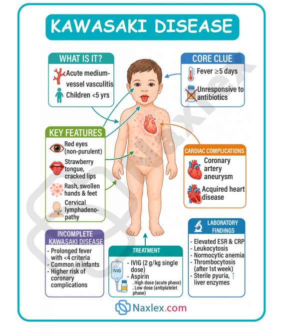
Epidemiology, Etiology, Risk Factors, And Pathophysiology
Epidemiology
- Age predilection: KD is primarily a disease of early childhood. Approximately 80% to 90% of cases occur in children under 5 years of age, with a peak incidence between 6 months and 2 years. It is relatively uncommon in infants under 3 months (due to maternal antibodies) and in adolescents.
- Sex bias: Male children are more commonly affected than females, with a male-to-female ratio of roughly 1.5:1.
- Geographic and ethnic distribution: Although KD occurs globally, children of Asian and Pacific Islander descent (particularly Japanese and Korean) experience the highest incidence rates. In Japan, the incidence is more than ten times higher than in Western countries.
- Seasonality: In temperate climates, cases peak in late winter and early spring, pointing to a possible environmental or infectious trigger.
Etiology and Risk Factors
The precise etiology of Kawasaki disease remains unknown. However, the prevailing scientific consensus points to an immunologically mediated response triggered by an infectious agent (viral or bacterial) in genetically susceptible children.
- Suspected infectious triggers: The seasonal presentation and epidemic-like waves suggest an infectious pathogen (e.g., RNA viruses, superantigens, or atypical bacteria). However, no single pathogen has been consistently isolated.
- Genetic susceptibility: Siblings of children with KD have a tenfold higher risk of developing the disease. Specific gene polymorphisms associated with immune system regulation (such as ITPKC and CASP3) have been linked to increased susceptibility and aneurysm formation.
Pathophysiology
The hallmark of KD is systemic vasculitis, which is the inflammation of blood vessels throughout the entire body. The pathophysiological process unfolds in a structured sequence of vascular damage:
- Acute Phase (Days 1–12):
- Widespread inflammation begins in the adventitia (outer layer) of medium-sized arteries, quickly spreading to the media (middle muscular layer) and intima (inner lining).
- Neutrophils heavily infiltrate the vessel walls, followed rapidly by T-lymphocytes, IgA-producing plasma cells, and macrophages.
- Active inflammation causes edema and structural weakening of the vascular wall.
- Subacute Phase (Days 13–25):
- The inflammatory cells damage the elastin and collagen fibers that give arteries their structural resilience.
- The muscular media layer necroticizes, and the internal elastic lamina splits.
- Because of this structural failure, the high pressure of arterial blood causes the weakened vessel walls to balloon outward, creating coronary artery aneurysms (CAAs).
- Concurrent thrombocytosis (elevated platelet count) peaks during this phase, dramatically increasing blood viscosity and the risk of localized clot formation (thrombosis) within the newly formed aneurysms.
- Myocardial Implications:
- Active myocarditis (inflammation of the heart muscle) is present in almost all patients during the acute phase, which can lead to temporary left ventricular dysfunction, valvular regurgitation (especially mitral), and dysrhythmias.
- Chronic Phase/Healing (Convalescent to Years):
- Over months to years, active inflammation resolves and is replaced by progressive fibrosis (scarring) of the vessel walls.
- This remodeling can lead to stenosis (narrowing) or calcification of the coronary arteries, potentially causing myocardial ischemia, angina, or myocardial infarction later in life.
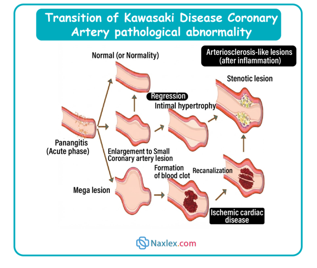
The diagnosis of classic Kawasaki disease is strictly clinical. There is no diagnostic laboratory test. Nurses use the classic "CRASH and BURN" mnemonic to recognize the cardinal signs:
To establish a diagnosis of classic (complete) Kawasaki disease, a child must present with a fever lasting 5 or more days (BURN) and meet at least 4 out of the 5 criteria (CRASH).
Breaking Down the Mnemonic
- C — Conjunctivitis: Bilateral, bulbar conjunctival injection (redness). It is non-purulent (no discharge, crusting, or pus) and typically spares the limbus (the ring around the iris). It is generally painless but photophobia (sensitivity to light) is common.
- R — Rash: Polymorphous exanthem (can take many forms, including maculopapular, scarlatiniform, or erythema multiforme-like). Appears primarily on the trunk and extremities, with a distinct accentuation in the perineal (diaper) area, where early desquamation (peeling) may occur. It is never vesicular (fluid-filled blisters) or bullous.
- A — Adenopathy: Cervical lymphadenopathy. Typically unilateral (one side of the neck), large (at least one node is greater than 1.5 cm in diameter), non-fluctuant, and moderately tender. It is the least common of the five classic criteria.
- S — Strawberry Tongue (and Oral Changes): Eradication of the top layer of cells on the tongue reveals prominent, bright red papillae, resembling a strawberry. Associated erythematous, dry, swollen, and cracked lips (fissuring) that may bleed easily, along with generalized redness of the oropharyngeal mucosa. No tonsillar exudate is present.
- H — Hands and Feet (Extremity Changes): Acute Phase: Erythema (intense redness) of the palms and soles, accompanied by firm, painful induration (swelling/edema). The child may refuse to bear weight. Subacute Phase (Late Sign): Periungual desquamation (peeling of the skin starting under the fingernails and toenails), occurring 2 to 3 weeks after the fever begins.
- BURN — Fever: High, spiking fever (typically greater than 102.2°F / 39°C to 104°F / 40°C) lasting for at least 5 days. It is characteristically remittent and unresponsive to standard doses of acetaminophen, ibuprofen, or antibiotics.
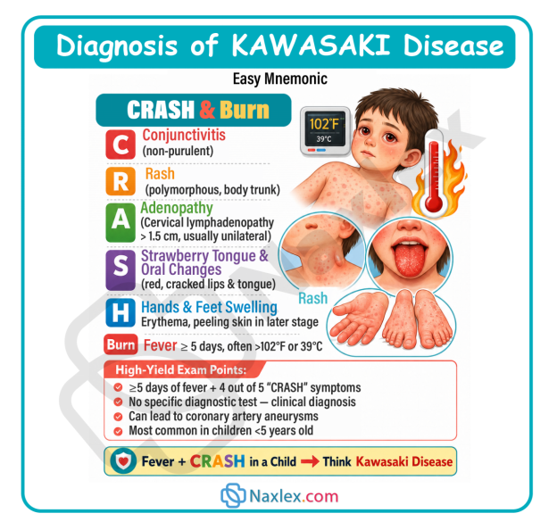
Nursing Insight: Incomplete (Atypical) Kawasaki disease
If a child has a persistent fever for 5 or more days but exhibits fewer than 4 classic CRASH criteria, they may have incomplete Kawasaki disease. This is especially common in infants under 12 months of age. Because young infants are at the highest risk for developing coronary artery aneurysms, any infant under 6 months old with an unexplained fever lasting 7 or more days must be evaluated for incomplete KD, even if they show no other symptoms. Evaluation includes inflammatory markers and an immediate echocardiogram.
Clinical Manifestations
The diagnosis of classic Kawasaki disease is strictly clinical. There is no diagnostic laboratory test. Nurses use the classic "CRASH and BURN" mnemonic to recognize the cardinal signs:
To establish a diagnosis of classic (complete) Kawasaki disease, a child must present with a fever lasting 5 or more days (BURN) and meet at least 4 out of the 5 criteria (CRASH).
Breaking Down the Mnemonic
- C — Conjunctivitis: Bilateral, bulbar conjunctival injection (redness). It is non-purulent (no discharge, crusting, or pus) and typically spares the limbus (the ring around the iris). It is generally painless but photophobia (sensitivity to light) is common.
- R — Rash: Polymorphous exanthem (can take many forms, including maculopapular, scarlatiniform, or erythema multiforme-like). Appears primarily on the trunk and extremities, with a distinct accentuation in the perineal (diaper) area, where early desquamation (peeling) may occur. It is never vesicular (fluid-filled blisters) or bullous.
- A — Adenopathy: Cervical lymphadenopathy. Typically unilateral (one side of the neck), large (at least one node is greater than 1.5 cm in diameter), non-fluctuant, and moderately tender. It is the least common of the five classic criteria.
- S — Strawberry Tongue (and Oral Changes): Eradication of the top layer of cells on the tongue reveals prominent, bright red papillae, resembling a strawberry. Associated erythematous, dry, swollen, and cracked lips (fissuring) that may bleed easily, along with generalized redness of the oropharyngeal mucosa. No tonsillar exudate is present.
- H — Hands and Feet (Extremity Changes): Acute Phase: Erythema (intense redness) of the palms and soles, accompanied by firm, painful induration (swelling/edema). The child may refuse to bear weight. Subacute Phase (Late Sign): Periungual desquamation (peeling of the skin starting under the fingernails and toenails), occurring 2 to 3 weeks after the fever begins.
- BURN — Fever: High, spiking fever (typically greater than 102.2°F / 39°C to 104°F / 40°C) lasting for at least 5 days. It is characteristically remittent and unresponsive to standard doses of acetaminophen, ibuprofen, or antibiotics.
Diagnostic Evaluation
Because there is no definitive diagnostic test for Kawasaki disease, the diagnosis remains clinical. However, laboratory findings and imaging are essential to support suspected incomplete cases, track systemic inflammation, and monitor for cardiac complications.
Laboratory Findings
The systemic vasculitis and intense inflammatory state trigger characteristic abnormalities across multiple organ systems:
|
Lab Test |
Typical Trend in KD |
Clinical Significance |
|
ESR (Erythrocyte Sedimentation Rate) |
Elevated (often greater than 40 mm/hr) |
Reflects systemic acute-phase inflammatory response. |
|
CRP (C-Reactive Protein) |
Elevated (often greater than 3.0 mg/dL) |
Rises and falls more rapidly than ESR; excellent for tracking treatment response. |
|
Complete Blood Count (CBC) |
Leukocytosis (WBC greater than 15,000/µL) with a left shift (neutrophil predominance). |
Confirms active, non-specific acute inflammation. |
|
Platelet Count |
Thrombocytosis (elevated platelets; often 500,000 to over 1,000,000/µL). |
Key subacute marker: Rises dramatically in weeks 2–3. Normal in the first week. |
|
Serum Albumin |
Hypoalbuminemia (less than 3.0 g/dL) |
Low levels correlate with severe, prolonged inflammation and capillary leak. |
|
Liver Transaminases (ALT/AST) |
Mildly elevated |
Indicates mild, transient hepatic inflammation (hepatitis). |
|
Urinalysis (UA) |
Sterile pyuria (greater than 10 WBC/hpf without bacteria) |
Caused by urethritis from localized mucosal inflammation; catheterized samples may be normal. |
Diagnostic Imaging: The Echocardiogram (Echo)
The echocardiogram is the gold standard for evaluating coronary artery involvement and cardiac function in patients with KD.
- Timing of Echo:
- At Diagnosis: Establishes a baseline and evaluates for early complications (myocarditis, pericardial effusion, or valvular dysfunction).
- At 1 to 2 Weeks: Monitors for early coronary dilation or aneurysm formation.
- At 4 to 6 Weeks: Performed during the convalescent phase, when subacute changes and aneurysms are most prominent.
- What the Echo Measures:
- Z-scores: Coronary artery internal diameters are adjusted for the child's body surface area (BSA) and expressed as Z-scores. A Z-score greater than 2 indicates coronary artery dilation; a score of 2.5 to 5 is a small aneurysm, and a score greater than 10 indicates a giant aneurysm.
- Other Parameters: Ventricular contractility, mitral/tricuspid valve regurgitation, and fluid around the heart (pericardial effusion).
Therapeutic Management
The primary goals of therapeutic management in Kawasaki disease are to reduce systemic inflammation and prevent coronary artery abnormalities. Standard therapy involves a dual-drug regimen, which must be initiated within the first 10 days of fever onset to maximize efficacy.
First-Line Pharmacological Management
Intravenous Immunoglobulin (IVIG)
- Dose: 2 g/kg, administered as a single, slow continuous intravenous infusion over 10 to 12 hours.
- Mechanism of Action: Highly concentrated antibodies from pooled human donor plasma reduce systemic vascular inflammation through mechanisms that are still not fully understood.
- Clinical Value: Reduces the risk of developing coronary artery aneurysms from 20–25% down to less than 5%.
- Administration Guidelines:
- Treat as a blood product. Obtain baseline vital signs.
- Start the infusion slowly (typically 0.01 mL/kg/min) and titrate up per facility protocol if tolerated.
- Monitor vital signs continuously (every 15 minutes for the first hour, then hourly) for signs of anaphylaxis or transfusion reaction (fever, chills, hypotension, respiratory distress).
- IVIG Resistance: Roughly 10% to 20% of patients fail to respond to the initial dose of IVIG (defined as persistent or recurrent fever 36 hours after the infusion ends). These patients are typically treated with a second dose of IVIG (2 g/kg) or systemic corticosteroids (e.g., Methylprednisolone).
Aspirin (ASA) Therapy
- High-Dose (Anti-inflammatory Phase):
- Dose: 80 to 100 mg/kg/day (in the US) divided into four equal doses (every 6 hours). In some international guidelines, a moderate dose of 30 to 50 mg/kg/day is used.
- Duration: Maintained until the child has been afebrile for 48 to 72 hours (or until day 14 of the illness).
- Low-Dose (Anti-platelet Phase):
- Dose: Decreased to 3 to 5 mg/kg/day, administered as a single daily dose.
- Duration: Continued for 6 to 8 weeks (until platelet counts normalize and follow-up echocardiograms confirm no coronary artery abnormalities).
- Special Clinical Note: If coronary artery aneurysms are detected, low-dose aspirin is continued indefinitely to prevent thrombosis.
Nursing Insight: Reye Syndrome Risk
In children, aspirin is generally strictly avoided due to its association with Reye syndrome (a rare but fatal condition causing acute encephalopathy and liver infiltration following a viral illness like influenza or varicella). However, because the risk of coronary artery aneurysm and myocardial infarction in KD far outweighs the risk of Reye Syndrome, aspirin is a critical, standard-of-care medication for KD. Educate parents to contact their provider immediately if the child is exposed to or exhibits symptoms of influenza or varicella. If exposure occurs, the aspirin may be temporarily transitioned to an alternative anti-platelet medication (such as Clopidogrel).
Second-Line Management for IVIG-Resistant Kawasaki Disease
High-Dose Corticosteroids
- Rationale: Used to rapidly suppress intense vascular inflammation when standard therapy fails or to prevent resistance in high-risk patients.
- Regimen:
- Intravenous Methylprednisolone Pulse Therapy: 30 mg/kg administered once daily over 2 to 3 hours for 1 to 3 consecutive days.
- Oral Prednisolone Taper: Administered at 2 mg/kg/day in 3 divided doses until C-reactive protein (CRP) normalizes, followed by a gradual taper over 2 to 3 weeks.
Biologic Therapies (TNF-Alpha Inhibitors)
- Infliximab: A chimeric monoclonal antibody targeting tumor necrosis factor-alpha (TNF-alpha).
- Regimen: A single intravenous dose of 5 mg/kg infused over 2 hours.
- Clinical Utility: Often preferred for its rapid action and relative safety profile compared to prolonged steroid regimens, particularly in reducing fever duration and systemic markers of inflammation.
- Etanercept: A soluble TNF receptor blocker, sometimes used as an alternative or adjunct in clinical trials.
Cyclosporine A
- Rationale: An immunosuppressant that inhibits calcineurin, suppressing T-cell activation and the nuclear factor of activated T-cells (NFAT) pathway, which is heavily implicated in the pathogenesis of Kawasaki disease.
- Regimen: Typically initiated orally at 4 to 8 mg/kg/day, adjusted based on clinical response and serum trough levels.
Combined Antithrombotic Therapy: When to Administer Clopidogrel with IVIG and Aspirin
Dual antiplatelet therapy (DAPT) combining aspirin and clopidogrel, occasionally paired with systemic anticoagulation, is strictly reserved for patients who develop severe structural coronary artery abnormalities.
Indications for Combined Therapy
Aspirin is introduced immediately during the acute phase alongside IVIG. Clopidogrel is added to the regimen under the following circumstances:
- Presence of Giant Coronary Artery Aneurysms: Defined as a coronary artery diameter z-score greater than or equal to 10, or an absolute internal diameter greater than or equal to 8 mm. These massive segments experience sluggish, turbulent blood flow and endothelial dysfunction, drastically raising the risk of localized thrombosis and subsequent myocardial infarction.
- Rapidly Expanding Aneurysms: Serial echocardiograms displaying rapid enlargement of coronary segments during the subacute phase.
- History of Coronary Thrombosis or Stenosis: Patients who have documented stenosis or a prior thrombotic event within a coronary artery segment.
Nursing Care Plan
A comprehensive nursing care plan for a pediatric patient with Kawasaki disease focuses on monitoring cardiac function, managing comfort, preventing skin breakdown, and providing essential family education.
Nursing Diagnoses, Interventions, and Rationales
Decreased Cardiac Output (Risk for)
- Related to: Coronary artery inflammation, myocardial ischemia, or impaired left ventricular contractility.
- Nursing Interventions:
- Continuous Cardiorespiratory Monitoring: Assess heart rate, rhythm, and quality of peripheral pulses. Report tachycardia or irregular rhythms.
- Frequent Vital Sign Assessment: Monitor blood pressure, respiratory rate, and oxygen saturation. Compare upper and lower extremity blood pressures if coarctation or severe vasculitis is suspected.
- Strict Intake and Output (I&O) Monitoring: Weigh all diapers (1 mL = 1 g). Report urine output less than 1 mL/kg/hour in infants or less than 0.5 mL/kg/hour in children.
- Assess for Fluid Overload: Monitor for tachypnea, crackles in the lungs, periorbital edema, or hepatomegaly, especially during the rapid administration of IVIG (which has high osmotic pull and can precipitate heart failure).
- Rationales:
- Tachycardia can be an early sign of heart failure or myocardial ischemia.
- Hypotension and tachypnea are systemic signs of failing perfusion.
- Inadequate urine output indicates poor renal perfusion, a primary consequence of decreased cardiac output.
- The large volume and high viscosity of IVIG put patients at high risk for acute fluid overload and congestive heart failure.
Impaired Skin and Mucous Membrane Integrity
- Related to: Intense systemic vasculitis, inflammatory desquamation, and mucosal dryness.
- Nursing Interventions:
- Oral Care: Offer small sips of cool water or ice chips. Apply a thin layer of petroleum-based ointment or lip balm to the dry, cracked lips every 2 hours. Avoid hard or dry foods.
- Skin Care: Keep skin clean and dry. Avoid using harsh soaps. Apply cool, moist compresses to areas of severe rash or peeling.
- Manage Desquamation: Instruct parents never to peel or pull off loose skin on the hands or feet, as this can tear the healthy tissue underneath and lead to infection. Let the skin shed naturally.
- Rationales:
- Gentle hydration prevents bleeding and infection in fragile oral tissues.
- Systemic irritation can be relieved by non-pharmacological, cooling interventions.
- Forced peeling exposes raw dermal layers, creating entry points for opportunistic pathogens.
Acute Pain and Hyperthermia
- Related to: Systemic inflammatory response, joint inflammation (arthralgia), and persistent high fever.
- Nursing Interventions:
- Fever Monitoring and Management: Document temperatures every 2 to 4 hours. Administer prescribed high-dose aspirin. Note that standard antipyretics are often ineffective.
- Environmental Comfort: Keep the child's room cool, quiet, and dimly lit to accommodate photophobia. Encourage loose, lightweight cotton clothing.
- Promote Rest: Group nursing activities to minimize disturbances and allow for long periods of uninterrupted sleep.
- Positioning for Joint Pain: Support inflamed joints with pillows. Keep joints in a neutral, slightly flexed position. Avoid passive range-of-motion exercises during the acute phase.
- Rationales:
- High fevers increase metabolic and myocardial oxygen demands.
- Systemic vasculitis makes the sensory nervous system highly sensitive; environmental triggers can escalate the child's irritability.
- Rest helps lower myocardial oxygen demands.
- Arthralgia and arthritis are common in KD; proper positioning reduces tension on inflamed synovial joints.
Family and Discharge Education (Including CPR)
Comprehensive discharge planning is critical to ensure safety at home and prevent long-term cardiac complications.
- Medication Compliance:
- Ensure parents understand the exact schedule for low-dose aspirin therapy (usually continued for 6–8 weeks, or indefinitely if aneurysms are present).
- Emphasize the importance of never skipping doses or stopping aspirin abruptly unless instructed by the cardiologist.
- Reye Syndrome Awareness:
- Teach parents the signs of Reye syndrome (persistent vomiting, lethargy, confusion, rapid breathing).
- Advise them to contact their pediatrician immediately if the child is exposed to or develops symptoms of influenza or chickenpox.
- Cardiopulmonary Resuscitation (CPR) Training:
- The Mandate: Because children with coronary artery aneurysms are at risk for sudden myocardial infarction and cardiac arrest, all primary caregivers must receive hands-on, certified infant/child CPR training before discharge.
- Why CPR is Crucial: If a coronary thrombosis occurs, immediate, high-quality bystander CPR can be life-saving before emergency services arrive.
- Irritability Management:
- Reassure parents that extreme, unexplained irritability is a normal part of the disease process (often lasting up to several weeks) and will resolve as systemic inflammation subsides.
- Postponement of Live Vaccines:
- Crucial Instruction: Children who receive IVIG must postpone all live virus vaccines (specifically MMR and Varicella) for at least 11 months after the infusion.
- Rationale: The high concentration of passive antibodies in IVIG can neutralize live vaccines, preventing the child's immune system from building an adequate active response.
- Activity Restrictions:
- Children with known coronary artery aneurysms must avoid contact sports, high-impact activities, or strenuous exercise that increases intra-thoracic pressure, which could cause a weakened coronary vessel to rupture.
- When to Seek Immediate Medical Attention:
- Return of fever (may indicate a relapse).
- Signs of myocardial infarction in a child: sudden chest pain, extreme pale or bluish skin (cyanosis), difficulty breathing, unexplained vomiting, or cold sweats.
Complications
The complications of Kawasaki Disease are primarily cardiovascular and can have life-threatening implications.
- Coronary Artery Aneurysms (CAAs): Occur in roughly 20–25% of untreated children and less than 5% of those treated promptly with IVIG. The structural weakening of the arterial wall leads to dilation. Giant aneurysms (internal diameter greater than 8 mm) have the lowest rate of spontaneous regression and are at high risk for rupture or thrombosis.
- Coronary Artery Thrombosis: The combination of slow, turbulent blood flow through dilated aneurysms and marked thrombocytosis in the subacute phase creates a high-risk environment for clot formation.
- Myocardial Infarction (MI): Caused by an occlusion of a coronary artery by a thrombus within an aneurysm. Symptoms in young children: Often silent or non-specific, presenting as sudden crying, screaming, pallor, vomiting, shock, or severe abdominal pain.
- Myocarditis and Heart Failure: Acute myocardial inflammation can impair contractility, leading to congestive heart failure.
- Valvular Regurgitation: Inflammation of the valvular endocardium can cause mitral or aortic valve incompetence.
- Aneurysms of Other Medium-Sized Arteries: Can occur in the subclavian, brachial, axillary, iliac, or femoral arteries, though much less commonly than in the coronary arteries.
Prognosis
The overall prognosis for children diagnosed with Kawasaki Disease is highly favorable, provided they receive early, aggressive medical therapy.
- Overall Mortality: The mortality rate is less than 0.1% in developed nations when treated with IVIG. Death is almost always a result of coronary thrombosis, myocardial infarction, or ruptured aneurysms.
- Aneurysm Resolution:
- Approximately 50% to 60% of small-to-medium coronary aneurysms regress spontaneously within 1 to 2 years of the acute illness.
- Giant aneurysms (internal diameter greater than 8 mm) rarely regress and require lifelong anticoagulation (often aspirin combined with Warfarin or Low-Molecular-Weight Heparin) and regular cardiological evaluations.
- Long-Term Cardiovascular Risk:
- Even children whose coronary arteries remain normal on echocardiograms may have subtle endothelial damage that could predispose them to accelerated atherosclerosis or premature cardiovascular disease as adults.
- Healthy lifestyle habits (heart-healthy diet, regular aerobic exercise, and avoiding smoking) are strongly recommended throughout life.
Summary
- Kawasaki disease (KD) remains a critical, "can't-miss" clinical diagnosis in pediatric nursing, representing the leading cause of acquired pediatric heart disease in developed nations.
- This acute, self-limiting medium-vessel vasculitis predominantly affects infants and young children under 5 years of age.
- While its exact etiology remains elusive, it is understood to be a hyperactive, immunologically driven response triggered by an infectious pathogen in genetically predisposed children. The disease systematically compromises the structural integrity of vessel walls, presenting a grave risk of coronary artery aneurysm (CAA) development, thrombosis, and subsequent myocardial infarction.
- Because there is no definitive laboratory test, establishing a diagnosis of complete KD requires a thorough understanding of clinical criteria. Nurses rely on the "CRASH and BURN" mnemonic to recognize the key signs: Conjunctivitis (non-purulent, bilateral), Rash (polymorphous, truncal), Adenopathy (unilateral cervical node greater than 1.5 cm), Strawberry tongue (and dry, cracked red lips), Hand and foot changes (edema, erythema, late periungual peeling), BURNing, high, unrelenting fever lasting 5 or more days.
- A high index of suspicion must also be maintained for incomplete (atypical) cases, particularly in infants under 12 months of age, who may present with prolonged fever but fewer than 4 classic criteria.
- Management centers on halting inflammation rapidly within the first 10 days of fever onset. This is achieved through a combination of high-dose intravenous immunoglobulin (IVIG) and anti-inflammatory doses of aspirin, which is later transitioned to a low anti-platelet dose.
- Nursing care requires rigorous cardiac monitoring, assessing for signs of fluid overload during IVIG administration, comfort measures for extreme irritability, and detailed family discharge education.
- Empowering parents with hands-on Infant/Child CPR training, highlighting the need to delay live vaccines for 11 months, and teaching them to recognize the signs of Reye Syndrome and myocardial infarction are essential steps to ensure a safe transition home and promote long-term recovery.
Naxlex
Videos
Login to View Video
Click here to loginTake Notes on Acquired Cardiovascular Disorders: Kawasaki Disease
This filled cannot be empty
Join Naxlex Nursing for nursing questions & guides! Sign Up Now