





 Video Library


Please set your exam date
Acquired Cardiovascular Disorders: Pediatric Cardiomyopathies
Study Questions
Practice Exercise 1
A 6-month-old client with dilated cardiomyopathy secondary to acute viral myocarditis is admitted to the pediatric unit. Which pathophysiological mechanism should the nurse recognize as the primary cause of impaired forward cardiac output in this client?
Explanation
Dilated cardiomyopathy (DCM) secondary to acute viral myocarditis is characterized by viral-mediated myocyte destruction, cellular necrosis, and subsequent inflammatory fibrosis. The loss of functional, contracting myocytes weakens myocardial pump function, causing severe systolic dysfunction. To compensate for reduced ejection fraction and impaired forward cardiac output, the heart undergoes progressive ventricular dilation, increased end-diastolic volume, and pathological ventricular remodeling.
Rationale for correct answer:
2. Reduced systolic contractile force is the defining pathophysiological feature of dilated cardiomyopathy. Inflammatory destruction of myocytes weakens ventricular contraction (systolic dysfunction), leaving a large volume of blood in the left ventricle at the end of systole. To accommodate this excess residual blood and maintain stroke volume via the Frank-Starling mechanism, the ventricle progressively dilates, increasing end-diastolic volume and stretch on the ventricular walls. Over time, this compensatory dilation becomes maladaptive, worsening heart failure and diminishing forward cardiac output.
Rationale for incorrect answers:
1. Severe diastolic filling restriction secondary to rigid myocardial tissue describes restrictive cardiomyopathy (RCM). In RCM, the myocardium becomes stiff and non-compliant, impairing ventricular relaxation and diastolic filling while systolic contractile function often remains relatively preserved initially. This is not the primary mechanism of dilated cardiomyopathy.
3. Dynamic subaortic obstruction caused by asymmetric septal hypertrophy is the cardinal hallmark of hypertrophic cardiomyopathy (HCM). HCM involves disarrayed, hypertrophied cardiac myocytes, most prominently in the interventricular septum, which impedes left ventricular outflow during systole.
4. Replacement of right ventricular myocytes with fibrofatty adipose tissue describes arrhythmogenic right ventricular cardiomyopathy (ARVC). ARVC is a genetic disease of cell adhesion proteins (desmosomes) that leads to ventricular arrhythmias and right-sided heart failure, rather than post-viral dilated cardiomyopathy.
Test-taking strategy:
- Analyze the scenario/question: Identify the primary pathophysiological mechanism causing impaired forward cardiac output in a 6-month-old with dilated cardiomyopathy secondary to viral myocarditis.
- Correlate pathophysiology:
- Dilated = Pump failure / Systolic dysfunction resulting in dilated, stretched chamber with high end-diastolic volume.
- Evaluate option statements:
- Rule out Choice 1: Diastolic filling restriction in a rigid heart describes restrictive cardiomyopathy.
- Choice 2 accurately identifies reduced systolic contractile force leading to increased end-diastolic volume as the core driver of dilated cardiomyopathy.
- Rule out Choice 3: Asymmetric septal hypertrophy causing subaortic obstruction describes hypertrophic cardiomyopathy.
- Rule out Choice 4: Fibrofatty replacement of RV myocytes describes arrhythmogenic right ventricular cardiomyopathy.
Take home points
- Dilated cardiomyopathy (DCM) is primarily a disorder of systolic contractile failure.
- Post-viral myocarditis leads to myocyte damage, ventricular dilation, increased end-diastolic volume, and reduced ejection fraction.
- Compensatory ventricular stretching eventually fails, leading to clinical signs of infant heart failure (e.g., tachypnea, feeding intolerance, hepatomegaly).
- Distinguish the four main cardiomyopathies: Dilated (systolic pump failure), Hypertrophic (thick septum/outflow obstruction), Restrictive (rigid/diastolic filling failure), and ARVC (fibrofatty RV replacement).
The nurse cares for a 6-month-old client presenting with tachypnea, poor feeding, diaphoresis during feeding, and hepatomegaly. The child's history reveals recent exposure to a severe viral respiratory infection. Which condition should the nurse suspect?
Explanation
Acute viral myocarditis triggers severe myocardial inflammatory infiltration, interstitial edema, and direct virally mediated structural injury to functional myocytes. Widespread loss of viable contracting cardiac muscle severely impairs systolic pump function, resulting in progressive ventricular dilation, elevated end-diastolic filling pressures, and diminished forward stroke volume. This sequence drives pediatric heart failure, systemic venous congestion, severe pulmonary edema, and marked cardiomegaly.
Rationale for correct answer:
2. Acute viral myocarditis progresssing to dilated cardiomyopathy represents the primary clinical diagnosis matching this presentation. Enteroviruses (e.g., Coxsackie B), adenoviruses, and respiratory syncytial virus can trigger post-infectious inflammatory cellular degradation of the myocardium. The infant manifests cardinal signs of congestive heart failure: tachypnea and diaphoresis during feedings reflect increased sympathetic compensation and pulmonary venous congestion, poor feeding reflects low cardiac output fatigue, and hepatomegaly signifies systemic venous backpressure from right ventricular dysfunction.
Rationale for incorrect answers:
1. Restrictive cardiomyopathy is an exceptionally rare pediatric cardiomyopathy characterized primarily by severe diastolic filling impairment due to rigid, non-compliant ventricular walls, while systolic pump function remains relatively preserved until late stages. Metabolic storage diseases (such as glycogen storage disease or lysosomal disorders) typically present with progressive developmental delay, skeletal muscle hypotonia, organomegaly, and distinctive dysmorphic features rather than an acute post-viral onset of acute congestive heart failure.
3. Hypertrophic obstructive cardiomyopathy secondary to maternal diabetes presents in the early neonatal period (typically within the first few days to weeks of life) due to fetal hyperinsulinemia stimulating cardiac myocyte hypertrophy, particularly of the interventricular septum. While it causes dynamic left ventricular outflow tract obstruction, it is a congenital metabolic phenomenon observed immediately post-partum rather than an acquired heart failure presentation following a viral respiratory illness in a 6-month-old infant.
4. Left ventricular non-compaction is a genetic congenital cardiomyopathy characterized by deep intertrabecular recesses and a prominent two-layered myocardium resulting from arrested embryological compaction. While affected infants can develop heart failure, the clinical presentation is structural and congenital rather than triggered by a preceding acute viral illness. Furthermore, clients presenting with classic symptomatic infant heart failure demonstrate significantly impaired, rather than preserved, ventricular systolic function.
Test-taking strategy:
- Analyze the scenario/question: A 6-month-old presents with classic infant congestive heart failure signs (tachypnea, poor feeding, diaphoresis during feeding, hepatomegaly) following a recent viral respiratory infection.
- Correlate clinical context:
- Preceding viral respiratory illness + signs of acute heart failure = Acute viral myocarditis / Dilated cardiomyopathy.
- Tachypnea and diaphoresis during feeding reflect high metabolic demand and pulmonary congestion.
- Hepatomegaly indicates right-sided systemic venous backpressure.
- Evaluate option distractors:
- Rule out Choice 1: Metabolic restrictive cardiomyopathy presents with metabolic systemic signs and diastolic rigidity rather than an acute post-viral onset.
- Choice 2 accurately links the acute viral antecedent to myocarditis and secondary dilated cardiomyopathy.
- Rule out Choice 3: Hypertrophic cardiomyopathy from maternal diabetes presents in early neonates, not at 6 months following a viral illness.
- Rule out Choice 4: Non-compaction is a congenital morphological defect, not an acute post-viral condition, and heart failure presents with impaired function.
Take home points
- Acute viral myocarditis is a leading cause of acquired dilated cardiomyopathy and acute heart failure in infants and children.
- Infant heart failure typically manifests as poor feeding, diaphoresis with exertion (feeding), tachypnea, and hepatomegaly.
- Diaphoresis during feeding occurs because feeding represents the infant's primary physical exertion, triggering maximal sympathetic nervous system activation.
- Common viral etiologies include Coxsackie B virus, adenovirus, enteroviruses, and parvovirus B19.
A 10-year-old client with restrictive cardiomyopathy is admitted to the pediatric unit. The nurse understands that the primary pathophysiological defect in this client is which mechanism?
Explanation
Restrictive cardiomyopathy (RCM) is the rarest form of pediatric cardiomyopathy, characterized by abnormal stiffness and non-compliance of the ventricular myocardium. While ventricular size and systolic contractility often remain normal or near-normal initially, the non-compliant walls impede normal ventricular relaxation during diastole. This leads to marked diastolic dysfunction, severely elevated end-diastolic pressures, significant massive bi-atrial enlargement, and secondary pulmonary hypertension.
Rationale for correct answer:
3. Stiff, non-compliant ventricular walls causing severe diastolic filling dysfunction represent the defining pathophysiological hallmark of restrictive cardiomyopathy. Fibrosis, endomyocardial scarring, or infiltrative processes make the ventricular tissue rigid, restricting blood flow into the heart during diastole. Because the ventricles cannot relax or expand adequately to accommodate venous return, filling pressures spike rapidly, forcing blood back into the atria and pulmonary circulation.
Rationale for incorrect answers:
1. Severe impairment of myocardial systolic contractility with chamber dilation is the defining characteristic of dilated cardiomyopathy (DCM). In DCM, weakened myocytes fail to generate adequate stroke volume, leading to compensatory chamber enlargement and systolic pump failure, whereas early RCM exhibits preserved systolic squeezing with impaired diastolic filling.
2. Massive asymmetrical septal hypertrophy causing dynamic subaortic obstruction describes hypertrophic obstructive cardiomyopathy (HCM). In HCM, disarrayed myocytes and asymmetric thickening of the interventricular septum create an outflow tract obstruction during systole, which is a distinctly different structural anomaly than RCM.
4. Replacement of right ventricular myocardium with fibrofatty tissue is the hallmark of arrhythmogenic right ventricular cardiomyopathy (ARVC). ARVC is a genetic desmosomal disorder leading to localized right ventricular akinesis, aneurysms, and ventricular arrhythmias, rather than diffuse rigid ventricular diastolic dysfunction.
Test-taking strategy:
- Analyze the scenario/question: Identify the primary pathophysiological defect in a client with restrictive cardiomyopathy.
- Correlate pathophysiology:
- Restrictive = Restricted filling resulting in stiff, rigid myocardium causing diastolic dysfunction.
- Evaluate option distractors:
- Rule out Choice 1: Impaired contractility and chamber dilation describe dilated cardiomyopathy.
- Rule out Choice 2: Asymmetrical septal hypertrophy describes hypertrophic cardiomyopathy.
- Choice 3 accurately identifies rigid walls and severe diastolic filling failure as restrictive cardiomyopathy.
- Rule out Choice 4: Fibrofatty replacement describes arrhythmogenic right ventricular cardiomyopathy.
Take home points
- Restrictive cardiomyopathy (RCM) is primarily a disorder of diastolic dysfunction caused by stiff, non-compliant myocardium.
- Systolic function and chamber dimensions are often preserved until late stages, but diastolic filling pressures are markedly elevated.
- Elevated diastolic filling pressures lead to massive bi-atrial enlargement, systemic and pulmonary venous congestion, and pulmonary hypertension.
- RCM carries a guarded prognosis in pediatric clients, often requiring heart transplantation.
A 15-year-old client with a family history of sudden cardiac death is evaluated for hypertrophic cardiomyopathy (HCM). The nurse understands that the pathophysiological hallmark of this disorder involves which structural abnormality?
Explanation
Hypertrophic cardiomyopathy (HCM) is an autosomal dominant genetic disorder primarily affecting sarcomeric protein genes. The pathophysiological hallmark of HCM is asymmetric hypertrophy of the interventricular septum, paired with disarray of cardiac myocytes and myocardial fibrosis. This abnormal thickening reduces left ventricular chamber size, impairs ventricular relaxation (diastolic dysfunction), and can cause dynamic left ventricular outflow tract (LVOT) obstruction, placing adolescents at high risk for exertional syncope, lethal ventricular arrhythmias, and sudden cardiac death.
Rationale for correct answer:
2. Asymmetric hypertrophy of the interventricular septum paired with reduced diastolic compliance is the structural hallmark of hypertrophic cardiomyopathy. The localized thickening of the septum disproportionately narrows the left ventricular outflow tract during systole. In addition, the thickened, disorganized cardiac muscle fails to relax normally during diastole, markedly increasing filling pressures and limiting stroke volume during exertion.
Rationale for incorrect answers:
1. Diffuse dilation of all four cardiac chambers with reduced wall thickness describes dilated cardiomyopathy (DCM). DCM is characterized by thin, stretched ventricular walls and impaired systolic contractile force, resulting in a low ejection fraction rather than severe asymmetrical septal hypertrophy.
3. Marked bi-atrial enlargement paired with normal ventricular wall thickness is the characteristic structural finding of restrictive cardiomyopathy (RCM). In RCM, rigid ventricular walls severely restrict diastolic filling, forcing the atria to dilate significantly under elevated backpressure while ventricular wall thickness remains relatively normal.
4. Excessive non-compacted endocardial trabeculations with deep intertrabecular recesses describes left ventricular non-compaction (LVNC). LVNC is a distinct congenital cardiomyopathy resulting from arrested intrauterine endomyocardial morphogenesis, creating a porous, spongy appearance of the left ventricular myocardium.
Test-taking strategy:
- Analyze the scenario/question: Identify the structural hallmark of hypertrophic cardiomyopathy (HCM) in a 15-year-old with a family history of sudden cardiac death.
- Correlate Pathophysiology:
- Hypertrophic = Thickened muscle results in asymmetric septal hypertrophy leading to LVOT obstruction and diastolic dysfunction.
- Evaluate option distractors:
- Rule out Choice 1: Chamber dilation with thinned walls describes dilated cardiomyopathy.
- Choice 2 accurately identifies asymmetric septal hypertrophy and reduced diastolic compliance as HCM.
- Rule out Choice 3: Bi-atrial enlargement with normal wall thickness describes restrictive cardiomyopathy.
- Rule out Choice 4: Non-compacted trabeculations and deep recesses describe left ventricular non-compaction.
Take home points
- Hypertrophic cardiomyopathy (HCM) is characterized by asymmetric interventricular septal hypertrophy and myocyte disarray.
- HCM is a leading cause of sudden cardiac death in adolescent athletes due to fatal ventricular arrhythmias (such as ventricular tachycardia/fibrillation).
- The thickened septum can cause dynamic left ventricular outflow tract (LVOT) obstruction, exacerbated by exertion, dehydration, or increased inotropy.
- Key clinical manifestations include exertional chest pain, dyspnea, fatigue, and exertional syncope.
The nurse reviews the prenatal and medical history of a neonate presenting with signs of dynamic left ventricular outflow tract obstruction and ventricular septal hypertrophy. Which maternal risk factor in the history is directly associated with this condition?
Explanation
Infant of a diabetic mother often presents with transient hypertrophic cardiomyopathy secondary to fetal hyperinsulinemia. Fetal hyperinsulinemia triggers excess glycogen and fat deposition within the myocardium, leading to asymmetrical septal hypertrophy and dynamic left ventricular outflow tract obstruction. Clinical manifestations include tachypnea, respiratory distress, and a systolic ejection murmur, with fetal serum glucose levels exceeding 126 mg/dL promoting fetal metabolic alterations.
Rationale for correct answer:
1. Poorly controlled maternal gestational diabetes induces fetal hyperglycemia followed by severe fetal hyperinsulinemia. Insulin acts as an anabolic hormone stimulating protein synthesis and lipid deposition within the fetal ventricular septum. This structural enlargement restricts cardiac output during ventricular contraction. Symptoms typically resolve within 2 to 4 months as maternal hormone influences wane.
Rationale for incorrect answers:
2. Maternal folate deficiency during early gestation is linked to neural tube defects. Deficient folic acid impairs embryonic DNA synthesis and neurulation during 3 to 4 weeks post-conception. It does not cause hypertrophic cardiomyopathy or septal wall thickening. Supplementation of 400 mcg daily prevents cranioschisis and spina bifida.
3. Intrauterine varicella zoster virus infection leads to congenital varicella syndrome. Manifestations include cutaneous cicatricial scarring, limb hypoplasia, chorioretinitis, and microcephaly rather than structural cardiac hypertrophy. Exposure during the late third trimester poses risks for neonatal varicella pneumonia. It does not alter fetal myocardial glycogen stores.
4. Inadequate thyroid hormone during pregnancy typically correlates with congenital hypothyroidism. Levothyroxine therapy restores maternal euthyroid status, preventing fetal neurodevelopmental impairment and low birth weight. Maternal hypothyroidism does not cause asymmetric ventricular hypertrophy or muscular outflow obstruction. Unmanaged hypothyroidism primary increases preeclampsia risk.
Test-taking strategy:
- Analyze the scenario/question: The neonate presents with dynamic left ventricular outflow tract obstruction caused by ventricular septal hypertrophy. The nurse must identify the specific maternal antecedents responsible for this pathophysiological feature.
- Evaluate pathological mechanisms:
- Hyperglycemia-induced hyperinsulinemia stimulates tissue growth and selective myocardial glycogen accumulation.
- Link maternal metabolic dysregulation directly to hypertrophic cardiomyopathy in the offspring.
- Methodology to rule choices in or out:
- Choice 1 is correct because fetal hyperinsulinemia selectively drives asymmetric septal hypertrophy and subaortic stenosis.
- Rule out Choice 2: Folate deficiency impairs closure of the neural tube rather than cardiac septal development.
- Rule out Choice 3: Viral varicella infection causes limb hypoplasia and neurological damage, not cardiac septal hypertrophy.
- Rule out Choice 4: Thyroid dysregulation affects metabolic rate and cognitive development without inducing myocardial hypertrophy.
Take home points
- Maternal diabetes mellitus causes fetal hyperglycemia, leading to secondary hyperinsulinemia and asymmetrical ventricular septal hypertrophy.
- Neonatal hypertrophic cardiomyopathy often causes dynamic left ventricular outflow tract obstruction that presents with respiratory distress and heart murmur.
- Folate deficiency is primarily associated with neural tube defects such as spina bifida and anencephaly.
- Infant hypertrophic cardiomyopathy from maternal diabetes is typically transient and resolves spontaneously over several months as insulin levels normalize.
Practice Excercise 2
The nurse assesses a 15-year-old client with hypertrophic cardiomyopathy. While listening to the client's precordium, the nurse notes a harsh systolic murmur along the left sternal border. Which action by the client during examination will increase the intensity of this murmur?
Explanation
Hypertrophic cardiomyopathy features asymmetric septal hypertrophy causing dynamic left ventricular outflow tract obstruction. Reductions in left ventricular volume reduce the distance between the hypertrophied septum and anterior mitral valve leaflet, worsening obstruction. Interventions that lower preload or afterload intensify the resulting harsh systolic ejection murmur, whereas increased ventricular volume widens the outflow tract and softens murmur intensity.
Rationale for correct answer:
3. Performing a Valsalva maneuver or standing rapidly decreases venous return and reduces left ventricular preload. A smaller left ventricular cavity allows the hypertrophied interventricular septum to appose the anterior mitral valve leaflet more closely. This exacerbates subaortic outflow tract obstruction and significantly increases murmur intensity. The maneuver dynamically isolates hypertrophic cardiomyopathy from fixed structural stenosis.
Rationale for incorrect answers:
1. Moving from standing to deep squatting increases systemic vascular resistance and venous return. Higher afterload and preload expand left ventricular end-diastolic volume. The increased intraventricular volume separates the septum from the mitral valve, widening the outflow tract. Consequently, squatting decreases the intensity of the hypertrophic cardiomyopathy murmur.
2. Transitioning from a standing position to a supine position increases central blood volume. Enhanced venous return augments left ventricular preload and expands internal cavity dimensions. The increased volume pushes the hypertrophied septum away from the anterior mitral valve leaflet during systole. This physiological change reduces outflow obstruction and softens the murmur.
4. Passive elevation of the lower extremities mobilizes pooled venous blood toward the heart. This maneuver increases right and left ventricular preload, expanding end-diastolic volume. The increased ventricular volume physically widens the left ventricular outflow tract, diminishing dynamic subaortic obstruction. As a result, the intensity of the murmur decreases during leg elevation.
Test-taking strategy:
- Analyze the scenario/question: The client is a 15-year-old with hypertrophic cardiomyopathy presenting with a harsh systolic murmur. The nurse must identify which physical maneuver increases murmur intensity.
- Evaluate pathological mechanisms:
- Recall that hypertrophic cardiomyopathy murmur intensity varies inversely with left ventricular volume.
- Identify maneuvers that reduce preload or lower afterload as key triggers for increased obstruction.
- Methodology to rule choices in or out:
- Choice 3 is correct because Valsalva and standing decrease preload, decreasing cavity size and accentuating the murmur.
- Rule out Choice 1: Squatting increases preload and afterload, expanding cavity size and softening the murmur.
- Rule out Choice 2: The supine position increases venous return, which fills the ventricle and decreases the murmur.
- Rule out Choice 4: Leg elevation increases preload, widening the outflow tract and softening the murmur.
Take home points
- Hypertrophic cardiomyopathy produces a dynamic systolic ejection murmur that changes intensity based on left ventricular volume.
- Maneuvers that decrease preload or afterload, such as the Valsalva maneuver or rapid standing, decrease cavity size and increase murmur intensity.
- Maneuvers that increase preload or afterload, such as squatting, leg elevation, or handgrip, increase cavity size and decrease murmur intensity.
- Differentiating dynamic outflow obstruction from fixed aortic stenosis relies on physical bedside maneuvers that alter ventricular filling.
A 7-year-old client with dilated cardiomyopathy is evaluated in the pediatric cardiology clinic. Which echocardiographic finding should the nurse expect to observe in the medical record?
Explanation
Pediatric dilated cardiomyopathy is characterized by ventricular enlargement and impaired systolic contractility. The pathophysiological hallmark involves progressive cardiac chamber stretch leading to systolic dysfunction and reduced ejection fraction. Clinical manifestations typically include dyspnea, fatigue, cardiomegaly, and gallop rhythms, with pediatric normal fractional shortening values ranging between 28% and 44%.
Rationale for correct answer:
2. Dilated cardiomyopathy produces marked enlargement of the left ventricle and significant systolic dysfunction. Fractional shortening measures the change in ventricular lumen diameter during systole and drops well below normal in dilated hearts. This echocardiographic parameter reflects severely diminished myocardial contractility and stroke volume. Echocardiography confirms chamber dilation with thin, stretched ventricular walls.
Rationale for incorrect answers:
1. Asymmetric septal hypertrophy without dilation defines hypertrophic cardiomyopathy. This hereditary condition features disarrayed myocytes and narrowed subaortic outflow tracts without initial chamber enlargement. Left ventricular cavity size is characteristically normal or reduced. It represents a diastolic filling disorder rather than primary dilated systolic failure.
3. Small ventricular chambers accompanied by massive bi-atrial enlargement characterize restrictive cardiomyopathy. This rare pediatric condition involves severe endomyocardial stiffness and impaired ventricular filling during diastole. Systolic contractility and ventricular dimensions initially remain within normal limits. The elevated filling pressures drive secondary bi-atrial dilatation.
4. Non-compaction cardiomyopathy displays prominent trabeculations rather than an absent trabecular network. Affected hearts demonstrate deep intertrabecular recesses and a thick endocardial non-compacted layer. Epicardial hyper-compaction with absent trabeculations is not a feature of pediatric dilated cardiomyopathy. Echocardiography instead demonstrates endocardial spongy morphology when non-compaction occurs.
Test-taking strategy:
- Analyze the scenario/question: The client is a 7-year-old evaluated for dilated cardiomyopathy. The nurse must identify the pathognomonic echocardiographic feature consistent with this diagnosis.
- Evaluate pathological mechanisms:
- Define dilated cardiomyopathy as a primary disorder of systolic contractility with muscular stretching.
- Correlate chamber dilation directly with diminished fractional shortening values.
- Methodology to rule choices in or out:
- Choice 2 is correct because left ventricular dilation and reduced fractional shortening define dilated cardiomyopathy.
- Rule out Choice 1: Asymmetric septal hypertrophy is the hallmark of hypertrophic cardiomyopathy.
- Rule out Choice 3: Bi-atrial enlargement with normal or small ventricles points to restrictive cardiomyopathy.
- Rule out Choice 4: Non-compaction cardiomyopathy presents with prominent, deep trabeculations rather than absent trabeculations.
Take home points
- Dilated cardiomyopathy is defined by ventricular chamber enlargement and impaired systolic contractile function.
- Fractional shortening and ejection fraction are markedly decreased on the echocardiogram of a client with dilated cardiomyopathy.
- Hypertrophic cardiomyopathy is characterized by asymmetric septal thickening without primary chamber dilation.
- Restrictive cardiomyopathy features stiff ventricular walls, impaired diastolic filling, and secondary bi-atrial enlargement.
A 5-year-old client with end-stage restrictive cardiomyopathy exhibits signs of severe systemic venous congestion. Which physical assessment finding should the nurse document as supporting this diagnosis?
Explanation
Restrictive cardiomyopathy involves non-dilated, rigid ventricular walls that severely impair diastolic filling. Although systolic function remains preserved initially, the non-compliant ventricles cause rapid elevation of diastolic pressures, leading to profound systemic venous congestion. Clinical manifestations include jugular venous distension, hepatomegaly, ascites, and peripheral edema, with elevated central venous pressures often exceeding 15 mmHg.
Rationale for correct answer:
1. Severe systemic venous congestion manifests as jugular venous distension and pronounced hepatomegaly. Rigid ventricular myocardium prevents adequate diastolic filling, causing venous backpressure into the right atrium, vena cava, and systemic venous system. This backpressure leads to fluid accumulation in hepatic sinusoids and systemic vasculature. Physical examination confirms elevated central venous pressure and tender enlarged liver margins.
Rationale for incorrect answers:
2. Bounding peripheral pulses reflect a widened pulse pressure commonly observed in patent ductus arteriosus. Restrictive cardiomyopathy causes reduced stroke volume and diminished cardiac output due to restricted ventricular filling. Diminished ventricular filling results in weak, thready peripheral pulses rather than hyperdynamic bounding pulses. Pulse volume remains low because diastolic filling is severely compromised.
3. A continuous machinery murmur is the classic auscultatory finding for patent ductus arteriosus. Restrictive cardiomyopathy exhibits normal valve structures without continuous extracardiac left-to-right vascular shunting. Auscultation in restrictive cardiomyopathy often reveals gallop rhythms such as S3 or S4 due to sudden cessation of ventricular filling. It does not produce a continuous machinery murmur.
4. A subaortic pressure gradient exceeding 50 mmHg indicates severe hypertrophic outflow tract obstruction. Restrictive cardiomyopathy features non-hypertrophied, non-dilated ventricles without dynamic subaortic muscular narrowing. The primary hemodynamic pathology is elevated ventricular end-diastolic pressure rather than intraventricular peak systolic pressure gradients. Left ventricular outflow obstruction is characteristically absent.
Test-taking strategy:
- Analyze the scenario/question: The client is a 5-year-old with end-stage restrictive cardiomyopathy showing systemic venous congestion. The nurse must identify the assessment finding that directly confirms systemic venous backpressure.
- Evaluate pathological mechanisms:
- Define restrictive cardiomyopathy as impaired diastolic filling secondary to myocardial rigidity.
- Connect right-sided heart strain directly to systemic congestion indicators like hepatomegaly and neck vein distension.
- Methodology to rule choices in or out:
- Choice 1 is correct because elevated systemic venous pressure produces jugular venous distension and hepatomegaly.
- Rule out Choice 2: Bounding pulses indicate hyperdynamic states or patent ductus arteriosus, whereas restrictive cardiomyopathy causes weak pulses.
- Rule out Choice 3: Continuous machinery murmur is pathognomonic for patent ductus arteriosus, not restrictive cardiomyopathy.
- Rule out Choice 4: Subaortic pressure gradients reflect hypertrophic obstructive cardiomyopathy, not restrictive pathology.
Take home points
- Restrictive cardiomyopathy is characterized by rigid, non-compliant ventricular walls that severely impair diastolic filling.
- Impaired right ventricular filling leads to systemic venous congestion, manifesting as jugular venous distension, hepatomegaly, and peripheral edema.
- Systolic function and ejection fraction are often preserved in early stages despite severe diastolic dysfunction and elevated filling pressures.
- Restrictive cardiomyopathy differs from patent ductus arteriosus and hypertrophic cardiomyopathy by lacking machinery murmurs or subaortic pressure gradients.
The nurse administers intravenous furosemide to a 2-year-old client with cardiomyopathy and acute pulmonary congestion. Which laboratory parameter requires immediate evaluation following diuretic administration?
Explanation
Loop diuretics like furosemide inhibit the luminal sodium-potassium-chloride cotransporter in the thick ascending limb of Henle loop. Rapid diuresis promotes pronounced renal excretion of water along with key electrolytes, leading to acute hypokalemia and hyponatremia. Electrolyte imbalances increase risks for life-threatening cardiac dysrhythmias and impaired muscular contractility, requiring close surveillance when pediatric normal serum potassium levels range between 3.5 and 5.0 mEq/L.
Rationale for correct answer:
1. Administration of intravenous furosemide requires immediate monitoring of serum sodium and potassium. Furosemide blocks electrolyte reabsorption in the renal tubules, resulting in rapid loss of potassium and sodium. In pediatric cardiomyopathy, hypokalemia significantly increases the risk of dangerous cardiac dysrhythmias and dig-toxicity. Frequent monitoring prevents severe metabolic imbalances and lethal cardiac instability.
Rationale for incorrect answers:
2. White blood cell count and platelet count reflect immune function and coagulation status. Furosemide does not exert direct cytotoxic effects on bone marrow cell lines or acute thrombocyte destruction. While severe hemoconcentration from dehydration can mildly elevate cell counts, loop diuretics do not require immediate hematologic panel evaluation. Hematologic parameters are monitored for infectious or hematologic disorders.
3. Serum lipase and amylase levels serve as specific markers for pancreatic tissue injury. Loop diuretics can rarely induce drug-induced pancreatitis during prolonged high-dose chronic therapy. However, acute administration for emergency pulmonary congestion does not necessitate routine post-dose pancreatic enzyme tracking. These enzymes are evaluated when clients present with acute abdominal pain.
4. Total serum bilirubin and AST levels evaluate hepatic parenchymal and biliary function. Furosemide undergoes hepatic metabolism and renal elimination without causing direct hepatocyte necrosis. While severe right-sided heart failure causes passive hepatic congestion, acute diuretic therapy does not acute-stage alter liver enzymes. Transaminases are evaluated when monitoring chronic organ perfusion or primary liver disease.
Test-taking strategy:
- Analyze the scenario/question: A 2-year-old client with cardiomyopathy and pulmonary congestion received IV furosemide. The nurse must identify the critical laboratory parameter requiring immediate assessment post-administration.
- Evaluate pathological mechanisms:
- Recall that furosemide acts on the ascending loop of Henle to inhibit electrolyte reabsorption.
- Anticipate acute wasting of potassium and sodium following rapid diuresis.
- Methodology to rule choices in or out:
- Choice 1 is correct because loop diuretics directly cause rapid potassium and sodium loss, predisposing to dysrhythmias.
- Rule out Choice 2: WBC and platelet counts monitor hematologic/infectious processes, not acute fluid and electrolyte shifts.
- Rule out Choice 3: Amylase and lipase evaluate acute pancreatitis, which is not an immediate complication of IV diuretic push.
- Rule out Choice 4: Bilirubin and AST assess liver function and are not acutely altered by furosemide administration.
Take home points
- Furosemide is a potent loop diuretic that inhibits sodium, chloride, and potassium reabsorption in the loop of Henle.
- Hypokalemia and hyponatremia are major acute adverse effects of loop diuretic therapy in pediatric heart failure.
- Electrolyte disturbances, especially hypokalemia, increase the risk of lethal cardiac arrhythmias and digoxin toxicity.
- Serum electrolytes must be evaluated shortly after IV loop diuretic administration to ensure patient safety and guide replacement therapy.
A 13-year-old client with hypertrophic cardiomyopathy is scheduled for genetic testing. The client's parents ask why this testing is necessary. Which response by the nurse is correct?
Explanation
Hypertrophic cardiomyopathy is an autosomal dominant genetic disease caused by sarcomeric gene mutations. Genetic testing confirms diagnosis, guides targeted risk stratification, and facilitates cascade screening among first-degree family members. Identifying specific sarcomeric variants enables early surveillance for asymptomatic relatives, while normal clinical findings do not completely rule out future disease expression in mutation-positive individuals.
Rationale for correct answer:
2. Genetic testing identifies pathogenic mutations in sarcomeric proteins, helping guide risk stratification and cascade screening. First-degree relatives share a 50% probability of inheriting the identical mutated gene. Establishing a molecular diagnosis allows family members to undergo targeted clinical screening and ongoing cardiac evaluation. Identifying high-risk genetic variants also assists clinicians in tailoring personalized medical management and sudden cardiac death prevention strategies.
Rationale for incorrect answers:
1. Genetic test results do not dictate the immediate necessity of an urgent cardiac transplantation. Indications for heart transplantation depend on clinical severity, end-stage heart failure, or refractory arrhythmias. Advanced heart failure is managed based on hemodynamic status rather than isolated genetic mutation profiles. Transplantation decisions rely on cardiopulmonary exercise testing and clinical staging rather than single-gene identification.
3. Genetic testing identifies hereditary germline DNA mutations rather than post-natal viral pathogens. Viral infections like coxsackievirus lead to infectious myocarditis rather than inherited sarcomeric hypertrophic cardiomyopathy. Myocarditis involves inflammatory cellular infiltration rather than genetic sarcomeric protein disarray. DNA sequencing evaluates inherited genetic structure and cannot detect historical environmental or viral exposures.
4. Prescribing cardiac medications like beta-blockers does not legally require prior genetic testing. Pharmacotherapy decisions are based on clinical symptoms, outflow tract gradients, and dysrhythmic risks. Legal regulatory standards permit drug administration based on clinical diagnostic criteria rather than genetic sequencing results. Waiting for genetic panel confirmation would inappropriately delay essential therapeutic interventions for dynamic obstruction.
Test-taking strategy:
- Analyze the scenario/question: The parents of a 13-year-old with hypertrophic cardiomyopathy ask about the rationale for genetic testing. The nurse must select the response that accurately describes the purpose of genetic evaluation.
- Evaluate pathological mechanisms:
- Recognize hypertrophic cardiomyopathy as an autosomal dominant inherited disorder of sarcomeric proteins.
- Understand that genetic testing serves a dual purpose in risk management and cascade screening for relatives.
- Methodology to rule choices in or out:
- Choice 2 is correct because identifying genetic mutations directs management and identifies family members needing screening.
- Rule out Choice 1: Need for transplantation depends on advanced clinical staging and hemodynamic failure, not genetic testing alone.
- Rule out Choice 3: Genetic testing identifies inherited DNA variants, not acquired post-viral myocardial inflammation.
- Rule out Choice 4: Prescribing cardiac medications is clinically driven and does not legally mandate pre-test genetic results.
Take home points
- Hypertrophic cardiomyopathy is predominantly an autosomal dominant genetic condition involving sarcomeric gene mutations.
- Genetic testing facilitates cascade clinical screening for first-degree relatives who are at risk of inheriting the mutation.
- Test results help clinicians stratify sudden cardiac death risk and tailor specific medical or surgical interventions.
- Pharmacotherapy and transplant evaluations are guided by clinical presentation, symptoms, and hemodynamics rather than mandatory genetic testing.
Practice Excercise 3
The nurse reviews laboratory results for an 8-year-old client with dilated cardiomyopathy receiving oral digoxin and furosemide. The client's serum potassium level is 3.1 mEq/L. Which action should the nurse take immediately?
Explanation
Hypokalemia dramatically enhances myocardial sensitivity to digoxin by increasing drug binding to the cardiac cell membrane Na+/K+-ATPase pump. When serum potassium drops below normal, digoxin toxicity can occur even within therapeutic serum drug levels. Clinical manifestations of toxicity include dangerous dysrhythmias, bradycardia, nausea, and visual disturbances, requiring immediate cessation of the drug when serum potassium levels drop below 3.5 mEq/L.
Rationale for correct answer:
2. The nurse must withhold the digoxin dose and immediately notify the primary healthcare provider. Hypokalemia significantly increases the affinity of digoxin for cardiac myocytes, greatly enhancing the risk of lethal cardiac arrhythmias. Furosemide-induced potassium depletion combined with ongoing digoxin administration creates a life-threatening situation. Holding the dose and reporting the low potassium level prevents severe digitalis toxicity and permits timely potassium repletion.
Rationale for incorrect answers:
1. Administering the scheduled digoxin dose in the presence of uncorrected hypokalemia is extremely dangerous. Low serum potassium allows digoxin to bind more extensively to myocardial cells, precipitating acute digitalis toxicity. Proceeding with medication administration without addressing hypokalemia increases the risk of fatal ventricular dysrhythmias and complete heart block. Serum potassium must be corrected to normal levels before digoxin administration.
3. Administering an additional dose of furosemide will further deplete serum potassium and worsen hypokalemia. Loop diuretics promote renal excretion of potassium and sodium, exacerbating the existing electrolyte imbalance. Giving extra furosemide directly increases the severity of digoxin toxicity and worsens cardiac instability. Additional diuretic therapy is strictly contraindicated without concurrent potassium replacement.
4. Encouraging plain water intake dilutes serum electrolytes without correcting the absolute potassium deficit. Free water administration can cause dilutional hyponatremia without increasing serum potassium concentration. In clients with dilated cardiomyopathy, excessive fluid administration also increases preload and worsens heart failure symptoms. Electrolyte deficits require targeted potassium replacement rather than simple fluid ingestion.
Test-taking strategy:
- Analyze the scenario/question: An 8-year-old with dilated cardiomyopathy taking digoxin and furosemide has a serum potassium level of 3.1 mEq/L. The nurse must identify the priority safety intervention.
- Evaluate pathological mechanisms:
- Identify serum potassium 3.1 mEq/L as significant hypokalemia (normal 3.5 to 5.0 mEq/L).
- Recognize that hypokalemia potentiates cardiac digoxin toxicity by increasing myocardial receptor binding.
- Methodology to rule choices in or out:
- Choice 2 is correct because holding digoxin and notifying the provider prevents fatal digitalis toxicity in hypokalemic states.
- Rule out Choice 1: Giving digoxin while hypokalemic can trigger lethal dysrhythmias and severe toxicity.
- Rule out Choice 3: Extra furosemide increases urinary potassium loss, worsening the hypokalemia.
- Rule out Choice 4: Plain water dilutes serum sodium and worsens fluid overload without replenishing potassium.
Take home points
- Hypokalemia greatly increases the risk of digoxin toxicity because potassium and digoxin compete for the same binding sites on the Na+/K+-ATPase pump.
- Digoxin must be held and the healthcare provider notified whenever serum potassium is low (<3.5 mEq/L) or when signs of toxicity appear.
- Loop diuretics like furosemide cause renal potassium wasting and require routine electrolyte monitoring and supplementation.
- Fluid restriction, rather than excessive plain water intake, is typically indicated in pediatric patients with dilated cardiomyopathy and heart failure.
A 12-year-old client with restrictive cardiomyopathy is admitted with worsening dyspnea, orthopnea, periorbital edema, and a serum lactate of 4.2 mmol/L. The nurse recognizes that these clinical findings indicate which severe complication?
Explanation
Restrictive cardiomyopathy produces rigid ventricular walls that impair diastolic filling, elevating pulmonary and systemic venous pressures. When compensatory mechanisms fail, acute decompensated heart failure develops, leading to pulmonary congestion and systemic hypoperfusion. Elevated serum lactate reflects widespread tissue ischemia and anaerobic metabolism secondary to cardiogenic shock, with normal pediatric serum lactate levels remaining under 2.0 mmol/L.
Rationale for correct answer
1. Dyspnea, orthopnea, periorbital edema, and elevated serum lactate signify acute decompensated heart failure progressing to cardiogenic shock. Impaired diastolic filling causes acute fluid backpressure into pulmonary capillaries and systemic vasculature, generating severe respiratory distress and peripheral edema. Concurrent elevation of serum lactate above 4.0 mmol/L indicates widespread end-organ hypoperfusion and tissue hypoxia. Immediate hemodynamic support and intensive care monitoring are required to prevent circulatory collapse.
Rationale for incorrect answers:
2. Transient outflow tract occlusion occurs in hypertrophic cardiomyopathy due to dynamic subaortic obstruction by the septum. Restrictive cardiomyopathy features rigid, non-hypertrophied ventricular walls without dynamic intraventricular pressure gradients. While outflow tract narrowing causes exercise intolerance, it does not primary produce systemic venous congestion with high serum lactate. The primary defect here is non-compliant filling rather than muscular outflow obstruction.
3. Respiratory syncytial virus bronchiolitis is a lower respiratory tract infection predominantly affecting infants. While it causes wheezing and respiratory distress, it does not account for systemic edema or severe lactic acidosis. A 12-year-old client with underlying restrictive cardiomyopathy exhibits primary cardiogenic failure rather than a benign viral airway infection. Attributing these critical hemodynamic findings to simple bronchiolitis delays life-saving cardiovascular interventions.
4. Left-to-right interatrial shunts represent structural defects like atrial septal defects that increase pulmonary blood flow. Shunting typically leads to gradual right ventricular volume overload rather than acute systemic tissue hypoxia. In restrictive cardiomyopathy, elevated atrial pressures may cause right-to-left shunting if a patent foramen ovale is present. Interatrial shunts do not directly produce acute lactic acidosis or rapid cardiogenic shock.
Test-taking strategy:
- Analyze the scenario/question: A 12-year-old with restrictive cardiomyopathy presents with dyspnea, orthopnea, edema, and a elevated serum lactate of 4.2 mmol/L. The nurse must identify the severe complication represented by these findings.
- Evaluate pathological mechanisms:
- Connect dyspnea and edema to acute decompensated heart failure and fluid overload.
- Interpret elevated lactate (>2.0 mmol/L) as a definitive marker of tissue hypoperfusion and shock.
- Methodology to rule choices in or out:
- Choice 1 is correct because pulmonary congestion combined with tissue hypoperfusion defines decompensated heart failure with cardiogenic shock.
- Rule out Choice 2: Dynamic outflow obstruction is pathognomonic for hypertrophic, not restrictive, cardiomyopathy.
- Rule out Choice 3: Viral bronchiolitis affects young infants and does not cause systemic venous congestion or hyperlactatemia.
- Rule out Choice 4: Atrial shunts cause volume changes over time rather than acute systemic shock with elevated lactate.
Take home points
- Acute decompensated heart failure in restrictive cardiomyopathy results from severe diastolic dysfunction and elevated filling pressures.
- Elevated serum lactate levels (>2.0 mmol/L) indicate systemic hypoperfusion, anaerobic metabolism, and impending cardiogenic shock.
- Periorbital edema and orthopnea reflect elevated systemic and pulmonary venous pressures secondary to rigid ventricular non-compliance.
- Cardiogenic shock requires immediate escalation of care, including hemodynamic support, oxygenation, and targeted inotropic therapy.
The nurse provides nutritional counseling to the parents of an infant with heart failure secondary to cardiomyopathy. Which recommendation by the nurse is appropriate?
Explanation
Infants with heart failure experience significantly elevated metabolic demands and increased work of breathing, requiring high caloric intake despite poor feeding tolerance. Compensatory mechanisms lead to rapid exhaustion during feedings, necessitating interventions that maximize caloric density while minimizing energy expenditure. Standard infant formulas providing 20 kcal/oz are frequently concentrated to 24 or 28 kcal/oz, combined with structured feeding schedules that prevent metabolic fatigue.
Rationale for correct answer:
3. Utilizing high-calorie formula density with small, frequent feedings optimizes caloric intake. Infants with heart failure become easily fatigued during feedings due to dyspnea and increased respiratory work. Increasing formula caloric density from 20 to 24 or 28 kcal/oz delivers essential nutrients within a smaller volume. Smaller, frequent feedings prevent gastric distension, reduce metabolic exhaustion, and lower the risk of pulmonary aspiration.
Rationale for incorrect answers:
1. Diluting infant formula with extra water leads to severe nutritional deficits. Excess water intake causes hyponatremia and fluid overload, worsening cardiac workload. Infants with heart failure require concentrated calories and volume restrictions rather than free water dilution. Diluting formula compromises growth and causes dangerous electrolyte imbalances.
2. Limiting feedings to twice daily causes severe hypoglycemic instability. Infants have minimal glycogen stores and require frequent caloric delivery to sustain basal metabolism. Infrequent, large-volume feedings overdistend the stomach, compromise diaphragmatic movement, and increase metabolic distress. Scheduled feedings should occur every 3 hours to balance rest and nutrition.
4. Substituting cow's milk for breast milk is contraindicated in infants under 12 months. Unmodified cow's milk contains high protein and mineral concentrations that stress immature renal tubules. It lacks essential fatty acids and iron, predisposing the infant to gastrointestinal bleeding and iron-deficiency anemia. Breast milk or specialized infant formulas remain the preferred nutritional sources.
Test-taking strategy:
- Analyze the scenario/question: The nurse is providing nutritional counseling for an infant with heart failure secondary to cardiomyopathy. The goal is to select the intervention that supports growth without increasing cardiac workload.
- Evaluate pathological mechanisms:
- Recognize that heart failure creates a high hypermetabolic state with low fatigue thresholds.
- Focus on strategies that maximize caloric density while minimizing fluid volume and exertion.
- Methodology to rule choices in or out:
- Choice 3 is correct because high-calorie, small-volume feedings increase nutrition while preventing exhaustion and fluid overload.
- Rule out Choice 1: Diluting formula causes hyponatremia and calorie deprivation, worsening heart failure.
- Rule out Choice 2: Feeding only twice daily leads to severe hypoglycemia and gastric distension during massive meals.
- Rule out Choice 4: Plain cow's milk is strictly contraindicated under 1 year due to renal solute load and anemia risk.
Take home points
- Infants with heart failure have higher caloric requirements due to tachypnea, increased cardiac work, and hypermetabolism.
- High-calorie formula concentration (24–28 kcal/oz) provides maximum calories with reduced fluid volume to prevent fluid overload.
- Small, frequent feedings (every 3 hours lasting no longer than 30 minutes) prevent infant exhaustion and conserve metabolic energy.
- Undiluted breast milk or specialized infant formulas must be used; plain cow's milk and excess free water are strictly avoided.
The nurse evaluates a 12-year-old client with arrhythmogenic right ventricular cardiomyopathy (ARVC). Which instruction regarding physical activity is vital for the nurse to emphasize?
Explanation
Arrhythmogenic right ventricular cardiomyopathy (ARVC) is an inherited myocardial disease characterized by fibrofatty replacement of the right ventricular myocardium. Mechanical stretch during strenuous physical activity accelerates desmosomal disruption, leading to structural progression and life-threatening ventricular arrhythmias. Strenuous exercise significantly increases the risk of sudden cardiac death in affected clients, making physical activity restriction a primary management strategy.
Rationale for correct answer:
2. The nurse must emphasize avoiding strenuous exercise and high-intensity competitive athletics. High wall stress and mechanical stretching during vigorous activity accelerate fibrofatty myocardial degeneration. Physical exertion directly triggers dangerous ventricular tachyarrhythmias and sudden cardiac arrest in ARVC. Restricting physical activity to low-intensity recreational pursuits slows disease progression and reduces life-threatening arrhythmic events.
Rationale for incorrect answers:
1. Competitive sprinting involves high-intensity burst exertion that dramatically increases myocardial stress. Rapid surges in catecholamines combined with right ventricular strain trigger dangerous ventricular tachycardia. Sprinting does not strengthen diseased right ventricular myocardium; instead, it accelerates fibrofatty tissue replacement. High-intensity competitive running is strictly contraindicated in clients diagnosed with ARVC.
3. Isometric weightlifting generates marked elevations in systemic vascular resistance and intraventricular pressure. Sudden pressure spikes increase mechanical stretch on compromised desmosomal junctions. This wall stress accelerates disease progression and precipitates malignant ventricular arrhythmias. High-resistance isometric exercises must be avoided in favor of light non-strenuous activities.
4. Intense endurance cycling causes prolonged cardiac volume overload and repetitive ventricular stretching. Sustained high cardiac output accelerates right ventricular structural remodelling and fibrofatty infiltration. Adding warm-up periods does not protect against exercise-induced arrhythmic risk or mechanical myocardial damage. Endurance sports are strongly associated with early onset of heart failure and sudden death in ARVC.
Test-taking strategy:
- Analyze the scenario/question: A 12-year-old client has arrhythmogenic right ventricular cardiomyopathy. The nurse must identify the essential instruction regarding physical activity.
- Evaluate pathological mechanisms:
- Recognize ARVC as a disease of desmosomal proteins leading to fibrofatty tissue replacement.
- Understand that mechanical stretch from vigorous exercise accelerates myocardial degeneration and triggers fatal arrhythmias.
- Methodology to rule choices in or out:
- Choice 2 is correct because avoiding strenuous exercise reduces mechanical wall stress and lowers the risk of sudden cardiac death.
- Rule out Choice 1: Competitive sprinting increases catecholamines and wall strain, triggering lethal ventricular dysrhythmias.
- Rule out Choice 3: Isometric weightlifting creates high wall pressure spikes that accelerate desmosomal detachment.
- Rule out Choice 4: Endurance cycling causes sustained ventricular stretch, accelerating fibrofatty remodeling despite warm-up periods.
Take home points
- Arrhythmogenic right ventricular cardiomyopathy is characterized by fibrofatty replacement of right ventricular myocytes, predisposing to ventricular arrhythmias.
- Strenuous physical exercise and competitive athletics accelerate disease progression and significantly increase the risk of sudden cardiac death.
- High-intensity burst activities, heavy isometric weightlifting, and endurance sports are strictly contraindicated in clients with ARVC.
- Physical activity should be limited to low-intensity recreational movements to reduce right ventricular mechanical wall stress.
The nurse designs a plan of care for a 4-year-old client with severe dilated cardiomyopathy and volume overload. Which intervention should the nurse include to accurately evaluate changes in the client's fluid volume status?
Explanation
Evaluating fluid volume status in pediatric clients with heart failure requires sensitive, objective, and reproducible measurements. Fluctuations in body weight directly reflect acute gains or losses of intravascular and interstitial fluid. Small changes in total body water significantly alter cardiac hemodynamics, making accurate daily weight tracking the gold standard for assessing fluid balance, where 1 kilogram of weight gain equals approximately 1 liter of retained fluid.
Rationale for correct answer:
2. Obtaining accurate daily body weights at the same time each morning using the same scale is the single most sensitive indicator of fluid retention. Consistent measurement conditions eliminate confounding variables and detect rapid fluid volume shifts before clinical edema becomes visible. Weight changes provide an immediate, objective assessment of diuretic efficacy and cardiovascular fluid status. Nursing care plans prioritize daily weight monitoring to guide clinical decision-making.
Rationale for incorrect answers:
1. Measuring head circumference evaluates brain growth and intracranial structure alterations in infants. Cranial sutures fuse around 18 months of age, making head circumference measurements non-diagnostic for fluid shifts in a 4-year-old child. Head circumference changes do not reflect systemic volume overload or intravascular fluid status in young children. This intervention is irrelevant for evaluating heart failure and systemic congestion.
3. Measuring abdominal girth can indicate ascites, but it is less sensitive and less precise than daily weights. Variations in tape placement, examiner technique, and intestinal gas content introduce significant measurement error. While abdominal distension occurs with right-sided heart failure, relying solely on girth measurements misses acute systemic fluid shifts. It serves as a secondary assessment tool rather than the primary metric for volume status.
4. Omitting intravenous or medication flush volumes creates inaccurate intake and output records. Precise fluid monitoring requires accounting for every milliliter of fluid ingested, infused, or excreted over a specific timeframe. Excluding flush volumes leads to underestimating intake, masking fluid retention, and compromising diuretic therapy adjustments. Intake and output must be recorded meticulously and evaluated more frequently than every 24 hours in acute heart failure.
Test-taking strategy:
- Analyze the scenario/question: The nurse is planning care for a 4-year-old with dilated cardiomyopathy and volume overload. The goal is to identify the most accurate intervention for evaluating fluid volume status.
- Evaluate pathological mechanisms:
- Identify body weight changes as the most rapid, accurate marker of acute fluid shifts.
- Recognize the importance of standardized protocol execution (same time, same scale, same clothing) for diagnostic reliability.
- Methodology to rule choices in or out:
- Choice 2 is correct because daily weights measured consistently provide the most sensitive and objective assessment of fluid balance.
- Rule out Choice 1: Head circumference is monitored in infants under 2 years to assess neural development, not fluid overload in a 4-year-old.
- Rule out Choice 3: Abdominal girth measurements lack precision due to intestinal gas and variable tape placement.
- Rule out Choice 4: Failing to record flush volumes produces inaccurate intake totals and invalidates fluid balance calculations.
Take home points
- Daily weight measurement using the same scale, at the same time each morning, and with similar clothing is the gold standard for evaluating fluid volume changes.
- A weight change of 1 kilogram corresponds directly to approximately 1 liter of fluid gain or loss.
- Head circumference measurements are invalid for fluid tracking in children over 2 years of age due to cranial suture closure.
- Strict intake and output monitoring requires recording all fluid sources, including medication flushes and oral intake, to ensure balance accuracy.
Comprehensive Questions
The nurse develops a plan of care for a 4-month-old infant with dilated cardiomyopathy experiencing failure to thrive. Which feeding intervention should the nurse implement to optimize caloric intake while minimizing cardiac workload?
Explanation
Infants with dilated cardiomyopathy expend excessive energy due to persistent sympathetic stimulation and increased work of breathing. Prolonged feeding efforts trigger metabolic exhaustion, while volume overload risks acute pulmonary congestion. Interventions must increase caloric intake via nutrient-dense formulas while restricting feeding durations to prevent muscular fatigue and conserve myocardial energy.
Rationale for correct answer:
2. Restricting feeding times prevents muscular exhaustion and excessive oxygen consumption, while concentrated formulas increase daily energy density without expanding total fluid volume. This strategy directly addresses failure to thrive while mitigating systemic volume overload. Therefore, implementing fortified formula with time-limited oral feedings optimizes nutritional delivery while conserving cardiac energy.
Rationale for incorrect answers:
1. Infant stomach capacity is limited, and large fluid volumes overextend the myocardium and trigger acute fluid overload. Feeding every 6 hours also causes extended periods of hypoglycemia and severe distress in young infants. Frequent, small-volume feedings are required to prevent myocardial stress.
3. Positioning an infant flat increases venous return to an already compromised right ventricle and elevates diaphragmatic pressure from abdominal contents. This position increases pulmonary vascular congestion and significantly elevates the risk of aspiration during feeding. An upright position lowers diaphragmatic pressure and improves lung expansion.
4. Unrestricted water supplementation dilutes serum sodium, leading to severe hyponatremia, cellular edema, and potential seizure activity in infants. Furthermore, infants with dilated cardiomyopathy require strict fluid balance management due to compromised renal perfusion and aldosterone-mediated fluid retention. Free water supplies zero caloric value while worsening fluid retention.
Test-taking strategy:
- Analyze the scenario/question: A 4-month-old infant with dilated cardiomyopathy has failure to thrive; the plan of care requires optimizing calories while reducing cardiac workload.
- Apply pathophysiological principles:
- Increased work of breathing consumes calories faster than the infant can consume them.
- Prolonged feeding causes fatigue, sweating, and rapid exhaustion.
- Fluid overload must be prevented by controlling overall fluid volume.
- Evaluate clinical options:
- Rule in Choice 2: Concentrating calories and capping feed times at 20–30 minutes prevents caloric depletion.
- Rule out Choice 1: Large-volume meals cause fluid overload and myocardial strain.
- Rule out Choice 3: A flat supine position increases pulmonary congestion and aspiration risk.
- Rule out Choice 4: Excess water causes fluid retention and offers no nutritional benefit.
Take home points
- Oral feeding times for infants with heart failure should be limited to 20 to 30 minutes to prevent excessive calorie expenditure.
- Fortifying breast milk or formula increases caloric density from standard 20 kcal/oz to 24 or 30 kcal/oz, allowing higher caloric intake within restricted fluid limits.
- Infants with cardiac compromise should be fed in an upright or semi-Fowler position to decrease diaphragmatic pressure and reduce respiratory effort.
- Supplemental plain water is strictly contraindicated in young infants due to the risk of dilutional hyponatremia and fluid overload.
A 13-year-old client with restrictive cardiomyopathy presents with severe right-sided heart failure. Which physical assessment finding should the nurse document as a direct manifestation of systemic venous congestion?
Explanation
Restrictive cardiomyopathy features rigid ventricular walls that impair diastolic filling, elevating right atrial and systemic venous pressures. Backpressure behind the non-compliant right ventricle impairs venous return from the superior and inferior vena cavae. Transudative fluid accumulation in dependent tissues and organs results in classic manifestations of systemic venous congestion, where central venous pressure frequently exceeds 15 mmHg.
Rationale for correct answer:
2. Hepatomegaly, jugular venous distension, and peripheral edema directly reflect severe systemic venous congestion. Impaired right ventricular filling creates venous backpressure into the systemic vascular bed. Systemic venous congestion leads to fluid extravasation into hepatic tissue, internal organs, and subcutaneous dependent structures. Physical assessment confirms elevated central venous pressure, liver enlargement, and lower extremity fluid accumulation.
Rationale for incorrect answers:
1. Pulmonary crackles and hemoptysis represent clinical manifestations of pulmonary venous congestion. Left-sided cardiac failure elevates left atrial and pulmonary capillary hydrostatic pressures, causing fluid transudation into alveoli. While restrictive cardiomyopathy can affect both ventricles, pulmonary crackles reflect left ventricular filling failure rather than direct systemic right-sided congestion.
3. Bounding radial pulses and widened pulse pressure indicate hyperdynamic circulation or aortic regurgitation. Restrictive cardiomyopathy limits ventricular filling volume, causing reduced stroke volume and diminished cardiac output. In severe right-sided heart failure, peripheral pulses become weak and thready rather than bounding. Low stroke volume prevents high pulse pressures from developing.
4. A systolic ejection click at the upper right sternal border indicates valvular aortic stenosis. Restrictive cardiomyopathy features non-dilated ventricles without primary semilunar valve structural deformities. Pathophysiological alterations occur within the non-compliant myocardium rather than the aortic root. Systolic ejection clicks are not indicative of systemic venous congestion or right heart failure.
Test-taking strategy:
- Analyze the scenario/question: A 13-year-old client with restrictive cardiomyopathy presents with severe right-sided heart failure. The nurse must identify the assessment finding directly indicating systemic venous congestion.
- Evaluate pathological mechanisms:
- Define right-sided heart failure as an inability to clear venous return effectively.
- Connect impaired right ventricular compliance to systemic backpressure signs like hepatomegaly and neck vein distension.
- Methodology to rule choices in or out:
- Choice 2 is correct because hepatomegaly, jugular venous distension, and peripheral edema are direct indicators of systemic venous backpressure.
- Rule out Choice 1: Pulmonary crackles and hemoptysis represent left-sided heart failure and pulmonary venous congestion.
- Rule out Choice 3: Bounding pulses occur in hyperdynamic states, whereas right heart failure decreases stroke volume and weakens pulses.
- Rule out Choice 4: A systolic ejection click indicates semilunar valve pathology such as aortic stenosis, not systemic venous congestion.
Take home points
- Severe right-sided heart failure in restrictive cardiomyopathy impairs right ventricular filling, causing backpressure in the systemic venous system.
- Direct manifestations of systemic venous congestion include jugular venous distension, hepatomegaly, splenomegaly, ascites, and peripheral edema.
- Pulmonary crackles and hemoptysis reflect left-sided heart failure and pulmonary capillary venous congestion rather than systemic venous overload.
- Inadequate right-sided filling reduces left-sided preload, resulting in low stroke volume and weak peripheral pulses rather than bounding pulses.
A 2-month-old client is admitted with suspected dilated cardiomyopathy secondary to viral myocarditis. The nurse notes tachypnea, intercostal retractions, hepatomegaly, and diaphoresis during feeding. Which nursing intervention should be prioritized?
Explanation
Infants with dilated cardiomyopathy and heart failure exhibit marked hypermetabolism and reduced cardiac reserve. Tachycardia, tachypnea, and diaphoresis during feedings reflect compensatory sympathetic activation driven by severe systolic dysfunction. Excess physical exertion elevates myocardial oxygen demand, accelerating cardiac decompensation and respiratory failure, making energy conservation and oxygen demand reduction top clinical priorities.
Rationale for correct answer:
2. Clustering nursing activities minimizes handling stress and conserves vital infant energy. Reducing metabolic expenditure directly decreases myocardial oxygen consumption and prevents rapid exhaustion in decompensated hearts. Conserving energy helps stabilize heart and respiratory rates while mitigating oxygen desaturation episodes. Uninterrupted rest periods protect fragile cardiac reserve in young infants with acute viral myocarditis.
Rationale for incorrect answers:
1. Prolonging bottle-feeding sessions beyond 20 to 30 minutes increases metabolic exhaustion. Extended oral effort elevates oxygen consumption, triggers severe tachypnea, and worsens respiratory distress. Feedings should be limited in duration, with supplemental nasogastric feedings utilized if the infant cannot finish within the safe timeframe. Forcing prolonged oral feeding risks aspiration and precipitates acute cardiovascular collapse.
3. Placing an infant with heart failure in a flat supine position compromises diaphragmatic excursion. Upright or semi-Fowler positioning decreases venous return and reduces pulmonary capillary congestion. Elevating the head of the bed optimizes lung expansion and relieves dyspnea caused by cardiomegaly and pulmonary edema. Supine positioning increases pulmonary venous congestion and worsens respiratory distress.
4. Administering rapid intravenous fluid boluses worsens volume overload and pulmonary edema. Dilated cardiomyopathy involves severely compromised ventricular systolic function and elevated end-diastolic pressures. Rapid fluid administration rapidly overwhelms the failing left ventricle, triggering acute pulmonary congestion and cardiogenic shock. Fluid management instead mandates fluid restriction and prompt diuretic administration.
Test-taking strategy:
- Analyze the scenario/question: A 2-month-old infant with dilated cardiomyopathy and viral myocarditis displays respiratory distress, hepatomegaly, and feeding diaphoresis. The nurse must select the priority intervention.
- Evaluate pathological mechanisms:
- Identify clinical signs of acute heart failure and hypermetabolic exhaustion.
- Recognize energy conservation as critical to reducing myocardial oxygen demand in severe cardiac failure.
- Methodology to rule choices in or out:
- Choice 2 is correct because clustering care conserves metabolic energy and reduces cardiac workload.
- Rule out Choice 1: Prolonged feedings cause exhaustion and increase oxygen consumption; feedings must be limited to 20-30 minutes.
- Rule out Choice 3: Flat positioning impairs breathing and increases venous return; semi-Fowler positioning is preferred.
- Rule out Choice 4: Rapid fluid boluses precipitate pulmonary edema and cardiogenic shock in a client with compromised systolic function.
Take home points
- Infants with acute dilated cardiomyopathy have minimal cardiac reserve and exhaust quickly during routine care or feedings.
- Clustering nursing activities and maintaining a quiet environment reduces metabolic demand and conserves myocardial oxygen supply.
- Oral feedings should be limited to 20 to 30 minutes per session to prevent severe fatigue and excessive calorie expenditure.
- Semi-Fowler positioning elevates the head of the bed, promoting diaphragmatic excursion and easing pulmonary congestion.
The nurse assesses a 3-year-old client with left ventricular non-compaction (LVNC). The nurse selects which interventions to include in the care plan based on the client's elevated risk for thromboembolism? Select all that apply
Explanation
Left ventricular non-compaction is a genetic cardiomyopathy resulting from arrest of normal myocardial compaction during embryonic development. Deep intertrabecular recesses create prominent mesh-like trabeculations where stagnant blood flow promotes intraventricular thrombus formation. Thromboembolic events, heart failure, and ventricular arrhythmias constitute the clinical triad of non-compaction, with normal pediatric left ventricular ejection fraction values remaining above 55%.
Rationale for correct answers:
1. Prescribed prophylactic anticoagulation therapy prevents intraventricular thrombus formation within deep myocardial recesses. Deep intertrabecular trabeculations promote blood stasis, dramatically elevating the risk of systemic arterial embolization. Administering low-molecular-weight heparin or warfarin mitigates clot development in high-risk non-compacted myocardium. Anticoagulation remains a cornerstone therapy when left ventricular ejection fraction is depressed.
2. Routine extremity assessment detects early signs of peripheral arterial embolization. Emboli originating within the non-compacted left ventricle can dislodge into systemic circulation, causing acute limb ischemia. Monitoring extremity temperature, color, capillary refill, and peripheral pulses enables rapid recognition of vascular occlusion. Immediate identification allows prompt medical or surgical intervention to preserve tissue viability.
4. Serial neurological monitoring allows early detection of acute ischemic stroke secondary to cerebral embolization. Thrombi dislodged from the left ventricle frequently travel through carotid arteries into cerebral circulation. Sudden neurological changes, including focal weakness, altered consciousness, or seizure activity, signal acute cerebrovascular occlusion. Rapid identification triggers emergency thrombolytic or endovascular interventions.
Rationale for incorrect answers:
3. Strict bed rest with complete immobilization promotes venous stasis and increases deep vein thrombosis. Prolonged immobility compromises peripheral blood flow and exacerbates systemic hypercoagulable states. Passive and active range-of-motion exercises, along with age-appropriate activity, prevent venous stasis while minimizing excessive cardiac workload. Complete limb immobilization is strictly contraindicated due to elevated thrombotic risk.
5. High-volume intravenous fluid boluses induce acute fluid overload and precipitate pulmonary edema. Left ventricular non-compaction involves underlying systolic and diastolic dysfunction that impairs fluid tolerance. Excessive intravascular volume administration overburdens the failing ventricle, driving acute heart failure decompensation. Fluid management mandates cautious hydration rather than aggressive, high-volume fluid loading.
Test-taking strategy:
- Analyze the scenario/question: The client is a 3-year-old with left ventricular non-compaction at risk for thromboembolism. The nurse must select all interventions that address thromboembolic risk management.
- Evaluate pathological mechanisms:
- Connect deep trabeculations and stasis in non-compacted myocardium directly to systemic thromboembolism.
- Identify key management goals: prevent clot formation with anticoagulation and monitor for systemic or cerebral arterial occlusion.
- Methodology to rule choices in or out:
- Choice 1 is correct because anticoagulation inhibits clot formation within deep intertrabecular recesses.
- Choice 2 is correct because monitoring peripheral neurovascular status identifies acute peripheral embolic events.
- Rule out Choice 3: Strict immobilization causes venous stasis and increases DVT formation rather than preventing emboli.
- Choice 4 is correct because neurological assessments detect embolic cerebrovascular accidents early.
- Rule out Choice 5: Rapid fluid boluses cause volume overload and acute heart failure in non-compacted hearts.
Take home points
- Left ventricular non-compaction is characterized by prominent myocardial trabeculations and deep intertrabecular recesses that predispose to thrombus formation.
- Anticoagulation therapy is essential for clients with non-compaction, depressed systolic function, or a history of thromboembolism.
- Systemic embolic risks mandate frequent neurovascular assessments of extremities and routine monitoring for signs of acute ischemic stroke.
- Excessive fluid boluses and complete immobilization are contraindicated due to risks of fluid overload and venous stasis.
The nurse assesses a 14-year-old client with hypertrophic obstructive cardiomyopathy. Which auscultatory finding should the nurse expect when listening to the client’s precordium?
Explanation
Hypertrophic obstructive cardiomyopathy involves dynamic subaortic left ventricular outflow tract obstruction. Asymmetric septal hypertrophy and anterior motion of the mitral valve narrow the outflow tract during contraction, producing a characteristic harsh systolic ejection murmur. Maneuvers that decrease cardiac preload, such as rapid standing or Valsalva, reduce ventricular volume and bring the hypertrophied septum closer to the mitral valve, exacerbating obstruction and intensifying the murmur, where left ventricular outflow pressure gradients often exceed 30 mmHg.
Rationale for correct answer:
2. A harsh systolic ejection murmur heard best along the left sternal border is pathognomonic for hypertrophic obstructive cardiomyopathy. Standing rapidly reduces venous return to the heart, decreasing left ventricular end-diastolic volume. The reduced ventricular size allows the hypertrophied septum to appose the anterior mitral leaflet more tightly, increasing outflow tract turbulence. Consequently, dynamic obstruction worsens and murmur intensity significantly increases during standing.
Rationale for incorrect answers:
1. A continuous machinery-like murmur at the left infraclavicular area is characteristic of patent ductus arteriosus. Persistent fetal vascular communication allows continuous shunting of blood from the high-pressure aorta to the low-pressure pulmonary artery. It is an extracardiac shunt sound present throughout both systole and diastole. It is not associated with intraventricular dynamic septal hypertrophy or subaortic stenosis.
3. A diastolic rumble heard at the lower sternal border typically signifies tricuspid or mitral stenosis. Hypertrophic cardiomyopathy produces a systolic ejection murmur due to dynamic outflow tract obstruction during ventricular contraction. Diastolic rumbles occur during ventricular filling and represent structural AV valve narrowing or high flow states. They do not reflect dynamic subaortic muscular hypertrophy during systole.
4. A fixed, widely split second heart sound (S2) is classic for an atrial septal defect. Fixed splitting occurs because right ventricular overload delays pulmonic valve closure regardless of respiratory phase. Hypertrophic cardiomyopathy does not alter S2 split dynamics in this fixed pattern. Precordial auscultation in hypertrophic obstructive cardiomyopathy instead demonstrates a harsh systolic murmur and often an S4 gallop.
Test-taking strategy:
- Analyze the scenario/question: A 14-year-old client has hypertrophic obstructive cardiomyopathy. The nurse must identify the expected precordial auscultatory finding.
- Evaluate pathological mechanisms:
- Recall that hypertrophic obstructive cardiomyopathy creates a dynamic systolic outflow obstruction.
- Connect decreased preload maneuvers like standing to smaller ventricular volume and increased murmur intensity.
- Methodology to rule choices in or out:
- Choice 2 is correct because dynamic obstruction produces a harsh systolic ejection murmur along the left sternal border that intensifies with standing.
- Rule out Choice 1: Continuous machinery murmur is pathognomonic for patent ductus arteriosus, not hypertrophic cardiomyopathy.
- Rule out Choice 3: Diastolic rumbles indicate AV valve stenosis or high flow, whereas hypertrophic obstruction is a systolic phenomenon.
- Rule out Choice 4: Fixed wide splitting of S2 is characteristic of atrial septal defects rather than hypertrophic cardiomyopathy.
Take home points
- Hypertrophic obstructive cardiomyopathy presents with a harsh crescendo-decrescendo systolic ejection murmur heard along the left sternal border.
- Maneuvers that decrease left ventricular volume, such as standing or the Valsalva maneuver, increase dynamic obstruction and make the murmur louder.
- Patent ductus arteriosus is characterized by a continuous machinery-like murmur, while atrial septal defects feature a fixed, split S2.
- Dynamic auscultation is critical for differentiating hypertrophic obstructive cardiomyopathy from fixed structural defects like aortic stenosis.
A 17-year-old client with arrhythmogenic right ventricular cardiomyopathy (ARVC) asks the nurse why high-intensity competitive sports are restricted. Which response by the nurse is accurate?
Explanation
Arrhythmogenic right ventricular cardiomyopathy is an inherited disease of desmosomes characterized by progressive fibrofatty replacement of the right ventricular myocardium. Mechanical stretch during strenuous physical activity accelerates cell detachment and tissue fibrofatty degeneration. Increased catecholamine surges and structural remodeling during high-intensity sports directly trigger polymorphic ventricular tachycardia and sudden cardiac arrest, with symptomatic disease onset frequently occurring between 12 and 19 years of age.
Rationale for correct answer:
2. Strenuous physical exertion accelerates desmosomal disruption, leading to cell death and fibrofatty tissue replacement. Mechanical stretching of diseased myocytes during high-intensity competitive sports accelerates structural disease progression. Increased wall stress combined with catecholamine release creates an unstable substrate for lethal re-entrant ventricular arrhythmias and sudden cardiac death. Restricting competitive athletics decreases mechanical myocardial stress and mitigates life-threatening arrhythmic events.
Rationale for incorrect answers:
1. Arrhythmogenic right ventricular cardiomyopathy is an inherited disorder of desmosomal proteins rather than coronary arterial thrombosis. High-intensity physical exertion does not directly cause hypercoagulable intra-coronary clot formation. Myocardial injury in this condition stems from mechanical myocyte detachment rather than acute atherothrombotic vascular occlusion. Coronary artery thrombosis reflects ischemic heart disease, which is etiologically distinct from right ventricular fibrofatty dysplasia.
3. Rapid aortic valve regurgitation results from aortic root dilation, endocarditis, or structural leaflet deformities. Arrhythmogenic right ventricular cardiomyopathy primarily affects right ventricular myocytes and inter-cellular desmosomal junctions. It does not cause acute structural degradation or incompetence of the semilunar aortic valve apparatus. Athletic exertion in this client population carries arrhythmic risks rather than primary semilunar valve pathology.
4. High-intensity athletic exertion in young clients does not typically trigger acute systemic hypertension or intracerebral hemorrhage. While strenuous activity transiently raises systolic blood pressure, the primary threat in arrhythmogenic right ventricular cardiomyopathy is sudden cardiac arrest. Systemic hypertensive crises and intracranial bleeding are linked to vascular malformations or severe essential hypertension. The clinical priority focuses on preventing fatal ventricular dysrhythmias.
Test-taking strategy:
- Analyze the scenario/question: A 17-year-old with arrhythmogenic right ventricular cardiomyopathy asks why high-intensity competitive sports are restricted. The nurse must select the accurate physiological explanation.
- Evaluate pathological mechanisms:
- Recall that arrhythmogenic right ventricular cardiomyopathy involves defective desmosomes sensitive to mechanical stretching.
- Connect vigorous exercise to accelerated fibrofatty remodeling and malignant ventricular arrhythmias.
- Methodology to rule choices in or out:
- Choice 2 is correct because strenuous exercise drives myocyte fibrofatty replacement and precipitates lethal ventricular arrhythmias.
- Rule out Choice 1: The disease is non-ischemic and does not cause coronary artery thrombosis.
- Rule out Choice 3: Valvular structures like the aortic valve are unaffected by right ventricular desmosomal mutations.
- Rule out Choice 4: Primary risks are cardiac arrest and dysrhythmias, not systemic hypertensive emergency or brain hemorrhage.
Take home points
- High-intensity exercise increases mechanical wall stretch, accelerating fibrofatty myocardial replacement in arrhythmogenic right ventricular cardiomyopathy.
- Competitive sports and strenuous exertion significantly elevate the risk of polymorphic ventricular tachycardia, ventricular fibrillation, and sudden cardiac death.
- Restriction from competitive athletics and vigorous physical activity is a primary management strategy to slow disease progression.
- Arrhythmogenic right ventricular cardiomyopathy is a genetic disorder of desmosomes and is unrelated to coronary artery thrombosis or aortic valve disease.
The nurse reinforces discharge education for a 15-year-old client who had an implantable cardioverter-defibrillator (ICD) placed for hypertrophic cardiomyopathy. Which instructions should the nurse include? Select all that apply
Explanation
Hypertrophic cardiomyopathy carries a significant risk for lethal ventricular arrhythmias and sudden cardiac death, necessitating implantable cardioverter-defibrillator placement in high-risk adolescents. The device continuously monitors cardiac rhythm and delivers antitachycardia pacing or high-voltage electric shocks to terminate malignant ventricular fibrillation or tachycardia. Comprehensive discharge education focuses on preventing electromagnetic interference, ensuring proper identification, maintaining activity precautions, and establishing emergency response protocols for delivered shocks, with battery longevity typically ranging between 5 and 10 years.
Rationale for correct answers:
1. Avoiding proximity to strong industrial magnetic fields or unshielded heavy machinery prevents device malfunction. Strong electromagnetic interference can inhibit normal sensing mechanisms or inadvertently re-program internal ICD operational settings. Clients must maintain safe distances from high-voltage transformers, large magnets, and industrial arc welders. Recognizing environmental hazards protects against inappropriate shock inhibition or accidental therapy delivery.
2. Carrying an ICD medical identification card at all times ensures immediate identification during emergency healthcare encounters. The identification card contains essential details regarding device manufacturer, model number, lead type, and programmer settings. Emergency personnel rely on this information to manage acute device events and prevent dangerous interactions during medical procedures. Carrying formal identification is vital during travel, security screenings, and trauma management.
4. Notifying the cardiology team immediately following any ICD shock delivery ensures proper evaluation of therapy appropriateness. Immediate medical contact allows remote or in-clinic device interrogation to assess cardiac rhythm, lead integrity, and delivery voltage. Receiving a single shock requires urgent clinical evaluation, whereas receiving multiple shocks requires emergency department transport. Timely assessment identifies underlying arrhythmia triggers or potential device sensing errors.
Rationale for incorrect answers:
3. Permanently avoiding routine cellular telephone use is unnecessary because modern cell phones are generally safe when used properly. Cellular phones should simply be held to the ear opposite the ICD insertion site and kept at least 6 inches away from the generator pocket. Storing cell phones in shirt pockets directly over the device is contraindicated due to potential transient magnetic interference. Completely prohibiting cell phone use imposes unnecessary lifestyle restrictions.
5. Participating in contact sports is strictly contraindicated in clients with an ICD due to potential lead dislodgement. Direct physical impact can fracture generator leads, damage the subcutaneous device pocket, or cause blunt cardiac tissue trauma. Adolescents must avoid high-impact activities like football, wrestling, and hockey to preserve hardware structural integrity. Activity restrictions focus on non-contact recreational pursuits to ensure long-term device function.
Test-taking strategy:
- Analyze the scenario/question: A 15-year-old client with hypertrophic cardiomyopathy is receiving discharge education following ICD placement. The nurse must select all appropriate post-implantation instructions.
- Evaluate pathological mechanisms:
- Identify potential environmental hazards like strong magnetic fields that interfere with ICD telemetry.
- Emphasize safety protocols including carrying formal identification cards and contacting cardiology post-shock.
- Methodology to rule choices in or out:
- Choice 1 is correct because industrial magnets generate electromagnetic interference that disrupts ICD sensing.
- Choice 2 is correct because carrying a medical identification card ensures accurate emergency care and safety screening.
- Rule out Choice 3: Cell phones are safe if kept at least 6 inches away from the generator and held to the opposite ear.
- Choice 4 is correct because all shock events require prompt device interrogation and clinical assessment.
- Rule out Choice 5: Contact sports are permanently contraindicated due to severe risks of lead fracture and device damage.
Take home points
- Clients with an ICD must carry a medical identification card detailing device information and emergency contacts at all times.
- Any shock delivered by an ICD requires prompt notification of the cardiology team for device interrogation and rhythm assessment.
- Strong electromagnetic fields and high-voltage industrial equipment must be avoided to prevent device interference or re-programming.
- Contact sports are strictly prohibited to prevent lead displacement, pocket trauma, and physical damage to the ICD generator.
A 6-year-old client with dilated cardiomyopathy exhibits signs of severe systemic volume overload. Which clinical manifestation should the nurse anticipate during physical assessment?
Explanation
Dilated cardiomyopathy involves ventricular dilation and impaired systolic contraction leading to congestive heart failure. Elevated systemic venous pressure causes fluid transudation into tissue beds and visceral organs, manifesting as peripheral edema and organomegaly.
Rationale for correct answer:
1. Increased central venous pressure backs up into the inferior vena cava, causing fluid accumulation in liver parenchyma and soft facial tissues. The pediatric venous system readily demonstrates fluid shifting into dependent regions. This leads directly to marked hepatomegaly alongside prominent periorbital edema.
Rationale for incorrect answers:
2. Finger clubbing reflects chronic arterial hypoxemia seen in cyanotic congenital heart defects with right-to-left shunts. Dilated cardiomyopathy primarily causes impaired cardiac output rather than chronic desaturation. Central cyanosis occurs with severe ventilation-perfusion mismatching or right-to-left intra-cardiac shunts. Therefore, finger clubbing and central cyanosis are not primary indicators of systemic venous volume overload.
3. Raised right atrial pressure prevents normal venous emptying when the patient is in a horizontal position. Elevated systemic volume overload produces significant jugular venous distention rather than collapsed or flattened vessels. Flattened neck veins indicate intravascular volume depletion or severe hypovolemic state. Therefore, finding flattened veins in a supine position contradicts severe systemic volume overload.
4. Microcephaly represents an underlying neurodevelopmental or congenital brain growth disorder. The anterior fontanelle normally closes between 9 and 18 months of age. A 6-year-old child already has completely fused cranial sutures and closed fontanelles. Therefore, observing microcephaly or open anterior fontanelles is biologically impossible at 6 years of age.
Test-taking strategy:
- Analyze the scenario/question: The client is a 6-year-old with dilated cardiomyopathy presenting with severe systemic volume overload (right-sided heart failure signs).
- Apply pathophysiological principles:
- Systemic volume overload causes venous congestion upstream of the right atrium.
- Target tissues include the liver, subcutaneous tissue, and systemic veins.
- Evaluate clinical options:
- Rule in Choice 1: Systemic venous congestion leads to fluid accumulation in visceral organs and periorbital tissue.
- Rule out Choice 2: Central cyanosis indicates hypoxia or shunts, not isolated systemic volume overload.
- Rule out Choice 3: Elevated central venous pressure causes jugular venous distention, not flattened veins.
- Rule out Choice 4: Fontanelle closure occurs infancy, making this option developmentally inappropriate for a 6-year-old.
Take home points
- Systemic volume overload in pediatric heart failure presents primarily as hepatomegaly, ascites, and peripheral or periorbital edema.
- Jugular venous distention is a classic sign of right-sided fluid overload, whereas flattened neck veins signify hypovolemia.
- Cyanosis and clubbing indicate chronic hypoxemia from cyanotic heart disease rather than primary dilated cardiomyopathy.
- Anatomical landmarks such as fontanelles are fully closed long before age 6, making them non-viable physical assessment findings.
The nurse evaluates a 1-year-old client with suspected cardiomyopathy. Which underlying condition in the child's history is a known cause of secondary hypertrophic cardiomyopathy in neonates and infants?
Explanation
Maternal hyperglycemia causes hyperinsulinemia in the fetus, driving hyperplastic and hypertrophic cellular growth within cardiac myocytes. Hypertrophy predominantly affects the interventricular septum, producing asymmetric septal hypertrophy and transient outflow obstruction. This process leads to reduced diastolic compliance and impaired cardiac output, though it typically regresses spontaneously over the first months of life.
Rationale for correct answer:
1. Fetal hyperinsulinemia acts as a powerful anabolic growth hormone, increasing glycogen and fat deposition within the myocardial tissue. Asymmetric ventricular hypertrophy occurs, particularly affecting the interventricular septum and obstructing left ventricular outflow. This mechanism establishes maternal diabetes as a primary etiology for infant cardiomyopathy characterized by marked septal thickening.
Rationale for incorrect answers:
2. Mild iron deficiency anemia reduces hemoglobin concentration and oxygen delivery to systemic tissues, causing a compensatory baroreceptor-mediated increase in stroke volume and heart rate. Severe chronic anemia can produce high-output cardiac failure and eventual eccentric ventricular dilatation rather than asymmetric muscular wall hypertrophy. Therefore, iron deficiency leads to high-output states rather than hypertrophic remodeling.
3. Localized viral conjunctivitis is an isolated superficial ocular infection that does not cause systemic myocardial tissue inflammation or cellular hypertrophy. Systemic viral infections, such as enterovirus or adenovirus, can cause acute myocarditis and subsequent dilated cardiomyopathy.
4. Uncomplicated neonatal jaundice results from unconjugated hyperbilirubinemia due to immature hepatic conjugation and increased erythrocyte turnover. Treatment with phototherapy photo-isomerizes bilirubin to lumirubin for biliary and urinary excretion without affecting myocardial cell architecture or cardiac output.
Test-taking strategy:
- Analyze the scenario/question: The client is a 1-year-old evaluated for cardiomyopathy; the question asks for a historic risk factor for secondary hypertrophic cardiomyopathy in infants.
- Apply pathophysiological principles:
- Hypertrophic cardiomyopathy involves cardiac muscle enlargement driven by anabolic stimuli or structural mutations.
- Fetal hyperinsulinemia secondary to maternal diabetes stimulates fetal myocardial cell hypertrophy.
- Evaluate clinical options:
- Rule in Choice 1: Maternal diabetes causes fetal hyperinsulinemia, leading directly to asymmetric septal myocardial ventricular hypertrophy.
- Rule out Choice 2: Anemia causes high-output failure and cardiac chamber dilation, not primary muscle hypertrophy.
- Rule out Choice 3: Localized viral ocular infections lack systemic myocardial involvement or structural muscle impact.
- Rule out Choice 4: Hyperbilirubinemia and phototherapy involve hepatic bilirubin metabolism without structural cardiac effects.
Take home points
- Infant of a diabetic mother is a classic cause of transient secondary hypertrophic cardiomyopathy characterized by asymmetric septal hypertrophy.
- Hyperinsulinemia in utero functions as an anabolic hormone that promotes excessive glycogen storage and cellular growth within the infant's myocardium.
- Severe chronic anemia leads to high-output heart failure and dilated cardiomyopathy rather than muscular hypertrophy.
- Transient hypertrophic cardiomyopathy in infants of diabetic mothers usually resolves spontaneously within 2 to 6 months as maternal glucose influence wanes.
The nurse develops a plan of care for a 4-month-old infant with dilated cardiomyopathy experiencing failure to thrive. Which feeding intervention should the nurse implement to optimize caloric intake while minimizing cardiac workload?
Explanation
Infants with dilated cardiomyopathy expend excessive energy due to persistent sympathetic stimulation and increased work of breathing. Prolonged feeding efforts trigger metabolic exhaustion, while volume overload risks acute pulmonary congestion. Interventions must increase caloric intake via nutrient-dense formulas while restricting feeding durations to prevent muscular fatigue and conserve myocardial energy.
Rationale for correct answer:
2. Restricting feeding times prevents muscular exhaustion and excessive oxygen consumption, while concentrated formulas increase daily energy density without expanding total fluid volume. This strategy directly addresses failure to thrive while mitigating systemic volume overload. Therefore, implementing fortified formula with time-limited oral feedings optimizes nutritional delivery while conserving cardiac energy.
Rationale for incorrect answers:
1. Infant stomach capacity is limited, and large fluid volumes overextend the myocardium and trigger acute fluid overload. Feeding every 6 hours also causes extended periods of hypoglycemia and severe distress in young infants. Frequent, small-volume feedings are required to prevent myocardial stress.
3. Positioning an infant flat increases venous return to an already compromised right ventricle and elevates diaphragmatic pressure from abdominal contents. This position increases pulmonary vascular congestion and significantly elevates the risk of aspiration during feeding. An upright position lowers diaphragmatic pressure and improves lung expansion.
4. Unrestricted water supplementation dilutes serum sodium, leading to severe hyponatremia, cellular edema, and potential seizure activity in infants. Furthermore, infants with dilated cardiomyopathy require strict fluid balance management due to compromised renal perfusion and aldosterone-mediated fluid retention. Free water supplies zero caloric value while worsening fluid retention.
Test-taking strategy:
- Analyze the scenario/question: A 4-month-old infant with dilated cardiomyopathy has failure to thrive; the plan of care requires optimizing calories while reducing cardiac workload.
- Apply pathophysiological principles:
- Increased work of breathing consumes calories faster than the infant can consume them.
- Prolonged feeding causes fatigue, sweating, and rapid exhaustion.
- Fluid overload must be prevented by controlling overall fluid volume.
- Evaluate clinical options:
- Rule in Choice 2: Concentrating calories and capping feed times at 20–30 minutes prevents caloric depletion.
- Rule out Choice 1: Large-volume meals cause fluid overload and myocardial strain.
- Rule out Choice 3: A flat supine position increases pulmonary congestion and aspiration risk.
- Rule out Choice 4: Excess water causes fluid retention and offers no nutritional benefit.
Take home points
- Oral feeding times for infants with heart failure should be limited to 20 to 30 minutes to prevent excessive calorie expenditure.
- Fortifying breast milk or formula increases caloric density from standard 20 kcal/oz to 24 or 30 kcal/oz, allowing higher caloric intake within restricted fluid limits.
- Infants with cardiac compromise should be fed in an upright or semi-Fowler position to decrease diaphragmatic pressure and reduce respiratory effort.
- Supplemental plain water is strictly contraindicated in young infants due to the risk of dilutional hyponatremia and fluid overload.
Exams on Acquired Cardiovascular Disorders: Pediatric Cardiomyopathies
Custom Exams
Login to Create a Quiz
Click here to loginLessons
 Naxlex
Just Now
Naxlex
Just Now
Notes Highlighting is available once you sign in. Login Here.
Objectives
- Analyze the epidemiology, etiology, and genetic predispositions associated with pediatric cardiomyopathies including dilated, hypertrophic, restrictive, arrhythmogenic right ventricular, and left ventricular non-compaction subtypes.
- Compare and contrast the underlying pathophysiological mechanisms of ventricular dysfunction, remodeling, and altered hemodynamics across the major classifications of pediatric cardiomyopathy.
- Identify early and late clinical manifestations of heart failure, outflow tract obstruction, impaired diastolic filling, and lethal dysrhythmias in infants, children, and adolescents.
- Interpret key findings from diagnostic modalities including electrocardiography, chest radiography, echocardiography, cardiac magnetic resonance imaging, serum biomarkers, and genetic testing.
- Formulate evidence-based therapeutic management strategies encompassing pharmacological interventions, mechanical circulatory support, surgical procedures, implantable devices, and pediatric heart transplantation.
- Develop a prioritized, individualized nursing care plan utilizing the nursing process to manage fluid volume status, optimize cardiac output, promote energy conservation, meet nutritional demands, and deliver psychosocial support to pediatric clients and their families.
- Recognize and anticipate life-threatening complications associated with pediatric cardiomyopathies, such as sudden cardiac death, thromboembolism, refractory acute heart failure, and lethal ventricular dysrhythmias.
Introduction
- Pediatric cardiomyopathies represent a complex and heterogeneous group of acquired and inherited disorders affecting the myocardium, resulting in mechanical or electrical dysfunction of the heart.
- Although relatively rare compared to adult myocardial disease, pediatric cardiomyopathy is a leading cause of severe heart failure, morbidity, and cardiac transplantation in infants and children.
- These disorders disrupt the structural integrity of cardiac myocytes, leading to progressive impaired ventricular contractility, compromised diastolic relaxation, or mechanical obstruction to systemic blood flow.
- Unlike congenital structural heart defects that feature abnormal anatomical communications or misplaced blood vessels, cardiomyopathies primarily impair the functional capability of the cardiac muscle tissue itself, often in an structurally intact heart.
- The etiology of pediatric cardiomyopathy encompasses a broad spectrum of clinical drivers, ranging from genetic mutations encoding sarcomeric or cytoskeletal proteins to acquired myocardial injury caused by viral myocarditis, anthracycline chemotherapy toxicity, metabolic storage diseases, or autoimmune inflammation.
- Pediatric clients present across a vast developmental spectrum, from neonates with severe, rapidly progressive acute pump failure to adolescents with silent hypertrophic changes who are at elevated risk for sudden cardiac death during strenuous physical exertion.
- Because infants and young children possess limited physiological reserve, compensatory mechanisms such as sinus tachycardia and myocardial hypertrophy can quickly become overwhelmed, rapidly transitioning a stable client into overt cardiogenic shock.
- From a pediatric nursing perspective, early clinical recognition requires high diagnostic vigilance and a subtle assessment approach.
- Early presentation in infants frequently mimics benign infant conditions, featuring subtle signs such as feeding intolerance, diaphoresis during bottle feeding, tachypnea, irritability, and failure to thrive.
- As myocardial compromise advances, children exhibit overt signs of systemic venous congestion and impaired forward tissue perfusion.
- Pediatric nurses play a critical role in early identification, managing continuous intravenous inotropic therapy, maintaining delicate fluid and electrolyte balance, preventing thromboembolic events, coordinating interdisciplinary care, providing end-of-life or transplant education, and supporting families navigating a unpredictable, life-altering diagnosis.
Epidemiology, Etiology, Risk Factors, And Pathophysiology
Epidemiology
Pediatric cardiomyopathy has an estimated incidence of 1.1 to 1.5 cases per 100,000 children under 18 years of age. The highest incidence occurs within the first year of life, primarily driven by severe dilated cardiomyopathy and metabolic or malformation syndromes. Dilated cardiomyopathy is the most frequent form encountered in pediatric practice, accounting for approximately 50% to 60% of all childhood cases, followed by hypertrophic cardiomyopathy, which accounts for roughly 35% to 40%. Restrictive cardiomyopathy, arrhythmogenic right ventricular cardiomyopathy, and left ventricular non-compaction represent the remaining small percentage of cases, with restrictive cardiomyopathy carrying the worst overall long-term prognosis and highest early transplantation rate.
Etiology
The causative origins of pediatric cardiomyopathy are divided into primary (idiopathic or genetic) and secondary (acquired, systemic, or toxic) categories:
- Idiopathic: No identifiable underlying cause is discovered after exhaustive diagnostic evaluation; accounts for nearly half of all presenting cases.
- Genetic and Inherited: Mutations in genes encoding sarcomeric proteins (such as MYH7, MYBPC3), cytoskeletal proteins (dystrophin), nuclear membrane proteins, or ion channels. Mitochondrial DNA mutations and inherited metabolic disorders (such as Pompe disease, fatty acid oxidation defects, and mucopolysaccharidoses) also directly impair myocyte energy production.
- Acquired Infectious and Inflammatory: Acute viral myocarditis caused by enteroviruses (Coxsackievirus B), adenovirus, parvovirus B19, human herpesvirus 6, or SARS-CoV-2 (including Multisystem Inflammatory Syndrome in Children [MIS-C]).
- Toxic and Infiltrative: Exposure to cardiotoxic antineoplastic agents, most notably anthracyclines (doxorubicin, daunorubicin), or iron overload secondary to chronic transfusion therapy in conditions such as thalassemia major.
- Autoimmune and Endocrine: Systemic lupus erythematosus, Kawasaki disease with coronary or myocardial involvement, maternal diabetes mellitus (causing transient hypertrophic cardiomyopathy in neonates), and thyroid dysfunction.
Risk Factors
- Family history of sudden unexplained cardiac death, early heart failure, or known cardiomyopathy in a first-degree relative.
- Personal history of severe viral illnesses, viral myocarditis, or systemic inflammatory conditions.
- Prior exposure to cardiotoxic chemotherapy regimens or mediastinal radiation therapy.
- Genetic syndromes such as Noonan syndrome, Friedrich ataxia, Duchenne or Becker muscular dystrophy, and Inborn Errors of Metabolism.
Neonatal exposure to maternal hyperinsulinemia or maternal autoimmune disease.
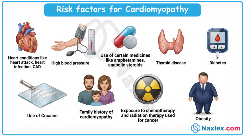
Pathophysiology
The central pathophysiological process in pediatric cardiomyopathy centers on structural failure of cardiac myocytes to effectively contract, relax, or maintain structural membrane integrity:
- In dilated cardiomyopathy, myocyte injury or genetic mutations impair systolic contractile force. To compensate for reduced stroke volume, the heart increases end-diastolic volume. Over time, progressive cardiac chamber dilation occurs with thin, stretched ventricular walls. This wall thinning increases ventricular wall stress according to Laplace's law, precipitating a downward spiral of increasing oxygen demand, progressive mitral or tricuspid valve regurgitation, elevated left ventricular end-diastolic pressure, pulmonary venous congestion, and diminished systemic cardiac output.
- In hypertrophic cardiomyopathy, disarray of cardiac muscle fibers and excessive myocardial hypertrophy occur without secondary cause. The thickened myocardium, particularly involving the interventricular septum, causes compliance failure (diastolic dysfunction) and elevated filling pressures. During exertion, dynamic left ventricular outflow tract obstruction can occur as the anterior leaflet of the mitral valve is pulled toward the hypertrophied septum during systole (systolic anterior motion [SAM]), drastically reducing forward cardiac output and increasing dynamic pressure gradients.
- In restrictive cardiomyopathy, severe myocardial fibrosis or infiltrative material creates a rigid, non-compliant ventricular myocardium. While systolic function and wall thickness initially remain near normal, impaired diastolic relaxation leads to elevated filling pressures, massive bi-atrial enlargement, systemic venous congestion, and pulmonary hypertension.
- In arrhythmogenic right ventricular cardiomyopathy, progressive fibrous and fatty tissue replacement of the right ventricular myocardium disrupts normal electrical conduction, predisposing the client to life-threatening ventricular tachyarrhythmias and right-sided heart failure.
- In left ventricular non-compaction, premature arrest of normal endomyocardial morphogenesis leaves prominent trabeculations and deep intertrabecular recess pockets within the ventricular apex, predisposing to localized thrombus formation, impaired contraction, and malignant dysrhythmias.
Classification
Pediatric cardiomyopathies are categorized based on structural appearance, myocardial wall architecture, and specific hemodynamic patterns:
- Dilated Cardiomyopathy (DCM)
- Structural Characteristics: Marked enlargement and dilation of the left ventricle (or both ventricles) accompanied by thinned ventricular walls.
- Hemodynamic Profile: Severe systolic dysfunction characterized by decreased ejection fraction (frequently below 40%), decreased stroke volume, increased end-diastolic volumes, and elevated left ventricular filling pressures. Mitral valve regurgitation commonly occurs secondary to annulus dilation.
- Hypertrophic Cardiomyopathy (HCM)
- Structural Characteristics: Inappropriate, non-dilated myocardial hypertrophy occurring in the absence of structural heart disease or systemic hypertension. Hypertrophy is often asymmetric, predominantly involving the interventricular septum.
- Hemodynamic Profile: Diastolic dysfunction due to a stiff, non-compliant left ventricle that resists passive filling. Dynamic left ventricular outflow tract (LVOT) obstruction can occur when the enlarged septum narrows the subaortic area, exacerbated by systolic anterior motion (SAM) of the anterior mitral valve leaflet.
- Restrictive Cardiomyopathy (RCM)
- Structural Characteristics: Normal or near-normal ventricular wall thickness and ventricular cavity dimensions, accompanied by massive, marked bi-atrial enlargement.
- Hemodynamic Profile: Severe diastolic dysfunction caused by rigid, unyielding endomyocardium. Systolic contractile function remains relatively intact early in the disease process. High filling pressures lead to pulmonary venous hypertension, right-sided heart failure, and systemic venous congestion.
- Arrhythmogenic Right Ventricular Cardiomyopathy (ARVC)
- Structural Characteristics: Progressive loss of normal right ventricular myocytes with replacement by fibrous and adipose (fatty) tissue. The right ventricle becomes dilated and thin-walled, often forming localized aneurysms.
- Hemodynamic Profile: Compromised right ventricular systolic function, right-sided heart failure, and severe electrical instability manifesting as re-entrant ventricular tachyarrhythmias.
- Left Ventricular Non-Compaction (LVNC)
- Structural Characteristics: Failure of normal myocardial compaction during intrauterine embryonic development, resulting in a two-layered myocardium: a thin, compacted epicardial layer and an excessively thick endocardial layer featuring deep intertrabecular recesses and prominent trabeculations.
- Hemodynamic Profile: Variable presentation including systolic pump failure, diastolic filling impairment, intraventricular thrombus formation within the deep myocardial recesses, and heightened risk for ventricular dysrhythmias.
IMAGE TITLE: PEDIATRIC CARDIOMYOPATHIES
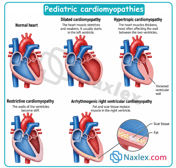
Clinical Manifestations
Clinical presentation varies depending on the subtype of cardiomyopathy, the child's developmental age, and the rapidity of disease onset:
Infants and Young Children
- Feeding Intolerance: Becoming easily fatigued, tachypneic, or diaphoretic during nipple feedings; taking only small volumes before stopping due to exhaustion.
- Respiratory Symptoms: Persistent resting tachypnea, intercostal and subcostal retractions, grunting, nasal flaring, wheezing, or chronic cough mistaken for reactive airway disease or bronchiolitis.
- Growth Failure: Failure to thrive, weight loss, or falling off growth percentiles due to combined inadequate caloric intake and elevated metabolic demands.
- Signs of Congestion: Periorbital edema, facial puffiness, hepatomegaly (palpable liver margin greater than 2 cm below the right costal margin), and rapid, unexplained weight gain from fluid retention.
- Poor Perfusion: Cool, pale, or mottled extremities, delayed capillary refill time (greater than 3 seconds), weak or thready peripheral pulses, and extreme lethargy or irritability.
Older Children and Adolescents
- Exercise Intolerance: Progressive fatigue, exertional dyspnea, and inability to keep up with peers during regular physical play or physical education classes.
- Left-Sided Congestive Symptoms: Orthopnea (requiring extra pillows to sleep), paroxysmal nocturnal dyspnea, exertional shortness of breath, and pulmonary crackles on auscultation.
- Right-Sided Congestive Symptoms: Jugular venous distension (in older children), peripheral lower extremity edema, right upper quadrant abdominal pain from hepatic congestion, ascites, and early satiety.
- Hemodynamic and Low Cardiac Output Symptoms: Dizziness, presyncope, syncope (particularly during or immediately following physical exertion), chest pain, palpitations, and decreased urinary output.
- Auscultatory Findings: S3 or S4 gallop rhythms; holosystolic murmur of mitral regurgitation at the apex (in DCM); harsh, systolic ejection murmur at the left sternal border that increases in intensity with standing or a Valsalva maneuver and decreases with squatting (in obstructive HCM).
Diagnostic Evaluation
Electrocardiography (ECG)
- Dilated Cardiomyopathy: Sinus tachycardia, left ventricular hypertrophy, ST-segment depressions, T-wave inversions, and left atrial enlargement.
- Hypertrophic Cardiomyopathy: Deep, narrow Q waves ("dagger Q waves") in lateral/inferior leads, striking voltage criteria for left ventricular hypertrophy, marked ST-T segment changes, and giant inverted T waves.
- Restrictive Cardiomyopathy: Prominent, high-voltage P waves indicating bi-atrial enlargement, along with non-specific ST-segment and T-wave abnormalities.
- ARVC: Inverted T waves in right precordial leads (V1 to V3), epsilon waves (a distinct low-voltage notch at the end of the QRS complex), and ventricular ectopy or ventricular tachycardia with left bundle branch block morphology.
Chest Radiography
- Dilated Cardiomyopathy: Marked cardiomegaly with an elevated cardiothoracic ratio (greater than 0.55 in children, greater than 0.60 in infants), pulmonary venous congestion, interstitial edema, and pleural effusions.
- Hypertrophic Cardiomyopathy: Heart size may appear normal or only mildly enlarged; cardiac contour may reveal prominent left ventricular enlargement.
- Restrictive Cardiomyopathy: Near-normal cardiac shadow size paired with marked enlargement of both atrial contours and prominent pulmonary vascular congestion.
Echocardiography (Transthoracic Echocardiogram)
- Establishes the definitive diagnosis by evaluating structural dimensions, wall thickness, ventricular volume, and ejection fraction.
- DCM demonstrates enlarged end-diastolic dimensions and depressed ejection fraction/fractional shortening.
- HCM displays asymmetric septal wall thickness, systolic anterior motion (SAM) of the mitral valve, and dynamic subaortic pressure gradients via Doppler imaging.
- RCM demonstrates normal-sized ventricles with severely dilated left and right atria and abnormal Doppler mitral inflow velocities (elevated E/A ratio).
- LVNC reveals a non-compacted to compacted myocardial thickness ratio greater than 2.0:1 at end-systole, with color Doppler demonstrating blood flow within the deep intertrabecular recesses.
Advanced Diagnostic Modalities
- Cardiac Magnetic Resonance Imaging (cMRI): Provides gold-standard visualization of myocardial mass, tissue characterization, non-compacted myocardium, right ventricular anatomy, and late gadolinium enhancement (LGE) to detect myocardial fibrosis and scar tissue.
- Serum Biomarkers: Elevated B-type Natriuretic Peptide (BNP) or NT-proBNP confirms heart failure; Cardiac Troponin I or T indicates active myocyte injury (such as acute myocarditis).
- Genetic Testing: Identifies specific gene mutations in sarcomeric, cytoskeletal, or mitochondrial proteins to establish etiology, guide targeted therapy, and direct cascade screening for first-degree relatives.
- Endomyocardial Biopsy: Invasive procedure reserved for cases of suspected acute viral myocarditis, storage diseases, infiltrative disorders, or unexplained rapid myocardial failure.
Therapeutic Management
Pharmacological Management
- Afterload Reduction (ACE Inhibitors / ARBs): Enalapril, captopril, or sacubitril/valsartan (ARNI) reduce systemic vascular resistance, decreasing the workload on the failing left ventricle in dilated cardiomyopathy.
- Beta-Adrenergic Blockers: Carvedilol, metoprolol, or propranolol. In DCM, beta-blockers prevent adverse adrenergic remodeling; in HCM, they reduce heart rate, prolong diastolic filling, and decrease dynamic outflow tract obstruction.
- Diuretics: Loop diuretics (furosemide) and thiazides promote fluid excretion to relieve pulmonary and systemic venous congestion. Aldosterone antagonists (spironolactone) offer mild diuresis while preventing myocardial fibrosis.
- Inotropic Support: Oral digoxin provides weak positive inotropic support and controls rate. In acute decompensated heart failure, continuous intravenous inotropes/lusitropes (milrinone, dobutamine) are administered to improve contractility and decrease systemic vascular resistance.
- Anticoagulation: Systemic anticoagulation (low-molecular-weight heparin, warfarin, or direct oral anticoagulants) is indicated in clients with severe systolic dysfunction (EF < 30%), left ventricular non-compaction, or a history of thromboembolism.
- Antiarrhythmic Therapy: Amiodarone or sotalol for controlling dangerous atrial or ventricular tachyarrhythmias.
Surgical and Interventional Management
- Implantable Cardioverter-Defibrillator (ICD): Indicated for primary or secondary prevention of sudden cardiac death in high-risk pediatric clients with HCM, ARVC, or severe DCM who exhibit malignant dysrhythmias or advanced fibrosis. Following placement, caretakers must keep the incision clean and dry, restricted arm movements above shoulder height for up to four weeks to prevent lead displacement, and monitor for signs of infection. Children should avoid contact sports and maintain a six-inch distance between the device and electronics like cell phones or magnetic fields. If a single shock occurs, keep the child calm and contact their cardiologist. If multiple shocks occur or the child becomes unresponsive, initiate emergency medical services and begin CPR immediately.
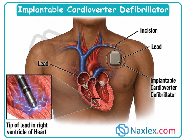
- Septal Myectomy: Surgical excision of hypertrophied septal muscle tissue performed in clients with hypertrophic obstructive cardiomyopathy who have severe LVOT obstruction unresponsive to medical therapy.
- Mechanical Circulatory Support (MCS): Ventricular Assist Devices (VADs, e.g., Berlin Heart EXCOR or HeartMate 3) used as a bridge to heart transplantation or bridge to recovery in children with refractory cardiogenic shock.
- Pediatric Cardiac Transplantation: The definitive treatment for end-stage pediatric cardiomyopathy refractory to medical and mechanical interventions.
Nursing Care Plan
Nursing Diagnosis 1: Decreased Cardiac Output related to impaired myocardial contractility, altered stroke volume, and dysrhythmias, as evidenced by resting tachycardia, oliguria, cold extremities, and delayed capillary refill.
- Expected Outcomes: The pediatric client will maintain adequate cardiac output as evidenced by age-appropriate heart rate, warm extremities, capillary refill time less than 3 seconds, and urine output greater than 1 mL/kg/hour.
- Nursing Interventions and Rationales:
- Assess baseline vital signs, peripheral pulse quality, skin temperature, and capillary refill time every 1 to 2 hours during acute instability. Rationale: Allows early detection of impending cardiogenic shock.
- Administer prescribed continuous intravenous inotropic infusions (such as milrinone) utilizing dedicated vascular access and smart infusion pumps. Rationale: Milrinone enhances myocardial contractility and decreases afterload through phosphodiesterase-3 inhibition.
- Continuously monitor telemetry for tachyarrhythmias, ectopy, or conduction blocks. Rationale: Impaired myocardium is highly arrhythmogenic, and sudden rhythm disturbances directly compromise cardiac output.
- Cluster nursing activities and provide a quiet, calm environment to reduce physiological stress and myocardial oxygen demand. Rationale: Energy conservation lowers metabolic demands on the failing heart.
Nursing Diagnosis 2: Excess Fluid Volume related to compromised ventricular pump function and sodium/water retention secondary to renin-angiotensin-aldosterone system activation, as evidenced by hepatomegaly, periorbital edema, pulmonary crackles, and rapid weight gain.
- Expected Outcomes: The pediatric client will demonstrate balanced fluid status as evidenced by clear lung sounds, decreasing hepatomegaly, stable daily weight, and absence of peripheral or periorbital edema.
- Nursing Interventions and Rationales:
- Obtain accurate daily weights at the same time each morning using the same scale. Rationale: Weight fluctuation is the most sensitive indicator of acute fluid retention or loss in pediatric clients.
- Monitor serum sodium, potassium, and chloride levels closely following loop diuretic administration. Rationale: Diuretics cause electrolyte depletion that can trigger dangerous dysrhythmias and digoxin toxicity.
- Maintain strict fluid intake and output measurement, including calculating total oral, enteral, and intravenous fluid volumes. Rationale: Prevents accidental fluid overload while ensuring adequate hydration.
- Elevate the head of the bed 30 to 45 degrees and position the child comfortably. Rationale: Decreases venous return to the heart, relieving pulmonary congestion and easing respiratory effort.
Nursing Diagnosis 3: Imbalanced Nutrition: Less Than Body Requirements related to elevated metabolic demands, respiratory distress during feeding, and poor intestinal absorption secondary to venous congestion, as evidenced by failure to thrive and fatigue during feedings.
- Expected Outcomes: The pediatric client will demonstrate adequate weight gain along an established growth curve without experiencing respiratory distress during enteral feedings.
- Nursing Interventions and Rationales:
- Provide high-calorie, concentrated infant formula or fortified breast milk as prescribed. Rationale: Maximizes caloric intake per unit volume, accommodating strict fluid restrictions.
- Limit oral nipple feeding attempts to a maximum of 20 to 30 minutes per feed. Rationale: Prolonged feeding sessions consume more calories in physical exertion than the infant ingests.
- Transition to supplemental nasogastric or gastrostomy tube feedings if the child exhibits tachypnea (respiratory rate greater than 60 breaths/min) or excessive tiredness during feeds. Rationale: Ensures full nutritional delivery while mitigating aspiration risk and working fatigue.
Complications
- Acute Decompensated Heart Failure and Cardiogenic Shock: Rapid failure of the pump mechanism resulting in severe systemic hypoperfusion, severe lactic acidosis, multi-organ dysfunction syndrome (MODS), and cardiac arrest.
- Sudden Cardiac Death (SCD): Triggered by malignant ventricular dysrhythmias (such as ventricular tachycardia or ventricular fibrillation). Extremely high risk in hypertrophic cardiomyopathy with severe obstruction and in arrhythmogenic right ventricular cardiomyopathy during physical exertion.
- Thromboembolism and Stroke: Blood stasis within dilated ventricles (DCM) or deep trabecular recesses (LVNC) promotes mural thrombi formation. Embolization can cause ischemic stroke, renal infarction, or peripheral vascular occlusion.
- Severe Pulmonary Hypertension: Chronic backpressure from elevated left ventricular end-diastolic pressures leads to structural remodeling of the pulmonary vascular bed, irreversible pulmonary hypertension, and eventual right ventricular failure.
- Malignant Dysrhythmias and Conduction Blocks: Electrical instability resulting from myocardial scarring, fibrosis, or cell disarray leads to atrial fibrillation, ventricular arrhythmias, high-grade AV blocks, or sinus node dysfunction.
- Graft Rejection and Post-Transplant Complications: In clients who undergo cardiac transplantation, primary graft dysfunction, acute cellular or antibody-mediated rejection, opportunistic infections secondary to immunosuppression, and accelerated cardiac allograft vasculopathy (CAV) remain major long-term risks.
Summary
- Pediatric cardiomyopathies represent a high-acuity spectrum of primary myocardial disorders characterized by structural, functional, and electrical abnormalities of the heart muscle.
- Categorized primarily into dilated, hypertrophic, restrictive, arrhythmogenic right ventricular, and left ventricular non-compaction subtypes, these conditions impair systolic contraction, diastolic relaxation, or electrical stability.
- Dilated cardiomyopathy remains the most common form in childhood, presenting with ventricular dilation, thin myocardial walls, and severe systolic failure often resulting from viral myocarditis or genetic mutations.
- Conversely, hypertrophic cardiomyopathy features inappropriate myocardial wall thickening, frequently involving the interventricular septum, leading to diastolic dysfunction, dynamic left ventricular outflow tract obstruction, and risk of exertional sudden cardiac death in young clients.
- Restrictive cardiomyopathy, though less common, carries a grave prognosis due to severe myocardial stiffness, elevated filling pressures, and bi-atrial enlargement.
- Diagnostic evaluation relies on a multidisciplinary approach combining electrocardiography, chest radiography, detailed transthoracic echocardiography, cardiac magnetic resonance imaging, serum biomarkers such as BNP and troponin, and specialized genetic testing.
- Clinical presentation varies by developmental age; infants present subtly with feeding fatigue, diaphoresis, poor weight gain, and tachypnea, whereas older children experience exercise intolerance, exertional chest pain, orthopnea, or exertional syncope.
- Therapeutic management is aimed at optimizing hemodynamics, controlling symptoms, delaying disease progression, and preventing life-threatening complications.
- Pharmacological strategies utilize afterload reducers, beta-blockers, targeted diuretics, inotropes, and anticoagulation.
- Invasive interventions include implantable cardioverter-defibrillators for arrhythmia control, surgical septal myectomy for obstructive hypertrophy, mechanical circulatory support as a bridge to therapy, and pediatric cardiac transplantation for end-stage heart failure.
- Pediatric nurses play a vital role in early clinical identification, managing complex intravenous infusions, enforcing meticulous fluid and electrolyte safety, maintaining adequate nutrition, preventing thromboembolic events, delivering specialized family education, and providing holistic support throughout the disease continuum.
Naxlex
Videos
Login to View Video
Click here to loginTake Notes on Acquired Cardiovascular Disorders: Pediatric Cardiomyopathies
This filled cannot be empty
Join Naxlex Nursing for nursing questions & guides! Sign Up Now