





 Video Library


Please set your exam date
Structural Anomalies Of The Gastrointestinal Tract
Study Questions
Practice Exercise 1
A nurse is educating the caregiver of an infant with a cleft lip. Which statement best describes the technique to feed this infant?
Explanation
Feeding an infant with a cleft lip requires specific compensatory techniques to ensure adequate nutritional intake while minimizing air swallowing and the risk of aspiration. Because a cleft lip affects the structural seal of the upper lip, infants often require specialized bottles, upright positioning, and paced feeding breaks.
Rationale for correct answer
B. Feeding the infant in an upright position (semi-upright or sitting upright) uses gravity to assist with swallowing and prevents milk from pooling in the nasal cavity or Eustachian tubes. Feeding slowly with frequent burping and rest breaks allows the infant to coordinate breathing and swallowing effectively, minimizing aerophagia (air swallowing).
Rationale for incorrect answers
A. Thickening feedings is generally unnecessary for an isolated cleft lip unless concurrent gastroesophageal reflux is present. Furthermore, enlarging the nipple opening arbitrarily can flood the oral cavity with too much milk too quickly, dramatically increasing the risk of choking and aspiration.
C. Laying an infant on their side during feeding does not prevent aspiration and is an unsafe feeding posture. Infants should always be held in an upright or semi-upright position during feeding to promote safe swallowing mechanics.
D. While giving smaller, more frequent meals can be helpful for infants with poor stamina or rapid fatigue, simply offering "small, frequent feedings" without specifying the crucial upright positioning and paced feeding technique makes Choice 2 the superior and more comprehensive answer.
Test-taking strategy:
- Analyze the scenario/question: The nurse is educating a caregiver on feeding techniques for an infant with a cleft lip. The nurse must identify the best feeding practice.
- Apply Pediatric Cleft Feeding Principles:
- Position infants upright to use gravity and protect the airway.
- Feed slowly and offer frequent breaks to prevent fatigue and air swallowing.
- Evaluate Choices:
- Rule out Choice 1: Thickening is unnecessary, and enlarging the hole causes rapid flooding and choking.
- Rule in Choice 2: Upright positioning, slow feeding, and frequent breaks optimize swallowing safety and efficiency.
- Rule out Choice 3: Side-lying is incorrect; infants must be upright when feeding.
- Rule out Choice 4: While small feedings help, Choice 2 provides the essential positioning and pacing details.
Take home points
- Always feed an infant with a cleft lip or palate in an upright or semi-upright position to reduce aspiration risks.
- Burp the infant frequently (every 15 to 30 mL) because infants with clefts swallow more air during feeding.
- Use specialized cleft bottles (such as the Haberman feeder or compressible squeeze bottles) that allow caregivers to control milk flow manually.
- Pate the feeding to match the infant's natural rhythm and breathing capacity.
A 3-month-old client returns to the pediatric unit following a cheiloplasty. Which post-operative nursing intervention is essential to protect the surgical site?
Explanation
Cheiloplasty requires strict post-operative protection to prevent suture line dehiscence. Cleft lip repair restores upper lip continuity and facial symmetry. Surgical integrity relies on minimizing local tension and preventing infant hands or hard objects from contacting the repair site.
Rationale for correct answer:
B. Soft elbow restraints, or elbow immobilizers, prevent the infant from bending the arms to touch the surgical site. This intervention shields the fragile facial suture line from accidental mechanical trauma and disruption. Restraints must be removed periodically to assess skin integrity and allow range of motion.
Rationale for incorrect answers:
A. Maintaining a prone position forces the infant's face against the mattress, causing direct pressure and friction on the fresh repair. This position increases the risk of suture breakdown and surgical site trauma. The infant must be placed supine or side-lying to keep the lip free from pressure.
C. Frequent routine suctioning with a rigid tip catheter can easily cause mechanical damage to the delicate oral tissue and suture line if the infant moves. Unnecessary oral instrumentation causes distress and physical tissue disruption. Suctioning should only occur for airway compromise using gentle, careful technique.
D. Pacifiers require active sucking, which creates significant mechanical tension across the newly repaired upper lip muscles. This suction force jeopardizes the surgical repair and risks suture displacement. Non-nutritive sucking devices are strictly contraindicated during the immediate recovery phase.
Test-taking strategy:
- Analyze the scenario/question: The infant is 3 months old and post-cheiloplasty. The primary nursing focus is protecting the surgical site from disruption.
- Apply Surgical Site Protection Principles:
- Rule in Choice 2: Applying elbow restraints physically prevents the infant from reaching the face while allowing free movement of other extremities.
- Rule out Choice 1: Prone positioning places direct friction on the face, which compromises the repair.
- Rule out Choice 3: Rigid catheters present a severe risk of accidental mechanical injury to the upper lip.
- Rule out Choice 4: Sucking creates internal muscle tension across the fresh lip suture line.
Take home points:
- Elbow restraints are essential post-cheiloplasty to prevent the infant from inserting fingers into the mouth or touching the facial sutures.
- Infants must be positioned supine or side-lying to keep the surgical site clear of mattress pressure and friction.
- Pacifiers, rigid eating utensils, and oral thermometry are strictly prohibited to avoid suture line tension and trauma.
- Elbow immobilizers require regular removal per institutional protocol to evaluate skin integrity, neurovascular status, and passive range of motion.
The nurse is caring for a 2-month-old client with isolated cleft palate. The nurse understands that this condition places the infant at high risk for which long-term complication?
Explanation
Isolated cleft palate creates an abnormal anatomical connection between the oral cavity and nasopharynx. Eustachian tube dysfunction prevents adequate middle ear drainage, causing persistent fluid accumulation. Affected infants experience impaired ventilation, frequent bacterial colonization, and structural middle ear damage leading to potential conductive hearing loss.
Rationale for correct answer:
B. Tensor veli palatini muscle dysfunction prevents proper Eustachian tube opening during swallowing. Impaired ventilation leads to negative pressure and fluid buildup within the middle ear tympanic cavity. Recurrent effusion increases the risk for chronic inflammation and permanent auditory deficits.
Rationale for incorrect answers:
A. Hypertrophic pyloric stenosis involves progressive hypertrophy of the gastric pyloric sphincter muscle leading to non-bilious projectile vomiting. It is a GI tract disorder completely unrelated to palatal development. This condition exhibits no structural or genetic link to primary orofacial clefts.
C. Intestinal malrotation results from failure of normal embryonic midgut rotation and fixation within the abdominal cavity. It predisposes infants to midgut volvulus and acute intestinal vascular occlusion. The embryological origins of gastrointestinal rotation are independent of cranial branchial arches.
D. Esophageal stricture formation develops secondary to severe gastroesophageal reflux disease, chemical ingestion, or congenital narrowing. It manifests with dysphagia and progressive esophageal obstruction. Cleft palate alters suction mechanics but does not cause anatomical luminal narrowing.
Test-taking strategy:
- Analyze the scenario/question: The infant is 2 months old with an isolated cleft palate. The question asks for a specific long-term complication associated with this structural defect.
- Apply Anatomical and Physiological Principles:
- Rule in Choice 2: Open palate defects directly impair tensor veli palatini muscle action, directly causing Eustachian tube collapse, fluid retention, and subsequent hearing impairment.
- Rule out Choice 1: Pyloric stenosis is an isolated pyloric sphincter malformation unrelated to head and neck structures.
- Rule out Choice 3: Malrotation involves embryonic midgut fixation failures with no pathogenic connection to the palate.
- Rule out Choice 4: Strictures represent esophageal lumen fibrotic changes rather than upper airway/palatal mechanics.
Take home points:
- Children with cleft palate require early audiologic evaluations due to persistent Eustachian tube malfunction and middle ear effusion.
- Myringotomy tube placement is commonly performed to relieve middle ear pressure and prevent permanent hearing deficits.
- Interprofessional management includes pediatric otolaryngologists, audiologists, and speech-language pathologists.
- Uncorrected hearing loss in infants with cleft palate significantly hinders early speech and language development milestones.
A 3-day-old client with cleft palate swallows excessive air during feedings. Which nursing action directly addresses this physiological problem?
Explanation
Cleft palate defects disrupt intraoral seal formation, causing significant aerophagia during liquid ingestion. Ingested air causes gastric distension, uncomfortable flatulence, and a high risk for emesis. Proactive clearance of trapped gas prevents gastric volumetric overload and reduces subsequent pulmonary aspiration risks.
Rationale for correct answer:
A. Frequent expulsion of air prevents significant intra-abdominal pressure buildup and uncomfortable gastric distension. Pausing every 15 to 30 mL allows timely clearance of swallowed air, stabilizing gastrointestinal mechanics. This simple intervention directly addresses excess aerophagia before reflux occurs.
Rationale for incorrect answers:
B. Large volume feedings expand an already air-filled stomach beyond physiological limits, provoking massive non-bilious emesis. Lengthening intervals between feeds causes extreme hunger, leading to uncoordinated, rapid swallowing efforts. This feeding schedule significantly worsens air ingestion.
C. Horizontally positioned infants lose gravitational assistance during swallowing, allowing liquid to pool in the nasopharynx. Flat positioning significantly elevates the risk of nasal regurgitation, coughing, and pulmonary aspiration. Feeding must always occur in an upright position.
D. Standard nipples demand high intraoral negative pressure, which infants with cleft palate cannot generate due to structural orofacial defects. Thickened formula requires larger nipple openings, creating high flow rates that increase choking hazards. Specialized feeders or compressible bottles are mandatory instead.
Test-taking strategy:
- Analyze the scenario/question: The client is a 3-day-old infant with a cleft palate who swallows excess air during feeds. The question asks for the specific nursing action to manage this issue.
- Apply Physiological and Airway Safety Rules:
- Rule in Choice 1: Frequent burping directly evacuates trapped air, relieving gastric pressure and reducing vomiting risks.
- Rule out Choice 2: Large, infrequent feeds overstretch the stomach, directly worsening gastric reflux.
- Rule out Choice 3: Flat positioning compromises airway clearance and increases aspiration risk.
- Rule out Choice 4: Standard nipples fail because the defect prevents intramural suction.
Take home points:
- Infants with cleft palate experience severe aerophagia and require burping after every 15 to 30 mL of liquid ingested.
- Feedings should always be conducted with the infant held upright to prevent nasal regurgitation and airway aspiration.
- Specialized cleft palate feeders or flexible squeeze bottles should be utilized rather than standard commercial nipples.
- Small, frequent feedings given over a period of 20 to 30 minutes prevent fatigue and severe abdominal distension.
The nurse is instructing the parent of an infant with cleft lip on feeding techniques. Which instruction should the nurse include?
Explanation
Cleft lip defects impair the infant's ability to maintain a tight seal around a nipple during feeding, causing excessive airway ingestion and fluid leakage. Directing the nipple toward intact oral structures leverages functional tissue to assist compression. Proper positioning improves fluid delivery, prevents tissue trauma, and minimizes the risk of nasal regurgitation.
Rationale for correct answer:
B. Positioning the nipple against intact tissue allows the infant to squeeze the nipple against the hard palate using their tongue. This technique maximizes milk extraction while preventing direct irritation to the delicate cleft margins. Utilizing intact structures provides a mechanical advantage that compensates for orofacial defects.
Rationale for incorrect answers:
A. Directing the nipple straight into the cleft causes friction and mechanical trauma to delicate, unjoined tissue borders. The nipple can easily become lodged within the defect, causing pain, mucosal injury, and severe choking episodes. Liquid directed at the cleft increases nasal passage irritation.
C. Horizontal positioning eliminates gravity-assisted fluid transit, allowing liquid to pool in the posterior oral cavity and nasopharynx. This posture significantly increases the risk for nasal regurgitation, severe coughing, and pulmonary aspiration. Infants must always be maintained in an upright, upright position during feeds.
D. Hard-rubber narrow nipples require high intraoral negative pressure that infants with cleft lip cannot generate effectively. The firm material leads to rapid fatigue, inadequate caloric intake, and severe feeding frustration. Pliable, soft nipples with wide bases are required to support compression.
Test-taking strategy:
- Analyze the scenario/question: The nurse is teaching a parent feeding techniques for an infant with a cleft lip. The objective is to identify the safest, most effective instruction.
- Apply Anatomical and Safety Principles:
- Rule in Choice 2: Placing the nipple against intact tissue allows the infant to compress it using the tongue and palate.
- Rule out Choice 1: Directing the nipple into the cleft causes local tissue trauma and airway compromise.
- Rule out Choice 3: Flat positioning increases fluid entry into the nasal cavity, elevating aspiration risk.
- Rule out Choice 4: Stiff nipples require high negative suction, which the defect prevents.
Take home points:
- Nipples should be directed toward the intact side or back of the mouth, avoiding direct contact with the cleft margin.
- Infants with cleft lip should be held in an upright or semi-upright position (at least 45 degrees) during all feedings.
- Soft, wide-base nipples or specialized cleft feeders should be selected to facilitate compression-based milk delivery.
- Burp the infant frequently throughout the feeding session due to increased air swallowing associated with an incomplete lip seal.
The nurse creates an educational plan for parents of a client with cleft palate. Which interdisciplinary specialist should the nurse inform the parents will be involved long-term? Select all that apply
Explanation
Cleft palate management requires coordinated long-term interdisciplinary care to address complex craniofacial, speech, and auditory structural deficits. Soft tissue and bone disruptions cause chronic middle ear effusion, altered dental arch alignment, and severe velopharyngeal insufficiency. Multidisciplinary team management prevents severe structural malocclusion, persistent hearing deficits, and profound articulation disorders.
Rationale for correct answers:
A. Audiologists provide continuous baseline and post-treatment monitoring for middle ear effusion and conductive hearing loss. Impaired Eustachian tube drainage secondary to tensor veli palatini dysfunction requires frequent audiometric testing. Early detection of hearing changes prevents downstream developmental speech delays.
B. Orthodontists manage complex maxillary arch collapse, alveolar cleft alignment, and severe dental malocclusion. They oversee palatal expansion devices and bone graft alignment as the facial craniofacial skeleton grows. Long-term care extends through adolescence to optimize masticatory function.
C. Speech-language pathologists evaluate and treat hypernasal speech, compensatory articulation errors, and velopharyngeal dysfunction. They guide early language milestones and provide specialized vocal rehabilitation therapy. Continuous intervention ensures proper palate closure mechanics during phonation.
Rationale for incorrect answers:
D. Nephrologists specialize in the management of renal system diseases and electrolyte disorders. Isolated cleft palate is an anatomical defect of the primary upper gastrointestinal tract and respiratory pathway, not the urinary system. Renal involvement is absent unless part of a distinct, severe chromosomal syndrome.
E. Neurologists evaluate and manage primary central and peripheral nervous system disorders. Cleft palate represents a localized structural failure of embryonic fusion rather than a primary neuropathic disease. Routine neurological oversight is unnecessary unless the infant exhibits concomitant intracranial anomalies.
Test-taking strategy:
- Analyze the scenario/question: The question asks for interdisciplinary specialists involved in the long-term management of a client with a cleft palate. This is a Select All That Apply (SATA) question.
- Apply Pathophysiological Connections to Interdisciplinary Roles:
- Rule in Choice 1: Cleft palate directly causes Eustachian tube dysfunction, making audiologist oversight essential for hearing preservation.
- Rule in Choice 2: Structural palatal defects alter dental and jaw development, requiring long-term orthodontist interventions.
- Rule in Choice 3: Soft palate involvement impairs air routing during speech, necessitating a speech-language pathologist.
- Rule out Choice 4: Renal pathology is completely unrelated to isolated orofacial clefts.
- Rule out Choice 5: Neurological pathology is absent in non-syndromic palatal defects.
Take home points:
- Cleft palate care requires an interdisciplinary team approach spanning from infancy through facial growth completion in early adulthood.
- Audiologists and otolaryngologists monitor for middle ear fluid accumulation, conductive hearing loss, and the need for tympanostomy tubes.
- Speech-language pathologists address velopharyngeal insufficiency and hypernasality to ensure normal communication development.
- Orthodontists and pediatric dentists correct maxillary alignment, bite malocclusion, and dental crowding as the child grows.
A newborn is born with a cleft lip and palate. Which of the following is the nurse's immediate priority?
Explanation
A newborn presenting with a cleft lip and palate experiences structural disruption of the primary and secondary palates. This anatomical defect impairs the infant's ability to create effective intraoral negative pressure needed for efficient feeding. While surgical restoration, long-term speech interventions, and family bonding are key components of overall care, establishing nutritional intake while safeguarding the airway is the most critical immediate priority in the initial post-birth assessment phase.
Rationale for correct answer:
C. Assessing the infant's ability to suck and swallow is the immediate clinical priority. The structural opening in the hard or soft palate prevents the creation of a vacuum, placing the infant at high risk for ineffective feeding, nasal regurgitation, fatigue, and aspiration. Evaluating oral-motor function allows the nurse to immediately implement specialized feeding equipment (such as a Haberman feeder or compressible bottle) and proper upright positioning to ensure safety and adequate nutrition.
Rationale for incorrect answers:
A. Surgical repair is not performed immediately at birth. Cleft lip repair is typically scheduled around 3 to 6 months of age (following the "Rule of 10s"), while cleft palate repair is usually performed between 9 and 18 months to allow for facial skeletal growth.
B. Promoting maternal-infant bonding is an essential psychosocial goal, especially given the emotional impact of a visible birth defect. However, physiological integrity, specifically airway protection and feeding safety, takes immediate priority over psychosocial outcomes during the initial newborn assessment.
D. Speech therapy education is an important long-term multidisciplinary consideration that becomes relevant as the child develops language skills post-palatoplasty. It is not an immediate post-birth priority.
Test-taking strategy:
- Analyze the scenario: The client is a newborn with a cleft lip and palate. The question asks for the nurse's immediate priority.
- Apply priority frameworks (Maslow's Hierarchy & ABCs):
- Rule in Choice 3: Assessing suck and swallow directly addresses airway safety (preventing aspiration) and essential physiological needs (nutrition).
- Rule out Choice 1: Surgical repair is delayed until specific physiological milestones are met.
- Rule out Choice 2: Psychosocial bonding is vital, but secondary to immediate physiological safety and feeding stability.
- Rule out Choice 4: Speech therapy is a future developmental concern, not an immediate newborn priority.
Take home points:
- The primary immediate nursing concern for an infant with a cleft lip/palate is preventing aspiration and ensuring adequate nutrition.
- Infants with cleft palate cannot generate negative intraoral suction; specialized squeeze bottles or wide-based nipples are required.
- Position the infant upright during feeds and burp frequently to prevent swallow-induced aerophagia and regurgitation.
- Cleft lip repair precedes cleft palate repair chronologically to facilitate proper facial development and feeding.
The pediatric nurse is caring for a 4-month-old infant who is 6 hours post cleft lip repair and has bilateral elbow restraints in place. Which nursing action is essential to maintain safety?
Explanation
Following a cleft lip repair (cheiloplasty), preventing the infant from touching, rubbing, or disrupting the delicate surgical suture line is paramount to achieving proper cosmetic healing and structural integrity. Elbow restraints (often called "welded" or "no-no" splints) immobilize the elbow joint to prevent flexion while allowing free movement of the hands and shoulders. However, because restraints carry risks of neurovascular impairment, skin breakdown, and psychological distress, strict safety monitoring guidelines must be followed.
Rationale for correct answer:
B. Regular direct observation and safety assessment, at least once every 1 to 2 hours (or per institutional restraint protocols), is essential to evaluate neurovascular status (pulses, capillary refill, skin temperature, and color), skin integrity beneath the splints, and joint mobility. During periodic assessment, the nurse should remove one restraint at a time to allow range-of-motion exercises and skin hygiene while directly supervising the infant.
Rationale for incorrect answers:
A. Securing ties to the crib frame or side rails is a major safety violation. If the side rail is lowered during an emergency or routine care, the restraint ties will pull tightly, risking injury or limb trauma. Restraints that require ties must be secured to the movable part of the crib mattress frame, never the side rails.
C. Allowing three fingerbreadths between the restraint and skin is too loose, rendering the restraint ineffective and allowing the infant to slip their arms out or flex their elbows to reach the surgical site. Proper fitting allows 1 to 2 fingerbreadths of space to ensure adequate circulation without compromising immobilization.
D. Restraint orders for children under 9 years of age generally require clinical re-evaluation and renewal every 1 hour (or up to 2 hours depending on facility policy for non-violent/medical-surgical restraints), not every 4 hours. A 4-hour renewal interval applies to adults aged 18 and older in behavioral management settings.
Test-taking strategy:
- Analyze the scenario: A 4-month-old is 6 hours post cleft lip repair with bilateral elbow restraints. The question asks for an essential safety action.
- Apply pediatric safety & restraint rules:
- Rule in Choice 2: Frequent assessment (at least every 1–2 hours) ensures neurovascular competence and protects skin integrity.
- Rule out Choice 1: Tying restraints to crib rails creates a severe mechanical injury risk when rails are moved.
- Rule out Choice 3: Three fingerbreadths is too loose, permitting joint flexion and suture disruption.
- Rule out Choice 4: Restraint renewal times for infants are far more frequent than 4 hours.
Take home points:
- Elbow restraints are used postoperatively following cleft lip/palate repair solely to prevent elbow flexion and protect the surgical site.
- Assess skin integrity and neurovascular status (temperature, color, capillary refill) under the restraint at least every 1 to 2 hours.
- Remove restraints periodically (one arm at a time) under direct nursing or parental supervision to perform gentle range-of-motion exercises.
- Ensure a proper fit allows 1 to 2 fingerbreadths between the device and skin; never tie restraints to movable crib side rails.
The nurse is caring for the child post cleft lip repair surgery. Which of the following orders should the nurse anticipate? Select all that apply
Explanation
Postoperative nursing care following a cleft lip repair (cheiloplasty) focuses on protecting the delicate facial suture line from mechanical tension, physical friction, and bacterial infection while maintaining a clear airway. Interventions such as applying protective topical ointments, securing tension-relieving devices (like a Logan bow or lip-strips), and performing gentle suture line cleansing are fundamental to achieving proper surgical healing.
Rationale for correct answers:
B. Applying a prescribed topical antibiotic/anti-infective ointment keeps the suture line moist, prevents crusting and scabbing, and creates a protective barrier against bacterial contamination.
D. Taping adhesive lip-straps or a Logan bow securely to both cheeks relieves muscular tension across the upper lip repair when the infant cries or moves, protecting the incision.
E. Gentle cleansing with sterile normal saline using a cotton-tipped applicator removes dried blood and serosanguinous crusts without disrupting the sutures.
Rationale for incorrect answers:
A. Prone positioning is strictly contraindicated post-cleft lip repair because it forces the facial suture line to rub directly against bed linens, risking trauma, friction, and suture disruption. The child must be placed supine or side-lying.
C. Elbow restraints ("no-no" splints) are required, not wrist restraints. Elbow immobilizers prevent the infant from flexing the arm and bringing hands up to touch or rub the face while still allowing free movement of the hands, wrists, and shoulders.
F. Standard immediate PACU and postoperative protocols require monitoring vital signs every 15 minutes for the first hour and every 30 minutes for the next 1 to 2 hours. Hourly vital sign checks during the initial 2 hours post-op are far too infrequent to detect early respiratory compromise or bleeding.
Test-taking strategy:
- Analyze the question: Select all anticipated orders for a child post-cleft lip repair.
- Evaluate each option:
- Rule out Choice 1: Prone position causes direct facial rubbing on linens.
- Rule in Choice 2: Ointment maintains site moisture and prevents infection.
- Rule out Choice 3: Wrist restraints restricts overall movement unnecessarily; elbow restraints are needed.
- Rule in Choice 4: Cheek tape relieves surgical line tension (e.g., Logan bow).
- Rule in Choice 5: Gentle cleansing with normal saline prevents crusting and maintains a clean surgical site. Avoiding harsh chemicals such as hydrogen peroxide prevents tissue damage and supports wound healing.
- Rule out Choice 6: Vital signs should be monitored every 15 minutes initially, then every 30 minutes for 2 hours, and then hourly thereafter. Early detection of airway compromise, bleeding, or pain is crucial.
Take home points:
- Position post-cleft lip repair infants supine or side-lying and never prone.
- Use elbow restraints instead of wrist restraints to prevent elbow flexion toward the face.
- Protect the incision line using adhesive cheek strips (Logan bow) to minimize tension.
- Clean the suture line gently with sterile normal saline and keep it moist with prescribed anti-infective ointment.
Practice Excercise 2
The nurse is caring for a newborn with esophageal atresia. When reviewing the mother’s history, which would the nurse expect to find?
Explanation
Esophageal atresia (EA) is a congenital anomaly characterized by the interruption of the esophageal lumen, preventing normal swallowing of amniotic fluid by the fetus in utero. During normal gestation, the fetus swallows substantial quantities of amniotic fluid, which is subsequently absorbed by the gastrointestinal tract and recycled via the placenta and fetal urinary system. When esophageal continuity is disrupted, amniotic fluid cannot pass into the stomach and intestines for absorption, leading to a significant excess of amniotic fluid accumulation within the amniotic sac.
Rationale for correct answer
A. Maternal polyhydramnios is a classic prenatal finding associated with congenital esophageal atresia. Because the fetus cannot swallow and absorb amniotic fluid normally due to the upper esophageal obstruction, fluid builds up rapidly, making polyhydramnios a key indicator that prompts prenatal suspicion or postnatal evaluation for foregut anomalies.
Rationale for incorrect answers
B. Pregnancies complicated by major congenital malformations such as esophageal atresia frequently result in preterm delivery or are unaffected by gestational age extension. Gestational age exceeding 38 weeks is not specifically correlated with the development of esophageal anomalies.
C. Poor maternal nutritional status during pregnancy is associated with various systemic conditions, intrauterine growth restriction, and neural tube defects, but it is not a direct etiologic factor or expected historical finding for esophageal atresia.
D. Maternal alcohol consumption during pregnancy causes fetal alcohol spectrum disorders, characterized by characteristic facial dysmorphism and central nervous system deficits. Alcohol intake is not established as a primary causative risk factor for esophageal atresia.
Test-taking strategy
- Analyze the scenario/question: A newborn has esophageal atresia. The nurse must identify which maternal history finding is expected.
- Apply Fetal Physiology Principles:
- Fetal swallowing recycles amniotic fluid.
- Obstruction of the esophagus prevents swallowing, causing fluid accumulation.
- Evaluate Choices:
- Rule in Choice 1: Polyhydramnios results directly from the fetus's inability to swallow amniotic fluid.
- Rule out Choice 2: Gestational length is variable and not directly linked to esophageal atresia.
- Rule out Choice 3: Maternal nutrition does not specifically cause esophageal malformations.
- Rule out Choice 4: Alcohol consumption causes fetal alcohol spectrum disorders, not foregut atresia.
Take home points
- Maternal polyhydramnios is a frequent prenatal clue indicating that the fetus is unable to swallow amniotic fluid normally.
- Esophageal atresia prevents the normal gastrointestinal clearance of amniotic fluid, resulting in excessive fluid volume in utero.
- Newborns presenting with copious frothy oral secretions and respiratory distress following a pregnancy complicated by polyhydramnios should be evaluated immediately for EA/TEF.
- Early identification and strict NPO status prevent aspiration pneumonia prior to surgical correction.
The nurse is caring for a newborn who has just been diagnosed with tracheoesophageal fistula and is scheduled for surgery. Which should the nurse expect to do in the preoperative period?
Explanation
Preoperative management for a newborn diagnosed with tracheoesophageal fistula (TEF) focuses on preventing respiratory complications, maintaining fluid and electrolyte balance, and preventing infection. Because the abnormal communication between the trachea and esophagus allows saliva, gastric secretions, and feeding fluids to enter the lower respiratory tract, immediate stabilization protocols require strict NPO status, continuous upper pouch suctioning, intravenous fluid support, and broad-spectrum antibiotic coverage to treat or prevent chemical pneumonitis and aspiration pneumonia.
Rationale for correct answer
B. Administering intravenous fluids maintains hydration and correct fluid balance while the infant is kept strictly NPO, and intravenous antibiotics treat or prevent pulmonary infections resulting from micro-aspirations of saliva or gastric contents through the fistula. This collaborative medical management is a foundational component of preoperative care.
Rationale for incorrect answers
A. While close monitoring and vital sign tracking are necessary, preventing parents from holding their infant is not a standard or required clinical protocol. Parents should be encouraged to interact with, touch, and comfort their infant within the parameters of safe incubators or radiant warmers.
C. Placing an infant on 100% continuous oxygen via a non-rebreather mask is unnecessary unless the infant is actively hypoxic or in severe respiratory distress. Routine administration of high-flow unhumidified oxygen can cause oxygen toxicity and mucosal drying, making targeted pulse oximetry monitoring and supplemental oxygen use strictly on an as-needed basis the appropriate clinical standard.
D. Feeding an infant with a tracheoesophageal fistula is strictly contraindicated. Any oral intake or feeding attempt will immediately flow through the fistula or overflow into the trachea, causing catastrophic pulmonary aspiration and severe respiratory arrest.
Test-taking strategy
- Analyze the scenario/question: A newborn with a tracheoesophageal fistula is awaiting surgical repair. The nurse must identify the correct preoperative intervention.
- Apply Preoperative Safety Principles:
- Maintain strict NPO status; never allow oral feedings.
- Provide supportive care via intravenous fluids, antibiotics, and respiratory monitoring.
- Evaluate Choices:
- Rule out Choice 1: Restricting parental holding unnecessarily hinders bonding; parents can interact safely.
- Rule in Choice 2: Intravenous fluids and antibiotics provide essential hydration, caloric support, and infection prevention.
- Rule out Choice 3: 100% non-rebreather oxygen is unnecessary unless hypoxemia is documented.
- Rule out Choice 4: Feeding an infant with TEF causes immediate aspiration and is dangerous.
Take home points
- Infants with tracheoesophageal fistula must be kept strictly NPO to prevent aspiration of food and fluids into the lungs.
- Intravenous access is vital for delivering maintenance fluids, total parenteral nutrition, and prophylactic antibiotics.
- Continuous or frequent intermittent suctioning of the blind upper pouch prevents the pooling and overflow of oral secretions.
- Preoperative nursing care requires elevating the head of the bed to 30 to 45 degrees to minimize gastric reflux through the fistula.
The nurse is assessing a newborn client 15 minutes after delivery. The infant displays excessive frothy salivation, coughing, and cyanosis. What initial nursing action is the priority?
Explanation
Excessive frothy salivation, coughing, and cyanosis in a newborn immediately following birth strongly indicate tracheoesophageal fistula or esophageal atresia. Congenital anatomical blind pouches cause immediate fluid accumulation within the pharynx. Continued secretions or enteral fluid intake quickly overflow directly into the tracheobronchial tree, causing severe respiratory distress and pulmonary aspiration.
Rationale for correct answer:
B. Immediate discontinuation of oral intake prevents fluid entry into the lungs, protecting airway patency. Suctioning secretions from the upper pouch clears the hypopharynx, relieving acute airway obstruction. Making the infant NPO stops progressive pulmonary contamination and stabilizes the neonate for immediate surgical evaluation.
Rationale for incorrect answers:
A. Administering oral glucose introduces liquid into an obstructed upper esophageal segment, forcing immediate pulmonary aspiration. Fluids spill over the larynx directly into the lungs, precipitating severe chemical pneumonitis and laryngospasm. Oral intake is strictly contraindicated when esophageal patency is absent.
C. Performing a heel stick delays critical immediate interventions required to clear a compromised respiratory airway. Hypoglycemia assessment is secondary to resolving active cyanosis, coughing, and impending airway asphyxiation. Primary airway stabilization must always precede diagnostic blood sampling.
D. Inserting a urinary catheter is completely inappropriate for an infant presenting with acute respiratory distension. Invasive urinary catheterization addresses fluid balance rather than life-threatening mechanical upper airway obstruction. This action delays critical pharyngeal suctioning and surgical stabilization.
Test-taking strategy:
- Analyze the scenario/question: A 15-minute-old newborn exhibits excessive frothy salivation, coughing, and cyanosis—the classic triad of esophageal atresia and tracheoesophageal fistula. The question asks for the priority initial nursing action.
- Apply Priority Nursing Principles (Airway, Breathing, Circulation):
- Rule in Choice 2: Making the infant NPO and suctioning directly protects the compromised airway and prevents severe aspiration.
- Rule out Choice 1: Administering oral fluids directly worsens pulmonary overflow and chemical pneumonitis.
- Rule out Choice 3: Checking blood glucose delays life-saving airway clearance interventions.
- Rule out Choice 4: Catheterization fails to address the immediate threat of asphyxiation.
Take home points:
- Excessive frothy oral secretions, choking, coughing, and cyanosis in a newborn are cardinal signs of tracheoesophageal fistula and esophageal atresia.
- The immediate priority nursing intervention is to withhold all oral feeds (NPO) and maintain airway patency through upper airway suctioning.
- The infant should be positioned with the head of the bed elevated (at least 30 degrees) to prevent gastric juice reflux into the lungs.
- Continuous or intermittent suction via a sump tube placed in the blind esophageal pouch is required until surgical repair is performed.
A client who is 1 hour old is suspected of having esophageal atresia. The nurse attempts to pass an 8-French nasogastric tube. Which finding confirms the suspected diagnosis?
Explanation
Esophageal atresia is a congenital anomaly where the proximal esophagus terminates in a blind-ending pouch, preventing continuous passage to the stomach. Attempting to insert an oro- or nasogastric catheter results in mechanical blind blockage within the upper esophagus. Radiographic visualization of a coiled radiopaque tube inside the upper pouch provides definitive anatomical confirmation.
Rationale for correct answer:
B. Resistance encountered at 10 cm indicates structural esophageal luminal obstruction. Radiographic confirmation of a coiled tube inside the blind upper pouch confirms esophageal atresia diagnosis. This classic diagnostic finding proves the absence of a continuous anatomical channel into the stomach.
Rationale for incorrect answers:
A. Obtaining acidic gastric aspirate requires direct passage into the stomach chamber, confirming full esophageal luminal continuity. A blindly terminating pouch prevents the tube from reaching acidic gastric secretions. Obtaining low pH fluid completely rules out proximal esophageal atresia.
C. Unobstructed tube insertion to 25 cm indicates normal anatomical passage directly through the gastroesophageal cardiac junction. Smooth catheter passage excludes a blind upper pouch and rules out mechanical esophageal obstruction. This finding confirms normal upper gastrointestinal tract structural continuity.
D. Drainage of dark green meconium signifies successful catheter passage into the lower gastrointestinal tract beyond the duodenal papilla. Bilious fluid cannot reach a blind-ending upper esophageal proximal pouch. This finding demonstrates patency throughout the upper gastrointestinal pathway.
Test-taking strategy:
- Analyze the scenario/question: A 1-hour-old infant is suspected of having esophageal atresia. The nurse attempts to insert an 8-French NG tube. The question asks for the specific finding that confirms this diagnosis.
- Apply Pathophysiological and Diagnostic Principles:
- Rule in Choice 2: Structural blockage at 10 cm with tube coiling on X-ray directly proves the existence of a blind esophageal pouch.
- Rule out Choice 1: Acidic gastric fluid indicates the tube reached the stomach cavity, ruling out atresia.
- Rule out Choice 3: Passing smoothly to 25 cm demonstrates intact esophageal patency.
- Rule out Choice 4: Bilious drainage requires entry into the duodenal segment, excluding a proximal blockage.
Take home points:
- Inability to pass a rigid nasogastric or orogastric tube into the stomach (meeting resistance at 10 to 12 cm) is the primary diagnostic sign of esophageal atresia.
- Anteroposterior chest and abdominal X-rays showing the radiopaque catheter coiled in the upper esophageal pouch provide definitive confirmation.
- Abdominal X-rays showing air in the stomach indicate the presence of an associated distal tracheoesophageal fistula (TEF).
- Once catheter coiling is noted, the tube should be connected to continuous low suction to decompress the blind pouch and protect the airway.
The nurse is caring for a 1-day-old client with esophageal atresia and a distal tracheoesophageal fistula pre-operatively. How should the nurse position the infant?
Explanation
Esophageal atresia with a distal tracheoesophageal fistula allows gastric juice to travel freely from the stomach through the fistulous tract directly into the trachea. Elevating the infant's upper body uses gravity to keep acidic stomach contents in the gastric lumen. Proper positioning prevents severe chemical pneumonitis, protects lung parenchyma, and minimizes the risk of fatal pulmonary aspiration.
Rationale for correct answer:
C. Elevating the head of the bed 30 to 45 degrees prevents gastric secretions from regurgitating through the distal fistula into the tracheobronchial tree. Supine positioning with head elevation maximizes lung expansion and optimizes continuous airway clearance. Gravity retains digestive juices in the lower stomach away from the respiratory tract.
Rationale for incorrect answers:
A. The Trendelenburg position elevates the stomach above the head, forcing acidic gastric contents upward through the distal fistula. Gravity propels stomach acid directly into the bronchial tree, precipitating immediate chemical pneumonitis. Positioning the head lower than the abdomen severely compromises respiratory function.
B. Prone positioning combined with foot elevation elevates the lower abdomen above the larynx, accelerating gastric regurgitation. Acidic fluid easily flows through the abnormal connection directly into the pulmonary tissue. This downward slope forces secretions into the lungs and increases aspiration risk.
D. Placing the head lower than the trunk eliminates gravitational resistance, allowing uninhibited flow of gastric acid through the fistula site. Declining positions fill the upper blind pouch with saliva, leading to severe asphyxiation risk. The head must always remain elevated above the stomach.
Test-taking strategy:
- Analyze the scenario/question: The client is a 1-day-old infant with esophageal atresia and a distal tracheoesophageal fistula awaiting surgery. The question asks for the correct pre-operative positioning strategy.
- Apply Gravity and Airway Protection Principles:
- Rule in Choice 3: Elevating the head of the bed utilizes gravitational force to hold gastric contents down and keep the airway clear.
- Rule out Choice 1: Trendelenburg position forces acidic stomach secretions directly into the tracheobronchial tree.
- Rule out Choice 2: Elevating the foot of the bed promotes pulmonary aspiration through the distal fistula.
- Rule out Choice 4: Lowering the head below the trunk causes rapid gastric reflux into the lungs.
Take home points:
- Infants with distal tracheoesophageal fistula must be placed in a supine position with the head of the bed elevated 30 to 45 degrees.
- Head elevation uses gravity to reduce the reflux of acidic gastric juice through the fistula into the lungs.
- Positions that lower the head below the waist (such as Trendelenburg) are strictly contraindicated due to severe aspiration risk.
- Continuous or intermittent suction must be maintained in the upper esophageal pouch alongside elevated positioning to prevent secretion overflow.
Practice Excercise 3
The nurse is attending the delivery of an infant with a prenatally diagnosed abdominal wall defect. Upon delivery, the nurse observes abdominal contents protruding through a right paraumbilical defect with no protective membrane covering the bowel. The nurse identifies this condition as:
Explanation
Gastroschisis is a full-thickness congenital abdominal wall defect typically occurring to the right of the umbilical cord insertion. Transposed abdominal viscera herniate directly into the amniotic cavity without a surrounding protective sac or membrane. Continuous intrauterine exposure to amniotic fluid leads to bowel wall thickening, severe edema, fibrous peel formation, and elevated post-delivery risks of hypothermia and fluid loss.
Rationale for correct answer:
B. Right-sided paraumbilical herniation without an enclosing protective membrane is the classic presentation of gastroschisis. Direct exposure of uncovered intestinal loops to the environment results in massive evaporative heat loss and fluid shifting. Immediate sterile protection and environmental control are required to preserve mesenteric blood flow.
Rationale for incorrect answers:
A. Omphalocele involves herniation of abdominal contents directly through the umbilical ring covered by a protective peritoneal sac. The protective membrane consists of peritoneum and amnion, with the umbilical cord inserting directly onto the hernia sac. Viscera in gastroschisis remain completely exposed without any membrane cover.
C. Umbilical hernia involves abdominal contents protruding through an incomplete closure of the umbilical ring, covered fully by intact skin. It presents as a soft, skin-covered bulge that typically resolves spontaneously without requiring surgical closure. Naked, exposed bowel loops do not occur in an umbilical hernia.
D. Meckel diverticulum is a congenital remnant of the vitelline duct located within the lumen of the ileum. It represents a vestigial gastrointestinal pouch rather than an external defect of the abdominal wall. This internal malformation remains contained entirely within the peritoneal cavity.
Test-taking strategy:
- Analyze the scenario/question: The infant presents at delivery with abdominal contents protruding through a right paraumbilical defect with no protective membrane covering the bowel. The question asks to identify the condition.
- Apply Pathophysiological Anatomical Rules:
- Rule in Choice 2: Evisceration through a right-sided defect lacking a protective membrane defines gastroschisis.
- Rule out Choice 1: Omphalocele features a midline defect covered by a peritoneal-amnion membrane.
- Rule out Choice 3: Umbilical hernias are covered by normal intact skin at the umbilicus.
- Rule out Choice 4: Meckel diverticulum is an internal vitelline duct anomaly, not an external defect.
Take home points:
- Gastroschisis presents as an unencapsulated, full-thickness abdominal wall defect, most commonly located to the right of an intact umbilical cord.
- The absence of a protective sac exposes the bowel directly to amniotic fluid, leading to edema, inflammation, and high evaporative fluid and heat loss at birth.
- Omphalocele differs from gastroschisis because the herniated organs are enclosed within a protective sac composed of peritoneum and amnion.
- Immediate post-delivery care for gastroschisis focuses on protecting the exposed bowel with sterile, warm, saline-soaked dressings and placing the lower torso in a sterile bowel bag.
A newborn client is born with a large omphalocele. Which immediate intervention should the nurse implement to protect the defect?
Explanation
An omphalocele is a congenital abdominal wall defect where viscera herniate through the umbilical ring enclosed within a translucent peritoneal membrane. The delicate sac is highly vulnerable to rupture, desiccation, and secondary bacterial peritonitis. Immediate care focuses on preserving membrane integrity, preventing massive evaporative heat and fluid losses, and avoiding direct pressure on mesenteric vessels.
Rationale for correct answer:
B. Covering the sac with warm, sterile saline-soaked non-adherent dressings keeps the membrane moist and prevents adherence to the fragile peritoneal sac. Applying an outer layer of plastic wrap creates a barrier against evaporative heat loss and severe fluid shifts. This intervention preserves sac integrity while preventing hypothermia and tissue necrosis.
Rationale for incorrect answers:
A. Applying dry cotton sponges causes dry fibers to stick directly to the delicate, moist visceral sac. Attempting to remove dry dressings pulls away delicate tissue layers, leading to membrane tears, rupture, and direct bowel exposure. Firm pressure also compromises underlying mesenteric arterial blood flow.
C. Applying topical alcohol solution causes severe tissue destruction, intense pain, and rapid absorption resulting in systemic toxicity. Alcohol dries out the membrane, accelerating desiccation, cracking, and eventual sac rupture. Harsh chemical solutions are strictly contraindicated on exposed embryonic membranes.
D. Placing the neonate prone subjects the herniated sac to direct body weight pressure and severe mechanical compression. Excess pressure compromises organ perfusion, triggers intestinal ischemia, and increases the risk of immediate sac rupture. The infant must always be placed supine to protect the defect.
Test-taking strategy:
- Analyze the scenario/question: The client is a newborn born with a large omphalocele. The nurse needs to identify the immediate action to protect the defect.
- Apply Tissue Protection and Thermal Principles:
- Rule in Choice 2: Moist, warm, sterile non-adherent dressings combined with plastic wrap preserve sac moisture, prevent temperature loss, and protect tissue.
- Rule out Choice 1: Dry cotton dressings adhere to delicate tissues, causing membrane tears upon removal.
- Rule out Choice 3: Alcohol causes severe chemical irritation, tissue desiccation, and sac breakdown.
- Rule out Choice 4: Prone positioning creates high pressure, risking ischemic injury and sac rupture.
Take home points:
- Protect the omphalocele sac immediately using warm, sterile saline-soaked non-adherent dressings covered with plastic wrap or a sterile plastic isolation bag.
- Position the infant strictly supine to avoid placing direct pressure or friction on the herniated abdominal viscera.
- Maintain strict NPO status and insert a decompressing nasogastric or orogastric tube to prevent bowel distension.
- Provide aggressive thermal management, as large abdominal defects significantly increase the risk for rapid neonatal hypothermia.
A client who is 2 hours old with an omphalocele is scheduled for an echocardiogram. The parent asks why a heart ultrasound is needed. What is the nurse's best response?
Explanation
Omphalocele arises from the failure of abdominal viscera to return to the abdominal cavity during the tenth week of embryonic development. Unlike gastroschisis, omphalocele carries a strong association with extra-gastrointestinal malformations and severe chromosomal trisomies. Preoperative identification of structural cardiac defects is essential to optimize anesthetic management, ensure hemodynamic stability, and prevent perioperative mortality.
Rationale for correct answer:
B. Up to 50 percent of infants born with an omphalocele have associated structural anomalies, most commonly congenital heart disease. Obtaining an echocardiogram preoperatively screens for underlying structural cardiac defects like tetralogy of Fallot or ventricular septal defects. Establishing cardiovascular stability is a critical requirement before undertaking complex abdominal wall repair.
Rationale for incorrect answers:
A. Surgical repair of an omphalocele utilizes standard pediatric anesthetic agents and broad-spectrum antibiotics that do not cause valvular damage. Attributing the diagnostic screening to drug-induced cardiotoxicity provides inaccurate information and causes unnecessary parental anxiety. The evaluation screens for preexisting structural malformations rather than drug toxicity.
C. Routine universal echocardiographic screening within 24 hours of birth is not standard practice for healthy asymptomatic newborns. Echocardiograms are targeted diagnostic tests reserved for infants demonstrating clinical cardiac symptoms or high-risk congenital anomalies. Stating that all newborns require cardiac ultrasound is a false generalization.
D. Abdominal viscera do not generate high blood pressure that transmits mechanically or vascularly to the cardiovascular system. Abdominal wall defects cause fluid loss, hypothermia, and third-spacing rather than direct mechanical hypertensive transmission. This statement presents incorrect vascular physiology to the parent.
Test-taking strategy:
- Analyze the scenario/question: The parent of a 2-hour-old infant with an omphalocele asks why an echocardiogram (heart ultrasound) is scheduled before surgery. The nurse must provide the accurate rationale.
- Apply Genetic and Association Principles:
- Rule in Choice 2: Omphalocele is strongly linked to congenital cardiac defects, making pre-operative cardiac evaluation mandatory.
- Rule out Choice 1: Surgical medications used during abdominal repair do not cause targeted heart valve damage.
- Rule out Choice 3: Universal screening with echocardiograms is not performed on all healthy neonates.
- Rule out Choice 4: Bowel tissue does not exert high pressure that directly elevates cardiac blood pressure.
Take home points:
- Omphalocele has a high rate of co-occurring congenital anomalies, particularly structural cardiac defects (such as VSD, ASD, or Tetralogy of Fallot).
- Preoperative echocardiograms are essential to identify underlying heart defects that could complicate anesthesia or surgical repair.
- Chromosomal abnormalities (such as Trisomy 13, 18, and 21) and syndromes like Beckwith-Wiedemann are frequently associated with omphalocele.
- Gastroschisis, in contrast to omphalocele, rarely presents with extra-gastrointestinal or cardiac anomalies.
A 1-day-old client with gastroschisis is receiving IV fluids. The nurse anticipates fluid maintenance requirements for this infant will be:
Explanation
Gastroschisis involves unencapsulated bowel loops directly exposed to the external environment, leading to massive evaporative loss. Exposed viscera experience severe serosal inflammation, third-spacing, and pronounced vascular shifts. These physiological alterations demand aggressive fluid resuscitation to prevent hypovolemic shock, restore circulating volume, and maintain tissue perfusion.
Rationale for correct answer:
A. Exposed abdominal contents cause massive evaporative fluid loss and rapid third-space fluid shifting into the interstitial space. Fluid requirements often reach 150 to 200 mL/kg/day, which is significantly higher than standard maintenance. Aggressive intravenous fluid administration is necessary to maintain adequate organ perfusion and systemic blood pressure.
Rationale for incorrect answers:
B. Restricting fluid intake to 25% of baseline causes catastrophic intravascular volume depletion and profound hypovolemic shock. Dehydration accelerates intestinal mucosal ischemia and leads to progressive systemic metabolic acidosis. Infants with gastroschisis require increased, rather than restricted, fluid volume to compensate for continuous losses.
C. Healthy term infants lose minimal fluid through intact skin and lungs, requiring standard baseline maintenance. Infants with gastroschisis lose substantial amounts of protein-rich fluid from the unencapsulated exposed viscera. Treating these fluid requirements as identical to a healthy infant results in severe fluid deficits.
D. Oral fluids are strictly contraindicated because the exposed, inflamed bowel exhibits severe paralytic intestinal ileus. Administering oral rehydration leads to massive gastric distension, severe vomiting, and pulmonary aspiration. All fluid resuscitation and maintenance must be delivered exclusively via central or peripheral intravenous access.
Test-taking strategy:
- Analyze the scenario/question: A 1-day-old infant with gastroschisis is receiving IV fluids. The question asks for the nurse's expectation regarding fluid maintenance requirements.
- Apply Pathophysiological and Fluid Balance Rules:
- Rule in Choice 1: Direct bowel exposure causes severe evaporative loss and third-spacing, demanding significantly higher fluid volumes.
- Rule out Choice 2: Restricting fluids rapidly leads to hypovolemic shock and tissue ischemia.
- Rule out Choice 3: Comparing an open abdominal defect to a healthy infant underestimates massive fluid shifts.
- Rule out Choice 4: Enteral intake is strictly prohibited due to functional paralytic ileus.
Take home points:
- Infants with gastroschisis require fluid maintenance rates significantly higher than normal (often 1.5 to 2 times baseline) due to continuous evaporative and third-space fluid losses.
- Isotonic crystalloid solutions (such as Normal Saline or Ringer's Lactate) and IV albumin are frequently required to restore circulating volume and oncotic pressure.
- Strict NPO status must be maintained, with gastric decompression initiated via a sump tube to prevent bowel distension and aspiration.
- Invasive monitoring, including hourly urine output tracking (aiming for ≥1 to 2 mL/kg/hr) and frequent electrolyte panels, is essential to guide fluid therapy.
The nurse is reviewing laboratory results for a pregnant client whose fetus was diagnosed with gastroschisis. Which laboratory abnormality was most likely detected during prenatal screening?
Explanation
Gastroschisis is an unencapsulated abdominal wall defect that permits direct contact between fetal serum, abdominal organs, and the surrounding amniotic fluid. Transudation of fetal proteins occurs across the exposed, hypervascular visceral serosa. This massive protein diffusion into amniotic fluid leads to significant clearance into the maternal circulation, producing a distinct elevation in alpha-fetoprotein levels.
Rationale for correct answer:
B. Markedly elevated maternal serum alpha-fetoprotein (MSAFP) occurs because the absence of an abdominal wall barrier allows fetal protein leakage into amniotic fluid. Fetal alpha-fetoprotein diffuses across membranes into maternal blood, generating values significantly higher than those seen in normal gestational age pregnancies. This finding serves as a primary biomarker during second-trimester screening.
Rationale for incorrect answers:
A. Human chorionic gonadotropin (hCG) is produced by placental trophoblastic tissue to maintain early corpus luteum progesterone production. Undetectable hCG levels indicate pregnancy loss or ectopic pregnancy failure rather than localized fetal abdominal defects. Gastroschisis does not halt or prevent normal placental trophoblastic hormone production.
C. Low maternal serum glucose reflects maternal metabolic states such as hyperinsulinemia, prolonged fasting, or hypoglycemia. Maternal circulating glucose levels are governed by pancreatic beta-cell function and insulin resistance, independent of fetal abdominal wall structural integrity. Gastroschisis does not lower maternal serum glucose.
D. Maternal serum calcium levels are tightly regulated by parathyroid hormone, calcitonin, and vitamin D endocrine pathways. Structural gastrointestinal anomalies like gastroschisis do not alter maternal bone metabolism or systemic calcium homeostasis. Maternal calcium remains within normal reference ranges during pregnancies affected by abdominal wall defects.
Test-taking strategy:
- Analyze the scenario/question: A pregnant client whose fetus has gastroschisis had prenatal laboratory screening. The question asks for the specific laboratory abnormality most likely detected during screening.
- Apply Biomarker and Pathophysiological Principles:
- Rule in Choice 2: Direct bowel exposure allows fetal protein to leak into amniotic fluid, causing marked MSAFP elevation.
- Rule out Choice 1: Undetectable hCG indicates non-viable pregnancy or trophoblastic collapse, not an isolated anatomical defect.
- Rule out Choice 3: Maternal glucose reflects metabolic status, which remains completely unaffected by fetal gastroschisis.
- Rule out Choice 4: Maternal calcium regulation depends on parathyroid pathways, completely independent of fetal structural malformations.
Take home points:
- Markedly elevated maternal serum alpha-fetoprotein (MSAFP) during second-trimester screening is a primary indicator of open fetal defects like gastroschisis and neural tube defects.
- Gastroschisis produces higher MSAFP elevations than omphalocele because the bowel is directly exposed without a protective sac barrier.
- An elevated MSAFP screen requires follow-up diagnostic evaluation via comprehensive target ultrasound to confirm the structural defect.
- MSAFP screening is routinely performed between 15 and 20 weeks gestation (optimally 16 to 18 weeks).
Practice Excercise 4
The nurse is providing discharge instructions to the parents of an infant who has had surgery to open a low imperforate anus. The nurse knows that the discharge instructions have been understood when the child’ s parents say:
Explanation
Postoperative management following surgical correction of a low imperforate anus (ano-rectal malformation) requires strict adherence to protective perineal care protocols, dietary modifications, and safety precautions. Reconstructed anal structures and surgical anoplasties must be protected from mechanical trauma, accidental laceration, and infection during the delicate healing phase.
Rationale for correct answer
A. Rectal temperatures, rectal tubes, and suppositories are strictly contraindicated following anorectal surgery. Using a rectal thermometer can easily puncture, lacerate, or disrupt the fresh surgical anoplasty, making the use of non-invasive oral, axillary, or temporal temperature routes an essential safety requirement.
Rationale for incorrect answers
B. Stool consistency naturally fluctuates in infants based on feeding changes, formula adjustments, and normal maturation. Unless the infant develops severe, persistent watery diarrhea or acute constipation, minor variations in stool consistency do not necessitate calling the healthcare provider.
C. Most children born with a low imperforate anus achieve normal bowel control and successful toilet training later in childhood because the external anal sphincter muscle complex is typically well-developed. Believing the child will never achieve continence is incorrect and unnecessarily discouraging.
D. Finding stool in the urine indicates the presence of a persistent rectourethral or rectovaginal fistula, which is a severe complication associated with high imperforate anuses rather than low lesions. Stool in the urine is never normal and must be reported immediately.
Test-taking strategy
- Analyze the scenario/question: The nurse is evaluating discharge instructions given to parents following surgical repair of a low imperforate anus. The nurse must identify which parent statement indicates correct understanding.
- Apply Postoperative Anorectal Safety Principles:
- Protect the delicate surgical anoplasty from trauma.
- Avoid inserting anything into the rectum (no rectal temps).
- Evaluate Choices:
- Rule in Choice 1: Oral/axillary thermometers must be used because rectal temperatures are contraindicated post-surgery.
- Rule out Choice 2: Normal stool consistency variations do not require constant provider calls.
- Rule out Choice 3: Children with low imperforate anus typically achieve good fecal continence.
- Rule out Choice 4: Stool in the urine indicates a fistula and is an abnormal, concerning finding.
Take home points
- Never take a rectal temperature or insert anything rectally following anorectal malformation surgery to protect the anoplasty.
- Children with low imperforate anus generally have a very favorable long-term prognosis for normal bowel control.
- Inspect the perineal and surgical site regularly for signs of infection, breakdown, or excessive swelling.
- Contact the pediatric surgeon immediately if signs of acute constipation, wound dehiscence, or unusual drainage occur.
The nurse caring for a neonate with an anorectal malformation notes that the infant has not passed any stool per rectum but that the infant’s urine contains meconium. The nurse can make which assumption?
Explanation
An anorectal malformation (ARM) encompasses a spectrum of congenital defects ranging from minor anal stenosis to complex cloacal anomalies. The presence of meconium in the urine (meconiuria) is a classic clinical indicator that an abnormal connection, a fistula, exists between the blind-ending rectum and the urinary tract (such as the urethra in male infants or the vagina in female infants). This anatomical finding is a hallmark characteristic of a high anorectal malformation.
Rationale for correct answer
B. The presence of meconium in the urine confirms the presence of a rectourethral or rectovesical fistula. Fistulas connecting the bowel above the level of the pubococcygeal line (levator ani muscle complex) are classified as high anorectal malformations, requiring multistage surgical interventions including an initial diverting colostomy.
Rationale for incorrect answers
A. A low anorectal malformation involves a rectum that terminates below the levator ani muscle sling. Low lesions may have perineal fistulas (such as a cutaneous or vestibule fistula), but they do not connect to the urinary tract, meaning meconium would not be present in the urine.
C. Because high anorectal malformations are complex and involve abnormal urinary communications and higher pelvic positioning, a diverting colostomy is almost always required initially to divert fecal stream away from the urinary tract and surgical repair site.
D. Surgical correction of a high anorectal malformation requires a complex posterior sagittal anorectoplasty (PSARP) or laparoscopic-assisted pull-through, combined with abdominal or pelvic dissection, rather than a simple nonoperative or minor procedure.
Test-taking strategy
- Analyze the scenario/question: A neonate with an anorectal malformation has not passed stool rectally, but meconium is present in the urine. The nurse must determine the clinical implication.
- Apply Embryonic and Anatomical Principles:
- Meconium in the urine proves a fistula exists between the bowel and the urinary tract.
- Urinary tract fistulas are characteristic of high anorectal malformations.
- Evaluate Choices:
- Rule out Choice 1: Low lesions do not connect to the urinary tract.
- Rule in Choice 2: Meconio-uria strongly indicates a high imperforate anus with a rectourethral/rectovesical fistula.
- Rule out Choice 3: High lesions do require a colostomy as a first stage.
- Rule out Choice 4: High malformations require complex multi-stage surgical pull-throughs, not nonoperative management.
Take home points
- The passage of meconium via the urethra is a diagnostic sign of a rectourethral or rectovesical fistula associated with high anorectal malformations.
- High anorectal malformations typically require a staged surgical approach: initial colostomy, definitive posterior sagittal anorectoplasty (PSARP), and later colostomy closure.
- Low anorectal malformations terminate below the pelvic floor muscles and often have more favorable primary repair outcomes without a colostomy.
- Careful perineal inspection in the nursery is essential to identify fistulous openings and abnormal anal placement.
The nurse is caring for a newborn with an anorectal malformation and a colostomy. The nurse knows that more education is needed when the infant’s parent states which of the following?
Explanation
Caring for an infant with a colostomy requires understanding stoma assessment, peristomal skin protection, and expected bowel output characteristics. A colostomy brings a section of the large intestine (colon) out to the abdominal wall to divert fecal contents. Unlike an ileostomy (which drains continuous, liquid, enzyme-rich small intestine output), a colostomy produces more formed or paste-like stool and does not continuously discharge irritating small bowel contents in the same caustic manner, though protective skin barriers are still essential.
Rationale for correct answer
C. This statement indicates a misunderstanding and requires further education. Colostomies involve the large intestine (colon), not the small intestine. Furthermore, depending on the type of colostomy (e.g., descending or sigmoid), stool becomes increasingly formed and predictable rather than draining continuously like small-bowel effluent, and management involves a pouching system rather than preventing "small intestine contents" from contacting the skin.
Rationale for incorrect answers
A. A healthy stoma should appear brick-red, moist, and well-perfused. A pale, pink, dusky, or dark purple/black stoma indicates compromised blood supply and is an emergency that must be reported immediately. This statement shows correct understanding.
B. Maintaining a healthy seal and protecting the skin means there should be no severe breakdown, raw irritation, or purulent discharge around the peristomal skin. Minor skin redness should be addressed promptly. This statement is correct.
D. As an infant matures and establishes regular feeding intervals, colostomy output typically follows a predictable pattern or routine regarding timing and consistency, making this a correct expectation.
Test-taking strategy
- Analyze the scenario/question: The parents of an infant with an anorectal malformation and a colostomy make several statements. The nurse must identify which statement indicates a need for further education (the incorrect statement).
- Apply Ostomy Physiology Principles:
- Colostomies involve the large intestine (colon), not the small intestine.
- Stomas should be red/moist; peristomal skin should be intact.
- Evaluate Choices:
- Rule out Choice 1: A red stoma is healthy and correct.
- Rule out Choice 2: Preventing irritation around the stoma is a correct goal.
- Rule in Choice 3: A colostomy involves the colon, not the small intestine, making this statement incorrect and requiring teaching.
- Rule out Choice 4: Developing a predictable bowel output pattern over time is correct.
Take home points
- A colostomy diverts stool through the large intestine, resulting in more formed output compared to an ileostomy.
- A healthy stoma is pink to brick-red, moist, and protrudes slightly above the abdominal wall.
- Peristomal skin must be kept clean, dry, and protected from effluent breakdown using a properly fitted pouching system.
- Report any changes in stoma color (pale, dusky, blue, or black), excessive bleeding, or severe skin breakdown immediately.
The nurse provides post-operative care for a 6-month-old client who underwent a posterior sagittal anorectoplasty (PSARP) for low imperforate anus. How should the nurse position the infant?
Explanation
Posterior sagittal anorectoplasty (PSARP) involves meticulous surgical reconstruction of the anorectal junction through a delicate perineal incision. Post-operative management focuses on preserving tissue perfusion, preventing suture breakdown, and eliminating direct mechanical pressure. Proper body positioning prevents wound dehiscence, minimizes localized tissue trauma, and promotes primary surgical healing.
Rationale for correct answer:
A. Positioned prone or side-lying with hips slightly elevated removes all mechanical forces from the fresh perineal wound. Eliminating surface friction prevents surgical suture tearing and maintains optimal microvascular perfusion. This specific posture keeps the surgical site clean and completely free from localized body weight pressure.
Rationale for incorrect answers:
B. Placing the infant in a hard plastic chair applies direct compression across the fresh perineal suture line. Upright seated forces compromise local tissue blood flow and increase the risk of severe wound dehiscence. Mechanical compression against a hard surface is strictly contraindicated following perineal reconstructive surgery.
C. Applying tight, heavy elastic diapers exerts continuous shear stress and friction against the fragile perineal repair. Confining the surgical site traps moisture, feces, and urine, leading to rapid maceration. Diapers must be left loose or completely open to keep the incision clean and dry.
D. Direct sitting forces the infant's entire lower body weight onto the newly reconstructed anal sphincter. Local pressure causes suture line breakdown, severe pain, and secondary wound infection. Direct weight-bearing on the perineum must be completely avoided during the initial healing period.
Test-taking strategy:
- Analyze the scenario/question: A 6-month-old infant is post-operative following a PSARP for imperforate anus. The question asks for the proper post-operative positioning strategy to protect the surgical site.
- Apply Surgical Site Protection and Tissue Integrity Principles:
- Rule in Choice 1: Prone or side-lying positions with hip elevation eliminate direct pressure and mechanical tension on the perineum.
- Rule out Choice 2: Hard plastic seats cause direct wound compression and tissue ischemia.
- Rule out Choice 3: Tight diapers exert shear stress and foster bacterial maceration.
- Rule out Choice 4: Direct sitting causes suture line breakdown and surgical failure.
Take home points:
- Following a PSARP, the infant must be positioned prone or side-lying to prevent any direct pressure on the perineal surgical site.
- Diapers should be left unfastened or loosely folded underneath the infant to avoid friction, shear, and trapped moisture on the suture line.
- The perineal incision must be kept meticulously clean and dry, with gentle cleansing after every bowel movement to prevent infection.
- Anal dilatations are typically initiated 2 weeks post-operatively using prescribed Peña dilators to prevent surgical site stricture formation.
The nurse is teaching the "Rule of 2s" for Meckel diverticulum to nursing students. Which statement should the nurse include?
Explanation
Meckel diverticulum is the most common congenital anomaly of the gastrointestinal tract, resulting from incomplete obliteration of the omphalomesenteric duct. This persistent embryonic remnant creates a true diverticulum containing all layers of the intestinal wall. Clinical manifestations primarily arise from ectopic gastric mucosa, which secretes acid and causes painless lower gastrointestinal ulceration.
Rationale for correct answer:
B. The classic "Rule of 2s" dictates that a Meckel diverticulum is located approximately 2 feet (60 cm) proximal to the ileocecal valve. Recognizing this specific anatomical landmark aids in rapid intraoperative identification during surgical exploration for lower gastrointestinal gastrointestinal bleeding. Anatomical proximity to the ileocecal junction is a defining characteristic of this congenital anomaly.
Rationale for incorrect answers:
A. Meckel diverticulum occurs in approximately 2 percent of the general population, not 20 percent. Stating a 20 percent incidence dramatically overstates its population prevalence. The "Rule of 2s" uses the digit two to denote a 2 percent prevalence rate across epidemiological studies.
C. The typical length of a Meckel diverticulum is approximately 2 inches (5 cm), not 2 meters. A length of 2 meters would exceed the total length of the small intestine. The "Rule of 2s" specifies 2 inches in length as a standard anatomical dimension.
D. Meckel diverticulum is diagnosed clinically or symptomatically twice as often in males compared to females. Stating a female predominance reverses the documented male-to-female ratio. Symptomatic presentation, particularly painless rectal bleeding, exhibits a 2-to-1 male predominance.
Test-taking strategy:
- Analyze the scenario/question: The nurse is teaching the epidemiological and anatomical "Rule of 2s" for Meckel diverticulum. The question asks which statement correctly accurately reflects this classic rule.
- Apply Epidemiological and Anatomical Principles:
- Rule in Choice 2: Located 2 feet proximal to the ileocecal valve is an accurate anatomical component of the rule.
- Rule out Choice 1: The prevalence is 2 percent, making 20 percent an incorrect overestimation.
- Rule out Choice 3: The structural length is 2 inches, whereas 2 meters is physiologically impossible.
- Rule out Choice 4: Symptomatic occurrence shows a 2-to-1 male predominance, not female.
Take home points:
- The "Rule of 2s" summarizes key features of Meckel diverticulum: 2% of the population, located 2 feet from the ileocecal valve, 2 inches long, 2 types of ectopic tissue (gastric/pancreatic), and symptomatic presentation typically by 2 years of age.
- Males are symptomatic twice as often as females (2:1 ratio).
- Ectopic gastric mucosa secretes acid, leading to painless, dark red or "currant jelly" lower gastrointestinal bleeding.
- The diagnostic test of choice for a symptomatic Meckel diverticulum is a Meckel scan (technetium-99m pertechnetate scintigraphy).
The nurse is caring for an 18-month-old client following a Meckel diverticulectomy. The client has a nasogastric tube attached to low intermittent suction. Which finding indicates return of GI function?
Explanation
Post-operative recovery following a Meckel diverticulectomy requires monitoring for the resolution of surgical paralytic ileus. Re-establishment of coordinated intestinal smooth muscle contraction restores enteric peristalsis. Functional recovery allows safe discontinuation of gastric decompression, advancement from strict fasting status, and re-introduction of enteral nutrition.
Rationale for correct answer:
B. Active bowel sounds indicate the return of gastrointestinal peristaltic motility. The passage of flatus or soft stool confirms complete intestinal patency throughout the lower gastrointestinal tract. These findings verify the resolution of post-operative ileus and signal that the gut can tolerate oral feedings.
Rationale for incorrect answers:
A. Absence of bowel sounds signifies persistent post-operative intestinal paralysis. A silent abdomen confirms the lack of active peristalsis and rules out functional recovery. Introducing fluids or removing gastric decompression during intestinal quiescence leads to severe nausea, vomiting, and abdominal distension.
C. Progressive abdominal distension and bilious green emesis indicate ongoing bowel obstruction or severe paralytic ileus. Accumulation of un-cleared digestive secretions increases intra-abdominal pressure and causes gastrointestinal distress. These findings demonstrate a failure of gastrointestinal recovery and require continued nasogastric decompression.
D. Persistent high-volume drainage of 100 mL/hour indicates severe fluid retention and upper gastrointestinal stasis. Continuous bilious output confirms that the stomach and small intestine are not actively moving gastrointestinal secretions downward. This finding requires ongoing gastric suction and delays enteral feeding.
Test-taking strategy:
- Analyze the scenario/question: An 18-month-old is post-op Meckel diverticulectomy with an NG tube to low intermittent suction. The nurse needs to identify the finding indicating the return of GI function.
- Apply Gastrointestinal Physiological Principles:
- Rule in Choice 2: Active bowel sounds combined with gas or stool expulsion directly confirm restored peristalsis.
- Rule out Choice 1: Absence of bowel sounds indicates ongoing paralytic ileus.
- Rule out Choice 3: Distension and bilious emesis signify gastrointestinal stasis or mechanical obstruction.
- Rule out Choice 4: High-volume gastric drainage proves a lack of downward transit.
Take home points:
- Return of gastrointestinal function post-diverticulectomy is evidenced by active bowel sounds, passage of flatus, and soft stool.
- Nasogastric suction is maintained post-operatively until bowel sounds return and NG tube output decreases and turns clear.
- Once GI function returns, the NG tube is clamped or removed, and clear liquids are initiated and advanced as tolerated.
- Increasing abdominal distension, bilious drainage, or vomiting indicates persistent ileus or surgical complications like adhesive bowel obstruction.
Practice Excercise 5
An expectant mother asks the nurse if her new baby will have an umbilical hernia. The nurse bases the response on the fact that it occurs:
Explanation
An umbilical hernia occurs due to an incomplete closure or weakness of the umbilical ring through which the abdominal contents (typically omentum or small bowel loops) protrude under the skin. This structural weakness is directly linked to immature or underdeveloped abdominal wall musculature, making premature infants and low-birth-weight neonates significantly more susceptible to developing the defect.
Rationale for correct answer
D. Premature infants have underdeveloped abdominal wall muscles and fascial structures surrounding the umbilical ring, leading to a markedly higher incidence of umbilical hernias compared to full-term infants.
Rationale for incorrect answers
A. Large infants (macrosomia or large-for-gestational-age newborns) do not have a natively higher predisposition for umbilical hernias; the condition is fundamentally driven by muscular immaturity and low birth weight rather than high birth weight.
B. While older medical literature historically noted varying racial frequencies (such as a higher reported prevalence in Black children), contemporary clinical consensus recognizes that umbilical hernias occur across all racial and ethnic groups, with prematurity serving as a much more definitive and reliable physiological risk factor. Furthermore, studies demonstrate that overall incidence between genders is relatively equal.
C. Umbilical hernias affect male and female infants at approximately equal rates, unlike congenital inguinal hernias, which are predominantly seen in male infants.
Test-taking strategy
- Analyze the scenario/question: An expectant mother asks about the likelihood of her baby developing an umbilical hernia. The nurse must identify the primary established risk factor.
- Apply Developmental and Physiological Principles:
- Recognize that umbilical hernias stem from fascial weakness at the umbilical ring.
- Recall that immature abdominal wall development strongly correlates with prematurity and low birth weight.
- Evaluate Choices:
- Rule out Choice 1: Large infants are not at increased risk.
- Rule out Choice 2: Racial generalizations are superseded by physiological risk factors like prematurity.
- Rule out Choice 3: Gender distribution for umbilical hernias is equal (unlike inguinal hernias).
- Rule in Choice 4: Prematurity is directly tied to abdominal wall immaturity and higher hernia rates.
Take home points
- Umbilical hernias are caused by an imperfect closure of the umbilical ring, allowing a small pouch of peritoneum to protrude.
- Premature infants and low-birth-weight neonates experience a significantly increased incidence due to immature musculature.
- The vast majority of pediatric umbilical hernias close spontaneously by 3 to 5 years of age without requiring surgical intervention.
- Parents should be reassured that unless the hernia becomes incarcerated, painful, or persistently enlarged past early childhood, conservative monitoring is the standard of care.
The nurse is providing discharge teaching to the parents of an infant with an umbilical hernia. Which should be included in the plan of care?
Explanation
An umbilical hernia is a common, benign congenital defect resulting from incomplete closure of the umbilical ring. Most pediatric umbilical hernias resolve spontaneously as the abdominal wall musculature strengthens during early childhood. Discharge teaching focuses on reassuring parents regarding expected natural closure timelines, educating them on dangerous complications such as incarceration, and dispelling outdated home remedies.
Rationale for correct answer
B. Increased swelling, discoloration, firm non-reducible bulges, tenderness, or persistent crying indicate that a loop of bowel has become incarcerated within the umbilical ring. Parents must be instructed to seek immediate medical evaluation for these signs to prevent bowel ischemia and necrosis.
Rationale for incorrect answers
A. Recommending surgery by 12 months of age is premature. Most pediatric umbilical hernias close spontaneously by 3 to 5 years of age. Surgical repair is typically deferred until the child reaches preschool age unless acute incarceration or strangulation occurs.
C. Applying pressure dressings, coins, belly bands, or adhesive tape over the hernia is strictly discouraged. These old-fashioned home remedies do not accelerate natural closure, and they frequently cause severe skin friction, breakdown, and contact dermatitis.
D. The recurrence rate following primary surgical repair (herniorrhaphy) for a pediatric umbilical hernia is exceptionally low. Once surgically closed, the defect rarely returns, making long-term prognosis extremely favorable.
Test-taking strategy
- Analyze the scenario/question: The nurse is providing discharge teaching for an infant with an umbilical hernia. The nurse must select the most accurate clinical instruction.
- Apply Pediatric Hernia Management Principles:
- Most hernias close on their own by age 3 to 5.
- Monitor for emergency complications (incarceration, tenderness).
- Avoid harmful folk remedies like taping or pressure coins.
- Evaluate Choices:
- Rule out Choice 1: Surgery is not indicated at 12 months; closure is expected by age 3 to 5.
- Rule in Choice 2: Swelling, tenderness, and non-reducibility signal incarceration, requiring urgent care.
- Rule out Choice 3: Pressure dressings damage the skin and do not help healing.
- Rule out Choice 4: Surgical recurrence rates are very low, not strong.
Take home points
- Umbilical hernias are common and usually resolve on their own by 3 to 5 years of age without intervention.
- Never tape, strap, or place coins over the hernia, as these methods cause skin breakdown without promoting closure.
- Teach parents to recognize signs of incarceration (pain, tenderness, discoloration, firm swelling), which require emergency treatment.
- Surgical repair is safe, effective, and rarely results in recurrence if needed later in childhood.
The nurse is assessing a 2-month-old male client during a well-child check. The nurse notes a smooth, painless swelling in the right groin that increases in size when the infant cries. The mass disappears when the infant is calm. The nurse suspects:
Explanation
An inguinal hernia in infants occurs due to a patent processus vaginalis, allowing abdominal viscera or fluid to protrude into the inguinal canal. Increased intra-abdominal pressure from crying, coughing, or straining forces the herniated sac outward, causing visible groin swelling. When the infant relaxes, the decreased pressure allows the sac to return to the peritoneal cavity, demonstrating classic intermittent reducibility.
Rationale for correct answer:
B. A smooth, painless right groin mass that enlarges during crying and spontaneously reduces when calm is the classic presentation of an inguinal hernia. Increased intra-abdominal pressure pushes abdominal contents through a persistent processus vaginalis. Prompt surgical evaluation is required to prevent progressive organ entrapment and subsequent ischemic strangulation.
Rationale for incorrect answers:
A. Isolated esophageal atresia is a congenital upper gastrointestinal defect characterized by a blind-ending proximal esophageal pouch. It presents immediately at birth with excessive frothy drooling, choking, coughing, and inability to pass an orogastric catheter. It produces no physical mass or localized swelling within the inguinal region.
C. Gastroschisis is an abdominal wall defect involving unencapsulated bowel loops protruding through a right paraumbilical defect. It is an obvious, dramatic birth defect present immediately at delivery rather than an intermittent mass appearing at a well-child visit. The herniated bowel remains continuously outside the body cavity.
D. Meckel diverticulum is a persistent remnant of the omphalomesenteric duct located on the ileal wall. It manifests primarily as painless, dark red lower gastrointestinal bleeding or intestinal obstruction within the abdominal cavity. It does not migrate into the inguinal canal to form a reducible subcutaneous mass.
Test-taking strategy:
- Analyze the scenario/question: A 2-month-old infant exhibits a smooth, painless right groin swelling that enlarges with crying and disappears when calm. The nurse must identify the condition.
- Apply Pathophysiological Presentation Principles:
- Rule in Choice 2: An intermittent groin mass influenced by intra-abdominal pressure defines a reducible inguinal hernia.
- Rule out Choice 1: Esophageal atresia involves upper gastrointestinal obstruction present at delivery.
- Rule out Choice 3: Gastroschisis is a congenital evisceration defect noted immediately at birth.
- Rule out Choice 4: Meckel diverticulum is an internal ileal remnant presenting with rectal bleeding.
Take home points:
- An intermittent, painless groin mass that enlarges with increased intra-abdominal pressure (crying, straining) and reduces at rest is diagnostic of an inguinal hernia.
- Pediatric inguinal hernias carry a high risk of incarceration and strangulation, requiring timely elective surgical repair (inguinal herniorrhaphy).
- Incarceration presents as an irreducible, firm, tender mass accompanied by irritability, severe pain, and signs of bowel obstruction (vomiting, distension).
- Parents must be taught manual reduction principles and instructed to seek immediate emergency care if the mass becomes hard, discolored, or painful.
A client who is 3 months old is brought to the clinic with an easily reducible, soft, skin-covered umbilical bulge that enlarges when crying. The parent asks when surgery will be scheduled. The nurse's best response is:
Explanation
An umbilical hernia results from an incomplete closure of the fascia at the umbilical ring. Increased intra-abdominal pressure from crying or straining forces intra-abdominal contents forward under intact skin, creating a classic reducible bulge. Most defects undergo progressive spontaneous closure as abdominal muscle wall strength increases with physical maturation.
Rationale for correct answer:
D. Most umbilical hernias resolve spontaneously without surgical intervention as the abdominal rectus muscles strengthen by age 3 to 5 years. Spontaneous closure occurs routinely in small defects measuring less than A.5 cm. Reassuring the parent avoids unnecessary surgical anxiety while establishing appropriate long-term clinical monitoring.
Rationale for incorrect answers:
A. Emergency surgery is reserved exclusively for acute complications such as incarceration, strangulation, or severe bowel obstruction. A soft, skin-covered, easily reducible hernia demonstrates no vascular compromise or organ tissue entrapment. Immediate emergency intervention is completely unnecessary for an uncomplicated, reducible defect.
B. Elective surgical repair is rarely scheduled at exactly 6 months because the vast majority resolve spontaneously as abdominal muscles develop. Standard guidelines recommend delaying surgical intervention until after age 3 to 4 years unless specific complications arise. Arbitrarily operating at 6 months subjects infants to unnecessary surgical risks.
C. Stating that umbilical hernias never close spontaneously provides false information and contradicts established pediatric epidemiological data. The overwhelming majority of congenital umbilical hernias undergo complete spontaneous fibrous closure. Permanent persistence requiring operative repair represents the minority of clinical cases.
Test-taking strategy:
- Analyze the scenario/question: A 3-month-old infant presents with a soft, skin-covered, easily reducible umbilical bulge that enlarges with crying. The parent asks when surgery will be performed. The nurse must select the best response.
- Apply Pediatric Growth and Surgical Principles:
- Rule in Choice 4: Most umbilical hernias close spontaneously by age 3 to 5 years as abdominal wall muscles strengthen.
- Rule out Choice 1: Emergency surgery is indicated only for non-reducible, strangulated hernias.
- Rule out Choice 2: Elective surgery is delayed beyond early infancy to allow time for spontaneous resolution.
- Rule out Choice 3: Stating they never close spontaneously contradicts known spontaneous closure rates.
Take home points:
- Pediatric umbilical hernias are common, usually painless, and typically undergo spontaneous resolution by age 3 to 5 years as abdominal wall strength increases.
- Incarceration and strangulation are extremely rare in umbilical hernias compared to inguinal hernias.
- Surgical intervention (umbilical herniorrhaphy) is generally deferred until age 4 or 5 unless the defect is exceptionally large (>2 cm), symptomatic, or becomes non-reducible.
- Parents should be taught to recognize signs of incarceration (a hard, discolored, or painful mass accompanied by severe crying or vomiting) and seek immediate care if they occur.
The nurse is caring for a 4-week-old client with an incarcerated inguinal hernia. Which clinical manifestation alerts the nurse to bowel strangulation?
Explanation
Incarcerated inguinal hernia occurs when herniated abdominal viscera become trapped within the inguinal canal. Progressive vascular compromise and ischemia lead directly to tissue strangulation. Entrapped bowel loops undergo venous stasis, hemorrhagic infarction, and full-thickness bowel necrosis, which manifests as severe localized inflammation, intractable pain, and acute gastrointestinal obstruction.
Rationale for correct answer:
B. A hernia site that becomes hard, dark red or purple, and extremely tender indicates acute tissue ischemia. Bilious vomiting confirms complete mechanical lower bowel obstruction. These cardinal manifestations signal impending gangrene, full-thickness perforation, and life-threatening peritonitis requiring immediate emergency surgical intervention.
Rationale for incorrect answers:
A. A soft, mobile, and non-tender mass indicates an uncomplicated, easily reducible hernia. Lack of tenderness and firm consistency confirms adequate tissue mesenteric perfusion. These benign characteristics Rule out acute incarceration, tissue ischemia, and bowel strangulation.
C. Gaining 20 grams of weight in 24 hours represents expected normal infant growth. Weight gain reflects adequate enteral caloric intake rather than acute intra-abdominal vascular compromise. Systemic ischemia and intestinal obstruction produce severe fluid loss, dehydration, and acute distress rather than normal weight gain.
D. Passing normal yellow soft stool demonstrates intact gastrointestinal luminal patency. Normal fecal transit confirms that bowel contents are moving unobstructed through the digestive tract. Intestinal strangulation halts downward transit, resulting in complete failure to pass gas or stool.
Test-taking strategy:
- Analyze the scenario/question: A 4-week-old infant has an incarcerated inguinal hernia. The nurse must identify the clinical manifestation alerting to bowel strangulation.
- Apply Ischemic and Obstructive Principles:
- Rule in Choice 2: Localized discoloration, extreme tenderness, and bilious vomiting directly indicate tissue necrosis and mechanical obstruction.
- Rule out Choice 1: Soft and non-tender characteristics define a benign, reducible mass.
- Rule out Choice 3: Daily weight gain reflects normal infant development, completely unrelated to strangulation.
- Rule out Choice 4: Normal stool passage confirms unobstructed transit, ruling out acute strangulation.
Take home points:
- Strangulation occurs when the blood supply to an incarcerated hernia is cut off, leading to rapid tissue ischemia and necrosis.
- Key indicators of strangulation include a hard, tense, discolored (red, purple, or blue) groin mass, extreme tenderness, high-pitched crying, and bilious emesis.
- Strangulated inguinal hernia is a surgical emergency requiring immediate operative intervention to prevent bowel gangrene, perforation, and peritonitis.
- Nurses must keep the infant strictly NPO, administer intravenous fluids and analgesics as ordered, and notify the surgical team immediately upon noting signs of strangulation.
Comprehensive Questions
A 2-week-old infant born with cleft lip and palate is being discharged from a hospital. The infant’s parents have each demonstrated the proper technique of feeding the infant with a special soft-sided bottle equipped with a cleft palate nipple. Which complication should a nurse inform the parents to monitor for with this type of feeding?
Explanation
Cleft lip and palate represent congenital craniofacial malformations disrupting structural integrity of the oral cavity and nasal septum. This anatomical defect prevents creation of negative intraoral pressure necessary for effective nutritive sucking mechanics. Affected infants swallow excessive ambient air during feedings, predisposing them to frequent gastrointestinal distension and heightened regurgitation risks requiring specialized Haberman or compressible feeding systems.
Rationale for correct answer
C. An anatomical defect in the primary palate compromises the oral seal, creating ineffective suction during feeding. Milk can easily enter the nasopharynx, leading to pulmonary contamination, coughing, or acute respiratory distress during feeds. Parents must maintain an upright posture and utilize special compressible bottles to mitigate these life-threatening events.
Rationale for incorrect answers
A. Overstimulation arises from environmental sensory overload rather than structural oral anomalies. It manifests as gated avoidance, color changes, or frantic motor activity during routine infant handling. This physiological response is entirely unrelated to the mechanical challenges associated with specialized infant feeding devices.
B. Overfeeding results from excess volumetric intake exceeding gastric capacity rather than altered oral mechanics. It typically produces fluid regurgitation or excessive daily weight gain without direct structural origin. Infants with clefts consume feeds slowly, making voluntary excess volume intake an unlikely outcome.
D. Hiccups occur secondary to transient diaphragmatic spasms caused by phrenic nerve irritation or gastric distension from swallowed air. While common in infants with cleft defects due to aerophagia, they remain a benign, self-limiting phenomenon rather than a major clinical complication.
Test-taking strategy
- Analyze the scenario/question: The infant has a cleft lip and palate and is feeding via a specialized cleft palate nipple. The nurse must identify the primary pathophysiological complication associated with this defect during oral intake.
- Apply Anatomical Principles:
- Failure of the soft and hard palate to fuse leaves a direct opening between the oral cavity and nasal cavity.
- During deglutition, liquid easily crosses this open boundary into the tracheobronchial tree.
- Evaluate Choices:
- Rule in Choice 3: Aspiration is the most severe, direct anatomical hazard during oral feedings for craniofacial defects due to compromised airway protection.
- Rule out Choice 1: Overstimulation is a behavioral response to environmental stress, not an anatomical feeding risk.
- Rule out Choice 2: Overfeeding relates to volume administration, whereas these infants typically struggle to achieve adequate caloric intake.
- Rule out Choice 4: Hiccups are minor and expected due to aerophagia, not a critical clinical complication.
Take home points
- Inability to generate negative intraoral pressure prevents effective suction and increases the risk of fluid misdirection into the respiratory tract.
- Positioning the infant upright at a 45 to 90 degree angle during feeds utilizes gravity to decrease the risk of nasopharyngeal reflux.
- Frequent burping every 15 to 30 mL is required to manage excessive air swallowing and prevent secondary gastric distension.
- Specialized cleft nipples allow manually controlled fluid flow timed with the infant's compression swallowing cues.
A nurse is completing discharge teaching with the parents of a 12-month-old child who has undergone a cleft palate repair. Which topics should be discussed in the discharge teaching? Select all that apply
Explanation
Postoperative care following palatoplasty focuses on maintaining surgical suture line integrity, preventing mucosal trauma, and ensuring effective analgesia to minimize crying-induced tissue tension. Surgical reconstruction exposes delicate granulation tissue to mechanical stress and thermal injury. The primary focus of discharge education involves enforcing soft diet restrictions, preventing mechanical disruption from hard objects or suction devices, utilizing non-pharmacological and pharmacological pain management strategies, and facilitating multidisciplinary follow-up for long-term speech, dental, and audiological management.
Rationale for correct answer
A. Surgical repair leaves delicate, healing palatal mucosa vulnerable to thermal trauma. Parents must verify that all liquid and soft foods are served warm or at room temperature to prevent mucosal burns or secondary tissue necrosis along the delicate surgical repair line.
B. Routine, around-the-clock administration of prescribed analgesics maintains a consistent therapeutic serum level to suppress pain. Uncontrolled pain leads to prolonged crying, which elevates intraoral pressure and applies excessive mechanical tension across the fresh palatal suture line, increasing dehiscence risks.
D. Soft elbow restraints (no-bow restraints) prevent the infant from placing fingers, toys, or hard objects into the oral cavity. They must be applied whenever the child is unmonitored to safeguard the palatal suture line from accidental mechanical trauma or disruption.
F. Cleft palate care extends well beyond the immediate surgical period, requiring a multidisciplinary team including speech therapy, orthodontics, and audiology. Long-term care imposes substantial financial stress, making early referral to social services and community assistance programs essential for family coping.
Rationale for incorrect answers
C. Feeding with a regular bottle nipple creates negative intraoral pressure and exerts direct mechanical pressure against the newly repaired hard and soft palate. Post-op infants must be fed using an open cup, syringe, or specialized side-douching spoon to prevent structural disruption of the surgical repair.
E. Inserting a bulb syringe or rigid suction tip into the mouth poses a severe risk of direct mechanical injury to the surgical site. Deep or routine oral suctioning is strictly contraindicated post-palatoplasty; secretions should be managed by positioning or gentle nasal suctioning if specifically indicated.
Test-taking strategy
- Analyze the scenario/question: A 12-month-old infant is being discharged following a palatoplasty. The nurse must select all appropriate discharge instructions aimed at protecting the surgical site, managing pain, preventing complications, and coordinating long-term care.
- Apply Surgical Protection Principles:
- Protect the palatal suture line from direct physical trauma, thermal injury, and stress caused by crying or negative intraoral suction.
- Avoid inserting any hard objects, bottle nipples, straws, or suction devices into the oral cavity.
- Evaluate Choices:
- Rule in Choice 1: Food temperature control prevents thermal injury to healing tissues.
- Rule in Choice 2: Scheduled analgesia prevents crying-induced suture tension.
- Rule out Choice 3: Standard bottle nipples create negative pressure and physical friction that can ruin the repair.
- Rule in Choice 4: Elbow restraints prevent the infant's hands from reaching the oral cavity.
- Rule out Choice 5: Suction devices inside the mouth risk direct suture disruption.
- Rule in Choice 6: Financial counseling addresses long-term multidisciplinary needs.
Take home points
- Nothing hard or sharp should enter the infant's mouth postoperatively, including spoons, straws, bottle nipples, pacifiers, or suction catheters.
- Elbow restraints should be used continuously when the child is unmonitored and removed periodically under direct supervision for range-of-motion exercises and skin checks.
- Analgesics should be administered on a regular schedule rather than strictly as needed to minimize crying and tension on the palatal suture line.
- Comprehensive post-repair care requires ongoing coordination with social work, speech therapy, audiology, and pediatric dentistry.
A nurse is observing a newly hired nurse providing care to an 11-month-old child who is 12 hours postoperative from a cleft palate repair. Which nursing action requires the experienced nurse to intervene?
Explanation
Postoperative care following palatoplasty focuses on maintaining surgical suture line integrity, preventing mucosal trauma, and avoiding any actions that increase intraoral pressure or cause direct mechanical disruption. Inserting hard objects or suction equipment into the oral cavity threatens the delicate reconstructed tissues. The immediate recovery phase requires non-invasive secretion management, strictly protecting the palatal repair, maintaining continuous elbow restraints with scheduled skin assessments, and providing proactive pain relief measures to prevent stress on the surgical site.
Rationale for correct answer
A. Inserting a rigid or flexible suction catheter into the mouth poses an immediate risk of direct mechanical disruption to the fresh palatal suture line. Deep or routine oral suctioning is strictly contraindicated post-palatoplasty; the nurse must intervene and instruct the newly hired nurse to manage secretions by positioning the child laterally or using gentle nasal suctioning if airway obstruction occurs.
Rationale for incorrect answers
B. Administering soft, blended foods or full liquids is appropriate for a child who has cleared an initial clear liquid trial post-op. Smooth, pureed textures do not cause mechanical friction against the healing oral mucosa, provided they are administered using a cup or the side of a spoon rather than standard bottle nipples or hard utensils.
C. Removing arm restraints individually to assess skin integrity, perform passive range-of-motion exercises, and evaluate capillary refill is a mandatory nursing action. Elbow restraints must be released periodically under direct nurse observation to prevent neurovascular impairment while ensuring the child does not place hands or objects into the healing mouth.
D. Administering prescribed analgesics maintains consistent serum drug levels to suppress postoperative pain and systemic distress. Effective analgesia prevents prolonged crying, which elevates intraoral pressure and places excessive mechanical tension on the delicate palatal repair, significantly reducing the likelihood of suture dehiscence.
Test-taking strategy
- Analyze the scenario/question: The experienced nurse is observing a newly hired nurse caring for an 11-month-old child 12 hours post-palatoplasty. The question asks for the action requiring immediate nurse intervention (identifying an unsafe practice).
- Apply Post-Palatoplasty Safety Rules:
- Protect the palatal suture line from direct physical trauma and mechanical irritation.
- Never insert hard or sharp objects, straws, pacifiers, or suction catheters into the oral cavity postoperatively.
- Evaluate Choices:
- Rule in Choice 1: Inserting a suction catheter into the mouth risks direct suture disruption and mucosal trauma, making this the incorrect action that requires intervention.
- Rule out Choice 2: Soft, blended foods are safe and appropriate for a post-op diet when offered without hard utensils.
- Rule out Choice 3: Removing restraints one at a time for skin and neurovascular checks is expected standard care.
- Rule out Choice 4: Analgesic administration prevents crying-induced suture line stress.
Take home points
- Suction catheters, straws, hard spoons, bottle nipples, and pacifiers are strictly contraindicated in the oral cavity following palatoplasty to prevent suture line disruption.
- Oral secretions should be cleared using gravity by positioning the child on their side or abdomen, reserving gentle nasal suctioning only for clear airway emergencies.
- Restraints must be removed at least every 2 hours, one arm at a time, to inspect skin integrity and provide joint range-of-motion exercises.
- Systemic analgesics must be administered regularly to manage pain effectively and prevent intense crying that strains the palatal repair.
A nurse is assessing a client, with a diagnosed inguinal hernia, at a scheduled clinic visit. The nurse suspects that the client’s hernia may be strangulated when which finding is noted on assessment?
Explanation
An inguinal hernia strangulation represents a life-threatening surgical emergency characterized by compromised blood supply to herniated intestinal tissue. Mechanical entrapment within the inguinal ring orifice leads to rapid venous congestion, localized tissue ischemia, and severe organ necrosis. Affected clients experience sudden, excruciating localized or generalized discomfort, accompanied by a firm, non-reducible protrusion, systemic inflammatory response activation, and progression toward intestinal perforation if immediate surgical revascularization is delayed.
Rationale for correct answer
B. Ischemia and impending tissue necrosis generate severe, localized abdominal and scrotal discomfort. Occlusion of the mesenteric vasculature triggers visceral pain receptors, producing intense, unyielding pain that signals tissue compromise and urgent need for emergency surgical repair.
Rationale for incorrect answers
A. Dyspnea reflects pulmonary pathology or severe metabolic acidosis rather than local tissue ischemia. While systemic septic shock secondary to late intestinal perforation can alter respiratory parameters, it remains an indirect, non-specific manifestation rather than a primary diagnostic indicator of hernia strangulation.
C. Constipation indicates general delayed gastrointestinal motility or mechanical bowel obstruction. While intestinal incarceration can impede stool passage, acute tissue vascular compromise is primarily heralded by severe pain rather than routine, non-emergent alterations in bowel elimination patterns.
D. Early mechanical obstruction produces high-pitched, hyperactive bowel sounds proximal to the blockage site. However, as strangulation progresses to ischemia and muscular bowel wall paralysis, intestinal motility ceases entirely, causing bowel sounds to become severely diminished or absent altogether.
Test-taking strategy
- Analyze the scenario/question: The nurse is assessing a client with a known inguinal hernia. The question asks for the specific physical assessment finding indicating that the hernia has progressed to vascular strangulation.
- Differentiate Hernia States:
- Reducible: Content slides easily back into the abdominal cavity.
- Incarcerated: Content is trapped, causing obstruction, but retains its blood supply.
- Strangulated: Blood supply is completely occluded, causing acute tissue ischemia.
- Evaluate Choices:
- Rule out Choice 1: Shortness of breath is unrelated to localized mesenteric ischemia.
- Rule in Choice 2: Sudden, intense abdominal pain directly signals acute tissue necrosis.
- Rule out Choice 3: Constipation reflects general motility slowing rather than immediate vascular compromise.
- Rule out Choice 4: Ischemic tissue leads to paralysis and silent abdomen, not hyperactive motility.
Take home points
- Severe, sudden abdominal or groin pain paired with a firm, tender, non-reducible mass is the cardinal hallmark of hernia strangulation.
- Vascular occlusion rapidly leads to tissue necrosis, intestinal perforation, peritonitis, and life-threatening systemic sepsis if untreated.
- Bowel sounds over a strangulated hernia shift from early high-pitched hyperactive sounds to absent sounds as tissue becomes ischemic and paralytic.
- Manual reduction of a suspected strangulated hernia is strictly contraindicated because returning necrotic tissue to the abdominal cavity triggers peritonitis.
A 3-year-old client is being admitted to a postsurgical unit following anorectal surgery. A nurse reviews the following postoperative orders from the surgeon. Which order should the nurse question?
Explanation
Postoperative management following anorectal surgical reconstruction (such as a posterior sagittal anorectoplasty) prioritizes maintaining surgical site integrity, avoiding direct mechanical shear stress, and reducing surgical site infection risks. Operative trauma leaves the perineal suture line vulnerable to breakdown from tissue tension, local contamination, and excessive pressure. Appropriate care focuses on aggressive systemic analgesia, early mobilization, dietary modification to prevent straining, and positioning strategies that eliminate direct pressure on the newly reconstructed anal sphincter and perineal structures.
Rationale for correct answer
D. Positioning an anorectal surgical client in a supine position directs the weight of the torso onto the sacral and perineal tissues. This exerts continuous pressure and shearing forces against the fresh anorectal suture line, increasing the risk of surgical site breakdown, flap failure, and severe local pain. The nurse must question this order and advocate for side-lying or prone positioning.
Rationale for incorrect answers
A. Defecation causes significant mechanical stretching of the compromised anal canal, triggering intense visceral and somatic pain. Pre-medicating with systemic opioid analgesics before the initial bowel movement blunts acute pain responses, reduces sphincter spasms, and prevents autonomic distress during early gastrointestinal elimination.
B. Warm sitz baths promote localized vasodilation, which accelerates tissue healing and enhances perineal blood flow. Applying warm water therapy after each bowel movement gently cleanses fecal residue from the surgical repair, reduces local bacterial colonization, and provides muscular spasm relief without introducing mechanical friction.
C. Initiating a high-fiber diet along with adequate hydration promotes soft, formed stool consistency. Soft stools pass easily through the reconstructed sphincter, preventing fecal impaction and eliminating excessive strain that could otherwise disrupt the delicate surgical suture line.
Test-taking strategy
- Analyze the scenario/question: A 3-year-old child is recovering from anorectal surgery. The nurse must identify which surgeon's order is unsafe or contraindicated for this specific surgical site.
- Apply Anorectal Postoperative Safeguards:
- Protect the perineal suture line from direct physical pressure, tension, friction, and contamination.
- Maintain soft stools to avoid mechanical trauma during bowel movements.
- Avoid positions that place direct body weight onto the gluteal and perineal region.
- Evaluate Choices:
- Rule out Choice 1: Administering pain medication before the first defecation prevents extreme anal pain.
- Rule out Choice 2: Sitz baths after defecation maintain perineal hygiene and reduce spasms.
- Rule out Choice 3: A high-fiber diet ensures soft stool, preventing suture disruption.
- Rule in Choice 4: The supine position places direct weight and pressure on the anorectal repair, making it an order the nurse must question.
Take home points
- Following anorectal surgery, clients should be positioned prone or side-lying to keep direct pressure off the delicate surgical site.
- Stool softeners and high-fiber diets are essential postoperatively to prevent constipation and avoid straining against the repaired sphincter.
- Warm sitz baths provide gentle cleansing and relief of anal sphincter spasms after each bowel movement.
- Analgesic pre-medication before the first postoperative defecation helps control predictable severe pain and prevents neurohumoral distress.
An infant has been tentatively diagnosed with esophageal atresia. What should be the priority nursing outcome?
Explanation
Esophageal atresia (EA) is a congenital anomaly where the esophagus terminates in a blind pouch rather than connecting to the stomach, often accompanied by a tracheoesophageal fistula (TEF). This structural abnormality prevents normal deglutition, causing oral secretions and milk feedings to accumulate in the upper esophageal pouch and spill over into the tracheobronchial tree. The primary immediate life-threatening risk is pulmonary contamination, leading to severe chemical pneumonitis, atelectasis, and acute respiratory failure. Nursing management focuses on maintaining airway patency, decompressing the blind pouch, and preventing pulmonary aspiration prior to surgical correction.
Rationale for correct answer
B. Compromised airway patency from aspirated oral secretions or gastric reflux via a fistula poses an immediate threat to life. Ensuring an effective breathing pattern takes absolute priority using basic life-support principles (Airway, Breathing, Circulation) to prevent severe hypoxemia, respiratory distress, and chemical pneumonitis secondary to aspiration.
Rationale for incorrect answers
A. Fluid volume balance is critical during the preoperative phase while the infant is maintained on strict NPO status. Intravenous hydration is provided to prevent systemic dehydration, but addressing fluid needs remains secondary to protecting the compromised airway.
C. Nutritional support is vital for growth and surgical healing, typically managed preoperatively via total parenteral nutrition or enteral tube feeds if a gastrostomy is placed. However, caloric intake is a secondary priority compared to preventing acute respiratory collapse.
D. Parental emotional health and anxiety management are important components of family-centered nursing care during a surgical crisis. Psychological support is an essential intervention, but physiological stability and airway protection must take precedence over psychosocial outcomes.
Test-taking strategy
- Analyze the scenario/question: An infant has a tentative diagnosis of esophageal atresia. The nurse must determine the highest priority nursing outcome among physiological and psychosocial options.
- Apply Prioritization Frameworks:
- Use the ABCs (Airway, Breathing, Circulation) framework: Airway and breathing always take precedence over fluid status, nutrition, and psychosocial needs.
- Recognize that blind esophageal pouches overflow directly into the lungs, leading to acute respiratory distress.
- Evaluate Choices:
- Rule out Choice 1: Fluid maintenance is important, but secondary to airway integrity.
- Rule in Choice 2: Effective breathing pattern directly addresses the life-threatening risk of aspiration pneumonitis.
- Rule out Choice 3: Maintaining nutrition is secondary to immediate respiratory stabilization.
- Rule out Choice 4: Psychosocial support for parents is secondary to physiological survival.
Take home points
- Airway patency and effective breathing patterns are the highest priority outcomes for infants with esophageal atresia due to high aspiration risks.
- The classic clinical manifestations of esophageal atresia and tracheoesophageal fistula are the "3 Cs": coughing, choking, and cyanosis during feeds.
- Infants must be placed on strict NPO status immediately upon suspicion, with the head of the bed elevated 30 to 45 degrees to prevent aspiration.
- Continuous or intermittent low suctioning of the blind upper esophageal pouch (via a double-lumen Replogle tube) is required preoperatively to clear secretions.
A nurse is assessing a newly born infant with respiratory distress and copious oral secretions. The nurse’s initial thought is tracheoesophageal atresia (TEA). Which nursing action should confirm this?
Explanation
Tracheoesophageal atresia (esophageal atresia with tracheoesophageal fistula) represents a congenital structural malformation where the proximal esophagus terminates in a blind-ending pouch. This anatomical interruption prevents normal esophageal continuity, causing swallowed saliva and blind pouch secretions to pool and spill over into the tracheobronchial tree. Attempts to pass a firm or soft enterogastric tube encounter blind pouch resistance, preventing advance into the gastric cavity. Confirmation at the bedside involves demonstrating obstruction to catheter passage, followed by radiographic visualization of the curled tube within the upper esophageal pouch to prevent severe aspiration pneumonitis.
Rationale for correct answer
D. Advancing a soft nasogastric or orogastric tube into a blind-ending upper esophageal pouch results in the tube coiling upon meeting anatomical resistance. The inability to advance the tube into the stomach or aspirate acidic gastric secretions confirms esophageal lumen discontinuity and supports the diagnosis of esophageal atresia.
Rationale for incorrect answers
A. Clearing pooled oral secretions temporarily relieves upper airway rattling, but respiratory distress quickly recurs as saliva continues to accumulate in the blind pouch. Transient improvement following suctioning does not confirm structural esophageal occlusion, as simple mucosal obstruction or transient tachypnea of the newborn exhibits a similar response.
B. Unimpeded advancement of an enterogastric tube directly into the gastric cavity confirms full esophageal patency. Obtaining acidic gastric contents or air via auscultation effectively excludes a diagnosis of complete esophageal atresia, making this finding inconsistent with the suspected anomaly.
C. Radiographic visualization of a catheter tip within the stomach verifies normal esophageal continuity and proper intragastric placement. Diagnostic x-ray confirmation of esophageal atresia instead demonstrates the radiopaque catheter curled harmlessly within the blind proximal pouch in the upper chest.
Test-taking strategy
- Analyze the scenario/question: A newborn presents with respiratory distress and copious oral secretions, raising suspicion for tracheoesophageal atresia. The nurse must select the clinical action/finding that confirms this anatomical obstruction.
- Apply Diagnostic Principles:
- Esophageal atresia means the esophagus ends in a blind pouch and does not connect to the stomach.
- Attempting to pass a nasogastric (NG) tube will meet resistance because the passage is structurally blocked.
- Evaluate Choices:
- Rule out Choice 1: Suctioning temporarily clears secretions in many conditions; it does not confirm a structural malformation.
- Rule out Choice 2: Easy advancement into the stomach proves the esophagus is open and rules out esophageal atresia.
- Rule out Choice 3: A catheter in the stomach on x-ray confirms normal anatomy and rules out esophageal occlusion.
- Rule in Choice 4: Tube advancement halting in the blind pouch without obtaining gastric contents confirms esophageal obstruction.
Take home points
- Inability to pass a nasogastric or orogastric tube into the stomach is the primary bedside clinical test to confirm suspected esophageal atresia.
- Copious, frothy oral secretions accompanied by the classic triad of coughing, choking, and cyanosis during early feeds strongly indicate esophageal anomalies.
- Definitive confirmation is established by a chest/abdominal radiograph showing the radiopaque catheter coiled inside the blind upper esophageal pouch.
- Once esophageal atresia is confirmed, the infant must be placed on immediate strict NPO status with continuous low suction applied to the upper pouch.
A 39-week-old infant is postoperative day 1 after emergency surgery for tracheoesophageal atresia. Which nursing action would be unsafe for the infant?
Explanation
Postoperative management following emergency repair of tracheoesophageal atresia (TEA) focuses on protecting the delicate tracheal and esophageal anastomotic suture lines, maintaining airway patency, and preventing surgical site disruption. Surgical reconstruction involves delicate mucosal reapproximation within a small operative field. Performing deep blind suctioning in the posterior pharynx or trachea risks direct mechanical trauma to fresh suture lines, leading to catastrophic anastomotic leak, tissue breakdown, severe tracheoesophageal refistulization, or fatal mediastinitis.
Rationale for correct answer
B. Routine or deep oral and tracheal suctioning poses a severe risk of direct physical disruption to the delicate esophageal anastomosis and repaired tracheal fistula site. Mechanical catheter irritation can tear fresh suture lines, causing fatal anastomotic leakage or mediastinitis. Blind suctioning beyond the pre-measured superficial depth is strictly unsafe and contraindicated.
Rationale for incorrect answers
A. Providing a non-nutritive pacifier during enteral gastrostomy feedings establishes a therapeutic association between sucking mechanics and satiation. Non-nutritive sucking promotes salivary secretion, enhances gastrointestinal hormone release, and minimizes oral aversion while keeping the esophageal repair completely resting and free from pressure.
C. Elevating and leaving the gastrostomy tube unclamped after feedings allows swallowed air to vent freely from the stomach. Venting reduces intragastric pressure, preventing gastric contents from refluxing upward through the newly repaired esophagus and protecting the fragile anastomotic site from acid irritation or mechanical tension.
D. Frequent gentle repositioning facilitates pulmonary secretion mobilization, enhances atelectasis prevention, and prevents localized tissue breakdown. Careful turning maintains optimal lung expansion and regional blood flow while maintaining strict support of the head and neck to avoid suture line traction.
Test-taking strategy
- Analyze the scenario/question: The question asks to identify an unsafe nursing action for a 39-week-old infant on postoperative day 1 following emergency repair of tracheoesophageal atresia.
- Apply Surgical Protection Principles:
- Protect the delicate tracheal and esophageal anastomotic suture lines from mechanical disruption, tension, and increased pressure.
- Avoid inserting rigid or deep catheter equipment into the oral cavity or airway that could contact surgical repairs.
- Evaluate Choices:
- Rule out Choice 1: Pacifier use provides safe non-nutritive sucking during G-tube feeds without stressing suture lines.
- Rule in Choice 2: Deep oral and tracheal suctioning risks catastrophic direct anastomotic disruption, making it unsafe.
- Rule out Choice 3: Venting the elevated G-tube relieves gastric pressure and protects the repair from acid reflux.
- Rule out Choice 4: Careful repositioning prevents pulmonary complications and promotes comfort.
Take home points
- Deep or unmeasured oral and tracheal suctioning is strictly contraindicated post-TEA repair to avoid tearing delicate tracheal and esophageal suture lines.
- If suctioning is essential for airway clearance, the catheter must be pre-measured to a safe, superficial depth to avoid reaching the surgical anastomosis.
- Venting an unclamped gastrostomy tube after feedings prevents elevated intragastric pressure and acid reflux against the newly repaired esophagus.
- Non-nutritive sucking during gastrostomy feeds satisfies normal infant oral drives and prevents oral aversion without compromising healing tissues.
An 18-month-old child with a history of cleft lip and palate has been admitted for palate surgery. The nurse should provide which explanation about why a tooth-brush should not be used immediately after surgery?
Explanation
Postoperative care following palatoplasty requires absolute physical protection of the fragile mucosal suture line against mechanical trauma, friction, and premature surgical dehiscence during early tissue healing phases.
Rationale for correct answer
C. A toothbrush introduces hard bristles and rigid plastic components directly into the oral cavity. These mechanical elements create significant friction against delicate tissue. Using a toothbrush can easily disrupt the fresh repairs. This action risks catastrophic suture line dehiscence.
Rationale for incorrect answers
A. Emotional distress represents a secondary clinical concern during initial recovery phases. While hospitalization frequently induces pediatric anxiety, tissue preservation remains the absolute priority. Psychological stress does not directly threaten structural integrity. Behavioral coping is managed separately.
B. Deciduous teeth are entirely normal at 18 months of age. Primary dentition is actively present in the oral cavity. Tooth presence does not contraindicate gentle cleaning. Dental anatomy remains unchanged by palate surgery.
D. Strict NPO status applies only during the immediate preoperative and recovery phase. Postoperative protocols advance rapidly from clear liquids to soft foods. Oral nutritional intake resumes very quickly. Enteral feeding does not preclude suture protection.
Test-taking strategy
- Analyze the scenario/question: An 18-month-old child is recovering from palatoplasty. The nurse must select the rationale explaining why toothbrushes are contraindicated postoperatively to prevent surgical complications.
- Apply Surgical Protection Principles:
- Evaluate mechanical hazards within the oral cavity.
- Protect delicate reconstructive tissue from abrasive contact.
- Evaluate Choices:
- Rule out Choice 1: Emotional stress is secondary to physical tissue disruption.
- Rule out Choice 2: Deciduous teeth are physiologically normal and present.
- Rule in Choice 3: Toothbrushes create mechanical friction that disrupts suture lines.
- Rule out Choice 4: NPO status is temporary and unrelated to toothbrush restriction.
Take home points
- Toothbrushes, cotton swabs, and hard utensils are strictly prohibited in the oral cavity following cleft palate repair.
- Oral hygiene is maintained via gentle water rinses or prescribed mouth care protocols without mechanical friction.
- Protecting the palatal suture line from mechanical disruption prevents costly and painful surgical dehiscence.
- Dietary advancement proceeds carefully from clear liquids to soft foods using non-traumatic feeding utensils like cups or spoons.
The nurse is caring for an infant with omphalocele. Immediately after the delivery of an infant with an omphalocele, the nurse should perform which intervention?
Explanation
Omphalocele represents a severe midline congenital abdominal wall defect characterized by the herniation of abdominal viscera into the base of the umbilical cord. The herniated organs remain enclosed within a delicate, translucent peritoneal sac. Immediate delivery room management focuses on preventing sac rupture, minimizing evaporative heat loss, and reducing fluid shifts. Encasing the lower half of the infant in a clear, sterile, warm saline-filled bowel bag isolates the exposed viscera, preserves core body temperature, prevents environmental contamination, and allows continuous visual monitoring of bowel perfusion prior to definitive staged surgical reduction.
Rationale for correct answer
D. Placing the infant's lower body into a sterile, warm saline bag up to the nipple line provides immediate protection to the herniated abdominal viscera. This critical intervention prevents severe evaporative heat loss, reduces insensible fluid losses, and maintains a sterile environment to safeguard the fragile peritoneal sac against rupture and infection.
Rationale for incorrect answers
A. Obtaining a baseline weight is an essential component of overall neonatal assessment and fluid balance management. However, exposing the uncontained abdominal defect to environmental air during prolonged weighing procedures causes rapid core hypothermia. Body weight measurement must be deferred until the herniated organs are viscerally protected.
B. Inserting an orogastric tube is indicated to decompress the stomach and prevent intestinal distension from swallowed air. However, an enterogastric tube is used strictly for gastric decompression, not for initiating enteral nutrition. Early enteral feeding is strictly contraindicated in the presence of an unrepaired omphalocele.
C. Immediate blood transfusion is rarely required upon initial stabilization unless severe fetal hemorrhage or umbilical cord disruption occurred during delivery. Preparing for massive blood administration is not a universal initial resuscitation priority compared to immediate visceral protection.
Test-taking strategy
- Analyze the scenario/question: An infant is born with an omphalocele. The nurse must determine the immediate delivery room priority intervention following birth.
- Apply Resuscitation and Protection Principles:
- Prioritize protecting exposed abdominal viscera from contamination, trauma, and rupture.
- Prevent rapid hypothermia and fluid loss caused by exposed abdominal contents.
- Evaluate Choices:
- Rule out Choice 1: Weighing the infant increases hypothermia risks before stabilizing the defect.
- Rule out Choice 2: Orogastric tubes decompress the stomach; they are not used for enteral nutrition.
- Rule out Choice 3: Routine blood transfusions are not indicated without active hemorrhagic shock.
- Rule in Choice 4: Encasing the infant in a sterile saline bag prevents heat loss and protects the sac.
Take home points
- Immediate delivery room care for an omphalocele focuses on protecting the peritoneal sac from rupture, contamination, and evaporative heat loss.
- Placing the infant's lower body in a sterile, warm, fluid-filled bag up to the nipple line maintains a sterile, humidified environment.
- Orogastric tubes attached to low intermittent suction are placed to decompress the stomach, not to deliver early nutrition.
- Routine baseline procedures like weighing must be delayed until visceral contents are safely covered to avoid severe neonatal hypothermia.
A pediatric client who underwent cleft palate repair has just returned from surgery with soft elbow immobilizers in place. The parents question why their child must have these. Which explanation to the parents by the nurse is best?
Explanation
Postoperative care following palatoplasty focuses on maintaining surgical suture line integrity, preventing mucosal trauma, and eliminating mechanical disruption during early healing phases. Reconstructed palatal tissue is exceptionally fragile, and accidental insertion of fingers, toys, or hard objects into the oral cavity can tear suture lines. Using elbow immobilizers serves as a critical safety intervention, preventing the infant from bending the elbows to reach the mouth while preserving mobility of the hands and shoulders for overall comfort management.
Rationale for correct answer
C. Soft elbow immobilizers physically restrict elbow flexion, preventing the child from placing their hands, fingers, or objects into the oral cavity. Protecting the delicate suture line from direct mechanical trauma is essential to prevent painful surgical dehiscence or structural repair failure.
Rationale for incorrect answers
A. Securing intravenous access sites is typically achieved using localized armboards or protective site wraps rather than bilateral elbow restraints. While immobilizers indirectly limit hand movement toward peripheral lines, their primary indication post-palatoplasty is suture line protection. Intravenous preservation represents a secondary, non-specific clinical benefit.
B. Maintaining musculoskeletal alignment relies on proper bed positioning, supportive pillows, and body mechanics rather than joint immobilization. Elbow restraints do not contribute to axial or appendicular body alignment. Anatomical positioning is managed separately through appropriate nursing care.
D. Preserving NPO status relies on healthcare provider orders, strict dietary supervision, and parental education rather than physical restraints. Restraints cannot prevent a child from swallowing if liquids are offered. Enforcing dietary restrictions involves environmental control rather than joint flexion prevention.
Test-taking strategy
- Analyze the scenario/question: A pediatric client returning from cleft palate repair has soft elbow immobilizers. The nurse must select the best explanation for the parents regarding the clinical necessity of these devices.
- Apply Post-Palatoplasty Safety Principles:
- Protect the delicate palatal suture line from mechanical trauma.
- Identify how elbow immobilizers specifically prevent hands from reaching the oral cavity.
- Evaluate Choices:
- Rule out Choice 1: Protecting IV sites is a secondary benefit, not the primary rationale.
- Rule out Choice 2: Joint immobilizers do not manage overall body alignment.
- Rule in Choice 3: Preventing hands from reaching the surgical site directly protects the repair.
- Rule out Choice 4: Restraints do not regulate dietary NPO status.
Take home points
- Elbow immobilizers are essential post-palatoplasty to prevent the child from bending their arms and inserting fingers or objects into the healing mouth.
- Immobilizers allow movement of the shoulders and hands, permitting freedom of motion while strictly preventing elbow flexion.
- Restraints must be removed individually every 2 hours under direct supervision to assess skin integrity, check circulation, and perform passive range of motion.
- Parental education should emphasize that elbow immobilizers directly safeguard the surgical repair from catastrophic suture line dehiscence.
A 2-month-old infant with a cleft lip is transferred to the pediatric floor immediately following surgical repair of the defect. Which of the following interventions should the nurse perform?
Explanation
Postoperative care following cheiloplasty (cleft lip repair) focuses on protecting the delicate facial suture line from mechanical disruption, tension, and local infection. Surgical reconstruction reapproximates the orbicularis oris muscle and labial mucosa, leaving fresh tension lines exposed across the upper lip. Immediate postoperative management prioritizes maintaining suture line integrity by preventing the infant from placing fingers or objects into the mouth, utilizing soft elbow restraints, applying prescribed topical antibiotic ointments, avoiding prone positioning, and managing pain to prevent intense crying-induced suture stress.
Rationale for correct answer
A. Soft elbow immobilizers physically restrict elbow flexion, preventing the infant from rubbing the facial suture line or inserting fingers into the oral cavity. Verifying proper restraint application is an immediate postoperative priority to prevent accidental mechanical disruption, tissue trauma, and catastrophic suture dehiscence.
Rationale for incorrect answers
B. Surgical repair of an isolated cleft lip does not involve gastrostomy tube placement, as infants retain full gastrointestinal continuity. Postoperative nutritional intake is provided orally using a specialized cup, syringe, or soft feeder. Checking enterostomy placement represents an irrelevant clinical intervention.
C. Isolated cheiloplasty does not cause significant fluid shifts or renal electrolyte wasting that would precipitate hypokalemia risks. Routine serum electrolyte monitoring is unnecessary unless persistent vomiting or severe dehydration develops. Metabolic surveillance remains a secondary, non-indicated monitoring measure.
D. Tarry stools (melena) indicate significant upper gastrointestinal hemorrhage, which is not an expected outcome of minor facial surgery. While small amounts of swallowed blood during repair may produce dark stools, monitoring for active gastrointestinal bleeding is not a primary cheiloplasty intervention.
Test-taking strategy
- Analyze the scenario/question: A 2-month-old infant arrives on the pediatric unit immediately following cheiloplasty. The nurse must select the most appropriate immediate nursing intervention to safeguard the surgical repair.
- Apply Surgical Protection Principles:
- Protect the facial suture line from direct physical friction, tension, and hands-to-mouth movements.
- Identify interventions that directly prevent the infant from disturbing the upper lip repair.
- Evaluate Choices:
- Rule in Choice 1: Evaluating elbow restraints prevents the infant's hands from reaching the fresh facial repair.
- Rule out Choice 2: Gastrostomy tubes are not placed for isolated cheiloplasty repair.
- Rule out Choice 3: Electrolyte imbalances like hypokalemia are not typical post-cheiloplasty complications.
- Rule out Choice 4: Monitoring for tarry stools is unrelated to routine post-lip repair care.
Take home points
- Elbow restraints must be assessed and maintained immediately post-cheiloplasty to prevent the infant from touching or rubbing the upper lip repair.
- Infants recovering from cleft lip repair should be positioned upright or on their back/side to avoid direct face-down contact with bed linens.
- Clean the facial suture line gently with sterile water or normal saline after feedings, applying prescribed petroleum or antibiotic ointment to prevent crusting.
- Avoid placing hard objects, standard bottle nipples, or pacifiers into the mouth during the immediate post-cheiloplasty recovery phase.
The nurse in the delivery room suspects that a newly birthed baby may have an esophageal atresia with tracheoesophageal fistula because the baby is exhibiting which of the following signs and symptoms?
Explanation
Esophageal atresia with tracheoesophageal fistula (EA/TEF) is a severe congenital anomaly characterized by a blind-ending proximal esophageal pouch and an abnormal communication between the trachea and distal esophagus. This structural disconnection prevents normal swallowing of salivary secretions, causing pooling in the upper pharynx. Newborns manifest classic clinical signs including excessive drooling, copious frothy oral mucus, and immediate respiratory distress accompanied by the diagnostic triad of coughing, choking, and cyanosis during initial feeding attempts.
Rationale for correct answer
D. The proximal esophagus ends blindly, preventing swallowed saliva from passing into the stomach. Consequently, saliva rapidly fills the upper pouch and overflows out of the mouth and nose, producing copious quantities of oral mucus and persistent frothing as an early hallmark sign of esophageal obstruction.
Rationale for incorrect answers
A. A palpable mass in the lower left abdominal quadrant is completely unrelated to upper gastrointestinal malformations. This physical finding is typically associated with lower bowel pathology, Hirschsprung disease, or renal anomalies, making it inconsistent with esophageal atresia.
B. Blood-tinged vomitus indicates mucosal irritation, gastritis, or upper gastrointestinal bleeding. While minor irritation can occur from gastric reflux through a tracheoesophageal fistula, blood-tinged emesis is not a primary diagnostic indicator for esophageal atresia compared to excessive mucus and choking.
C. Pseudostrabismus is a benign optical illusion in newborns caused by a prominent epicanthal fold giving the false appearance of crossed eyes. This developmental eye variation is entirely unrelated to gastrointestinal anomalies or congenital structural defects of the foregut.
Test-taking strategy
- Analyze the scenario/question: A newborn is suspected of having esophageal atresia with tracheoesophageal fistula. The nurse must identify the specific clinical sign presenting immediately after birth.
- Apply Embryonic and Anatomical Principles:
- The esophagus is blocked in a blind pouch, preventing swallowed saliva from moving downward.
- Saliva accumulates rapidly in the mouth and throat.
- Evaluate Choices:
- Rule out Choice 1: Abdominal masses are unrelated to upper esophageal defects.
- Rule out Choice 2: Blood-tinged emesis points to mucosal irritation, not primary esophageal atresia.
- Rule out Choice 3: Pseudostrabismus is an eye finding unrelated to foregut anomalies.
- Rule in Choice 4: Copious quantities of oral mucus result directly from saliva pooling in the blind proximal pouch.
Take home points
- Copious, frothy oral secretions and excessive drooling immediately after birth are cardinal signs of esophageal atresia.
- The classic presentation during feeding includes the 3 Cs: coughing, choking, and cyanosis.
- Infants with suspected EA/TEF must be placed on strict NPO status immediately to prevent aspiration pneumonia.
- Bedside confirmation involves gently attempting to pass a nasogastric tube, which will meet obstruction in the blind pouch.
A 12-hour-old neonate, in the neonatal intensive care unit, has been diagnosed with esophageal atresia with tracheoesophageal fistula. Which of the following assessments is highest priority for the nurse to make?
Explanation
Esophageal atresia with tracheoesophageal fistula (EA/TEF) is a severe congenital anomaly where the proximal esophagus ends in a blind pouch and the distal esophagus connects abnormally to the trachea. This structural defect allows pooled oral secretions and refluxed gastric acid to spill directly into the tracheobronchial tree. The immediate life-threatening risk is pulmonary aspiration, leading to chemical pneumonitis, atelectasis, and acute respiratory failure. Clinical nursing assessment must prioritize continuous respiratory monitoring to detect early hypoxemia and airway compromise before catastrophic collapse occurs.
Rationale for correct answer
B. Monitoring oxygen saturation levels is the highest priority assessment because infants with esophageal atresia face continuous risks of aspiration and acute respiratory distress. Tracking continuous pulse oximetry allows the nurse to detect rapid changes in pulmonary gas exchange, enabling immediate intervention to prevent severe hypoxemia and respiratory arrest.
Rationale for incorrect answers
A. Monitoring nasogastric or sump tube secretions is important for verifying proper suction function and estimating fluid output from the upper pouch. However, maintaining airway patency and tissue oxygenation takes absolute precedence over quantifying drainage volume.
C. Assessing apical heart rate is a standard vital sign parameter in neonatal intensive care to monitor cardiovascular stability. While bradycardia can occur secondary to severe hypoxia, primary respiratory status provides the earliest and most direct indicator of impending airway failure in this anomaly.
D. Tracking wet diaper weight is essential for evaluating renal perfusion and fluid balance during total parenteral nutrition. Fluid management is critical during preoperative stabilization, but metabolic monitoring remains secondary to immediate life-support prioritization (Airway, Breathing).
Test-taking strategy
- Analyze the scenario/question: A 12-hour-old neonate in the NICU has diagnosed esophageal atresia with tracheoesophageal fistula. The nurse must determine the highest priority assessment parameter.
- Apply ABC Prioritization Frameworks:
- Use the Airway, Breathing, Circulation (ABC) framework: Breathing and oxygenation parameters always take precedence over fluid output, drainage measurement, and routine vitals.
- Recognize that tracheoesophageal anomalies directly threaten pulmonary integrity through aspiration.
- Evaluate Choices:
- Rule out Choice 1: Quantifying nasogastric secretions is secondary to immediate pulmonary assessment.
- Rule in Choice 2: Monitoring oxygen saturation levels directly addresses the primary life-threatening risk of aspiration and respiratory failure.
- Rule out Choice 3: Apical heart rate is a standard vital sign, but oxygenation reflects respiratory stability more immediately.
- Rule out Choice 4: Measuring diaper weight evaluates renal fluid balance, which is secondary to breathing.
Take home points
- Oxygen saturation monitoring is the highest priority assessment for infants with esophageal atresia due to constant aspiration risks.
- The primary clinical danger stems from saliva and gastric secretions entering the trachea, causing chemical pneumonitis and acute hypoxia.
- Continuous low suctioning of the proximal esophageal pouch helps prevent the pooling of secretions that trigger respiratory distress.
- Preoperative care requires strict NPO status, intravenous fluid support, and an elevated head position to protect pulmonary function.
A 14-month-old child is in hospital post-op from repair of congenital esophageal atresia (anastomosis of the ends of the esophagus). It is important for the nurse to encourage the surgeon to order a referral for the child to which of the following health-care practitioners?
Explanation
Postoperative management following surgical repair of esophageal atresia (EA) with or without tracheoesophageal fistula involves addressing long-term gastrointestinal and functional complications. Reanastomosis of the esophageal ends frequently results in esophageal dysmotility, structural narrowing, and abnormal swallowing mechanics. Early multidisciplinary intervention is vital to monitor and support normal developmental feeding milestones, prevent pulmonary aspiration, and manage chronic swallowing difficulties effectively.
Rationale for correct answer
A. A speech therapist (or speech-language pathologist specializing in pediatric feeding and swallowing) plays a crucial role in evaluating and managing dysphagia, coordinating safe swallowing mechanics, and advancing oral textures post-esophageal anastomosis. Their expertise is essential for toddlers learning to tolerate varied diets while preventing chronic aspiration.
Rationale for incorrect answers
B. A stoma nurse (wound, ostomy, and continence nurse) specializes in managing fecal or urinary diversions, gastrostomies, and tracheostomies. While children with complex anomalies may occasionally require gastrostomy care, standard esophageal repair does not involve managing a fecal or urinary stoma, making this referral incorrect.
C. An otolaryngologist (ear, nose, and throat specialist) evaluates upper airway and vocal cord pathologies, which may be secondarily affected by congenital anomalies or surgical neck positioning. However, primary long-term functional rehabilitation following esophageal reanastomosis focuses directly on swallowing and feeding mechanics rather than routine otolaryngological care.
D. An occupational therapist focuses primarily on fine motor skills, sensory processing, and activities of daily living. While occupational therapists assist with general developmental delays, specialized feeding, swallowing therapy, and oral-motor rehabilitation for esophageal strictures fall squarely within the scope of speech-language pathology.
Test-taking strategy
- Analyze the scenario/question: A 14-month-old child is postoperative following a congenital esophageal atresia repair. The nurse must identify the most appropriate healthcare practitioner referral to manage ongoing functional recovery.
- Apply Esophageal Rehabilitation Principles:
- Evaluate the primary long-term physiological challenge following esophageal anastomosis: swallowing coordination and feeding safety.
- Match the complication to the appropriate specialist trained in oral-motor function and swallowing.
- Evaluate Choices:
- Rule in Choice 1: A speech therapist manages pediatric feeding difficulties and swallowing rehabilitation following esophageal surgery.
- Rule out Choice 2: Stoma nurses manage surgical waste diversions, not routine esophageal repairs.
- Rule out Choice 3: Otolaryngologists treat ear, nose, and throat structures, not primary esophageal motility.
- Rule out Choice 4: Occupational therapists address fine motor and sensory needs rather than primary swallowing rehabilitation.
Take home points
- Children who undergo surgical repair of esophageal atresia frequently experience long-term swallowing difficulties, esophageal dysmotility, and strictures.
- Speech-language pathologists specializing in pediatric feeding and swallowing are essential members of the multidisciplinary care team.
- Early feeding therapy helps prevent oral aversions, promotes safe textural advancement, and minimizes chronic aspiration risks.
- Comprehensive follow-up care also includes monitoring for gastroesophageal reflux disease and anastomotic strictures requiring dilation.
A 10-hour-old client was just born with a gastroschisis. Which of the following actions by the nurse is priority?
Explanation
Gastroschisis is a congenital abdominal wall defect characterized by evisceration of the intestines through a periumbilical opening, typically to the right of the umbilical cord, without a protective peritoneal sac. Because the exposed bowel loops are completely bare, neonates face immediate, life-threatening risks of severe evaporative heat loss, rapid fluid depletion, and chemical or bacterial trauma. Immediate delivery room nursing care must prioritize protecting the vulnerable bowel, preventing core hypothermia, and stabilizing the infant prior to emergency surgical intervention.
Rationale for correct answer
B. Covering the exposed, uncontained bowel with warm, moist sterile saline dressings wrapped in plastic film is the highest priority intervention. This crucial action prevents rapid mucosal drying, protects against physical contamination, minimizes insensible fluid loss, and preserves bowel viability before surgical reduction.
Rationale for incorrect answers
A. Discussing the etiology and genetic background of the defect is important for parental coping and education. However, psychological counseling and historical explanations are secondary priorities compared to immediate physiological stabilization and physical protection of the exposed viscera during initial resuscitation.
C. Administering prescribed intravenous antibiotics is necessary to prevent localized peritonitis and systemic infection resulting from bacterial translocation across the exposed bowel wall. While critical, infection prevention is initiated concurrently with, but remains secondary to, the immediate mechanical and thermal protection provided by sterile wound dressings.
D. Educating the parents regarding surgical repair procedures, staged silo reductions, and postoperative outcomes is an essential nursing responsibility. However, educational discussions must be deferred until the newborn’s condition is stabilized and immediate surgical site protection is successfully established.
Test-taking strategy
- Analyze the scenario/question: A 10-hour-old neonate is born with a gastroschisis. The nurse must determine the immediate priority nursing action.
- Apply Emergency Resuscitation Principles:
- Prioritize protecting exposed organs from physical trauma, drying, and heat loss.
- Recognize that gastroschisis lacks a protective sac, making immediate moisture and warmth vital.
- Evaluate Choices:
- Rule out Choice 1: Parental education regarding etiology is secondary to immediate physical stabilization.
- Rule in Choice 2: Covering the defect with moist sterile dressings prevents desiccation, hypothermia, and contamination.
- Rule out Choice 3: Antibiotic administration is important but secondary to immediate visceral protection.
- Rule out Choice 4: Surgical education is deferred until the infant's condition is fully stabilized.
Take home points
- Gastroschisis involves exposed abdominal viscera without a protective peritoneal sac, requiring immediate sterile, moist coverage.
- Preventing rapid evaporative fluid loss and core hypothermia is the primary physiological goal in the delivery room.
- The infant must be placed on strict NPO status with a decompressing nasogastric or orogastric tube to prevent bowel distension.
- Surgical management involves either primary closure or staged reduction using a spring-loaded silo pouch depending on the infant's stability.
The parents of a newborn diagnosed with a cleft lip and palate ask the nurse when their child’s lip and palate will most likely be repaired. Which is the nurse’s best response?
Explanation
Surgical correction of cleft lip and palate follows a staged reconstructive timeline designed to optimize facial aesthetics, restore anatomical function, and support normal speech development. Initial cheiloplasty focuses on early muscle and skin reapproximation, whereas subsequent palatoplasty balances facial growth preservation with adequate velopharyngeal closure prior to significant verbal articulation milestones.
Rationale for correct answer
D. Cleft lip repair (cheiloplasty) is typically performed during the first weeks to months of life, following the Rule of Tens (at age 10 weeks, 10 pounds, and hemoglobin of 10 g/dL). Conversely, cleft palate repair (palatoplasty) is delayed until approximately 9 to 18 months of age, before intensive speech development begins, allowing optimal palatal expansion during infant growth.
Rationale for incorrect answers
A. Repairing both defects in the first few weeks of life is clinically impractical and places excessive physiological stress on a newborn. Furthermore, immediate palate repair does not allow adequate maxillary growth, resulting in severe structural distortion and facial deformity.
B. Waiting until 6 months of age to repair a cleft lip delays the primary bonding and feeding improvements achieved by early labial reapproximation. Additionally, 6 months is generally too early for a complete, stable palatal reconstruction in many standard surgical protocols.
C. Delaying palate repair until 3 years of age is too late for optimal outcomes. Speech patterns and velopharyngeal habits are already well established by age 3, meaning delayed repair leads to persistent, irreversible hypernasal speech and articulation deficits.
Test-taking strategy
- Analyze the scenario/question: The parents ask when their child's cleft lip and palate will be repaired. The nurse must identify the correct multidisciplinary surgical timeline.
- Apply Pediatric Reconstructive Principles:
- Cleft lip is repaired early (first few months, Rule of Tens).
- Cleft palate is repaired later (9 to 18 months), before speech development.
- Evaluate Choices:
- Rule out Choice 1: Early repair of both defects compromises facial growth and infant stability.
- Rule out Choice 2: Waiting 6 months for the lip is unnecessarily delayed, and 6 months is early for palatoplasty.
- Rule out Choice 3: Waiting until age 3 for the palate causes severe, permanent speech defects.
- Rule in Choice 4: The lip is repaired in the first few weeks/months, and the palate is repaired by 18 months of age.
Take home points
- Cleft lip repair is typically performed within the first few months of life, frequently utilizing the Rule of Tens guidelines for surgical readiness.
- Cleft palate repair is usually scheduled between 9 and 18 months of age to support normal speech development and minimize structural maxillary growth restriction.
- Staged surgical intervention balances physiological safety, facial aesthetics, and long-term functional speech competency.
- Multidisciplinary care involves pediatric plastic surgery, speech-language pathology, orthodontics, and audiology.
The nurse is caring for a newborn with a cleft lip and palate. The mother states, “I will not be able to breastfeed my baby.” Which is the nurse’s best response?
Explanation
Caring for a newborn with craniofacial anomalies involves validating parental emotions while providing accurate clinical information regarding feeding options. An infant with a cleft lip and palate experiences difficulty generating negative intraoral pressure due to the structural opening between the oral and nasal cavities. However, successful breastfeeding remains possible in many cases, particularly with an isolated cleft lip or mild cleft palate, depending on the infant's latch capability, positioning, and maternal milk expression techniques.
Rationale for correct answer
B. This response is the best because it combines empathetic validation with accurate, encouraging clinical education. It respects the mother's initial concerns while informing her that breastfeeding is not automatically ruled out, offering further guidance to explore specialized positioning or lactation support.
Rationale for incorrect answers
A. While acknowledging the mother's feelings is therapeutic, stopping at emotional reflection without addressing the factual clinical misconception leaves her uninformed. A comprehensive nursing response should combine empathy with correct educational information.
C. Stating definitively that breastfeeding is "not an option" is incorrect and dismissive. Many mothers successfully pump breast milk, but direct breastfeeding can also be achieved with proper lactation consultation and adaptive feeding techniques.
D. Discouraging breastfeeding entirely under the blanket claim of increased aspiration risk is clinically inaccurate. Aspiration risks depend on milk flow rate and infant positioning rather than the method of milk delivery (breast vs. bottle). With proper guidance, direct nursing can be safely attempted.
Test-taking strategy
- Analyze the scenario/question: A mother expresses discouragement about not being able to breastfeed her infant with a cleft lip and palate. The nurse must select the most therapeutic and clinically accurate response.
- Apply Therapeutic Communication and Clinical Facts:
- Validate the mother's feelings (therapeutic communication).
- Provide accurate, non-restrictive medical facts (breastfeeding is often still possible with specialized support).
- Evaluate Choices:
- Rule out Choice 1: Offers empathy but fails to provide essential educational facts.
- Rule in Choice 2: Combines emotional support with accurate, encouraging clinical education.
- Rule out Choice 3: Incorrectly assumes breastfeeding is impossible and limits options prematurely.
- Rule out Choice 4: Gives overly restrictive and inaccurate clinical guidance regarding aspiration.
Take home points
- Breastfeeding is often still an option for infants with cleft lip and palate, depending on the severity of the defect and the infant's ability to create a seal.
- Lactation consultants and pediatric nurses can guide mothers on specialized nursing holds, positioning, and alternative feeding devices.
- Expressing breast milk and feeding via specialized bottles remains a wonderful, highly nutritious alternative if direct nursing is not fully achievable.
- Therapeutic communication requires balancing emotional empathy with accurate, supportive health education.
The nurse is caring for a 4-month-old who has just had an isolated cleft lip repaired. Select the best position for the child in the immediate post-operative period.
Explanation
Postoperative care following cheiloplasty (cleft lip repair) prioritizes protecting the fragile upper lip suture line from direct physical friction, pressure, and mechanical trauma. Reconstructed labial tissues must remain completely free from contact with bedding or equipment to prevent tension, suture dehiscence, and bleeding during early tissue healing.
Rationale for correct answer
C. Placing the infant in a supine position keeps the face completely clear of direct contact with the mattress or surrounding linens. This eliminates friction and mechanical pressure against the newly repaired upper lip, making it the safest position to preserve suture line integrity.
Rationale for incorrect answers
A. A right side-lying position can cause the infant's cheek and upper lip to press directly against the mattress or sheets, introducing friction and risking accidental trauma to the fresh facial repair.
B. A left side-lying position presents the same mechanical hazard as the right lateral position, allowing the infant's face to rub against the bedding and potentially disrupt the labial suture line.
D. The prone position is strictly contraindicated post-cheiloplasty. Placing an infant face-down on the mattress puts direct, continuous crushing pressure on the upper lip, which can instantly tear the suture line and cause catastrophic surgical failure.
Test-taking strategy
- Analyze the scenario/question: A 4-month-old infant is in the immediate postoperative period following an isolated cleft lip repair. The nurse must select the safest position to protect the surgical site.
- Apply Surgical Protection Principles:
- Protect facial repairs from direct contact with surfaces.
- Avoid any position that allows the face or mouth to rub against bedding.
- Evaluate Choices:
- Rule out Choice 1: Right side-lying risks facial rubbing against the mattress.
- Rule out Choice 2: Left side-lying presents the same friction hazard.
- Rule in Choice 3: Supine positioning keeps the face completely free from external contact and pressure.
- Rule out Choice 4: Prone positioning places direct, dangerous pressure on the repair site.
Take home points
- Infants must be placed in a supine position following cleft lip repair to protect the surgical site from friction and pressure.
- The prone position is never used post-cheiloplasty because it places direct body weight and pressure against the upper lip.
- Elbow restraints should be verified to prevent the infant's hands from rubbing the face while in the supine position.
- Gentle cleaning and ointment application keep the suture line moist and promote optimal healing.
A nurse is caring for a 14-month-old whose cleft palate was repaired 12 hours ago. Which of the following interventions should be included in the plan of care? Select all that apply
Explanation
Postoperative management following palatoplasty (cleft palate repair) requires meticulous adherence to safety protocols designed to protect the fragile mucosal suture line, ensure effective pain control, and eliminate mechanical trauma. Reconstructed palatal tissue is highly susceptible to tension, friction, and tearing during the early healing phase, necessitating strict dietary modifications, elimination of hard objects in the oral cavity, and scheduled analgesic administration to prevent intense crying-induced stress.
Rationale for correct answer
C. Administering pain medication on a scheduled (around-the-clock) basis maintains a consistent therapeutic blood level, effectively controlling postoperative discomfort. Preventing pain-induced crying is vital because intense crying places severe mechanical tension and stress across the delicate palatal suture line.
Rationale for incorrect answers
A. "Sippy" cups, pacifiers, straws, spoons, and hard stuffed toys or objects must be strictly prohibited in the oral cavity post-palatoplasty. Hard spouts and plastic edges can easily poke the palate and disrupt the fresh surgical repair. Fluids should be administered via an open cup or the side of a spoon.
B. Saltine crackers and other sharp, rough, or crunchy foods are strictly contraindicated because hard food particles create friction and can lacerate or dehisce the palatal suture line. The diet should advance from clear liquids to smooth, soft, blended foods only.
D. Yankauer suction catheters and rigid oral suction tips are strictly prohibited in the oral cavity following palate repair. The hard plastic tip can cause direct mechanical trauma and catastrophic tearing of the suture line. If suctioning is absolutely necessary, a soft catheter must be used with extreme caution.
E. Elbow restraints must remain in place continuously (except for supervised, one-at-a-time removal for skin care and range of motion) throughout the entire initial healing period, even upon discharge, until the surgeon confirms the palate is fully healed (typically 2 to 3 weeks). Removing them prematurely risks the child placing fingers or toys into the mouth and disrupting the surgical site.
Test-taking strategy
- Analyze the scenario/question: A 14-month-old child is 12 hours postoperative following cleft palate repair. The nurse must select the correct nursing interventions (Select All That Apply).
- Apply Post-Palatoplasty Safety Principles:
- Protect the palate from hard items, sharp foods, and rigid suction tips.
- Control pain to prevent crying-induced suture tension.
- Maintain elbow restraints to prevent hands-to-mouth contact.
- Evaluate Choices:
- Rule out Choice 1: "Sippy" cups and hard toys introduce mechanical trauma risks to the healing palate.
- Rule out Choice 2: Saltine crackers are sharp and crunchy, making them contraindicated for post-palatoplasty diets.
- Rule in Choice 3: Scheduled pain medication controls discomfort and prevents crying-induced suture stress.
- Rule out Choice 4: Yankauer suction catheters are rigid and can easily tear the palatal repair.
- Rule out Choice 5: Elbow restraints must be maintained post-discharge until the surgeon confirms complete wound healing.
Take home points
- Scheduled pain management prevents crying and minimizes mechanical stress on the palatal suture line.
- All hard objects, straws, pacifiers, sippy cups, and eating utensils (like spoons) must be kept out of the mouth to protect the repair.
- Dietary intake should progress slowly from clear liquids to soft, smooth foods, strictly avoiding crunchy or sharp items.
- Elbow restraints are maintained postoperatively and upon discharge to prevent the child from placing fingers or toys in the mouth.
Exams on Structural Anomalies Of The Gastrointestinal Tract
Custom Exams
Login to Create a Quiz
Click here to loginLessons
 Naxlex
Just Now
Naxlex
Just Now
- Objectives
- Introduction
- Cleft Lip And Palate
- Practice Exercise 1
- Esophageal Atresia And Tracheoesophageal Fistula
- Practice Excercise 2
- Omphalocele
- Gastroschisis
- Practice Excercise 3
- Anorectal Malformations
- Meckel Diverticulum
- Practice Excercise 4
- Inguinal Hernias
- Umbilical Hernias
- Practice Excercise 5
- Summary
- Comprehensive Questions
Notes Highlighting is available once you sign in. Login Here.
Objectives
- Classify key congenital and structural anomalies of the pediatric gastrointestinal tract, including craniofacial, foregut, abdominal wall, hindgut, and midgut defects.
- Describe the embryological mechanisms and anatomical defects associated with cleft lip/palate, esophageal atresia (EA), tracheoesophageal fistula (TEF), omphalocele, gastroschisis, anorectal malformations, Meckel diverticulum, and pediatric hernias.
- Identify critical clinical manifestations, including the classic "3 Cs" (coughing, choking, cyanosis) of TEF, painless rectal bleeding in Meckel diverticulum, and abdominal wall exposure in omphalocele versus gastroschisis.
- Differentiate between omphalocele and gastroschisis based on peritoneal membrane coverage, umbilical cord insertion, and associated congenital anomalies.
- Interpret diagnostic findings, including prenatal ultrasonography, plain abdominal radiographs, tube insertion resistance, and Technetium-99m pertechnetate scans.
- Outline surgical, medical, and interdisciplinary management strategies, prioritizing immediate airway protection, bowel preservation, and fluid replacement.
- Develop evidence-based pediatric nursing care plans addressing pre-operative stabilization, post-operative wound and drain care, specialized feeding techniques (e.g., Haberman feeder), and pain management.
- Recognize potential immediate and long-term surgical complications, including anastomotic leaks, esophageal strictures, bowel ischemia, short bowel syndrome, and compartment syndrome.
Introduction
- Congenital structural anomalies of the gastrointestinal (GI) tract encompass a diverse spectrum of anatomical malformations that arise during embryonic development, primarily between the third and twelfth weeks of gestation.
- These defects disrupt normal gut formation, elongation, rotation, and recanalization, ranging from minor midline fusion failures to life-threatening exposure or absence of vital abdominal organs.
- Because the GI tract is responsible for nutrient absorption, fluid homeostasis, and waste elimination, structural disruptions immediately compromise an infant's physiological stability.
- Early detection, specialized nursing assessment, and prompt surgical intervention are paramount to preventing catastrophic complications such as aspiration pneumonia, severe dehydration, sepsis, and permanent bowel loss.
- Pediatric GI anomalies present unique clinical challenges that require a highly coordinated, multidisciplinary nursing approach.
- Clinical presentations vary widely based on the specific defect: upper GI malformations like cleft lip/palate and esophageal atresia primarily threaten airway clearance and nutritional intake, whereas abdominal wall defects like omphalocele and gastroschisis present immediate risks of evaporative heat loss, fluid loss, and infection.
- Lower bowel anomalies, such as anorectal malformations and Meckel diverticulum, present with obstruction or lower GI hemorrhage.
- Nurses play a critical role in the trajectory of care for affected neonates and infants. Pre-operatively, nursing management centers on protecting exposed mucosal tissues, maintaining airway patency via continuous suctioning, optimizing fluid balance, and preventing hypothermia.
- Post-operatively, nurses monitor for systemic complications, support pain management, administer specialized nutrition, and facilitate surgical site healing.
- Beyond physical care, these visible or complex congenital conditions place immense emotional stress on parents.
- Nurses serve as primary educators and advocates, teaching specialized feeding techniques, stoma or wound management, and infection control to prepare families for discharge.
Cleft Lip And Palate
Definition
- Cleft Lip (CL): A facial malformation characterized by a fissure or separation in the upper lip resulting from the failure of the maxillary and median nasal processes to fuse. It ranges from a small notch in the vermilion border to a complete separation extending into the floor of the nose and maxillary alveolar ridge.
- Cleft Palate (CP): A midline fissure of the palate resulting from the failure of the palatal shelves to fuse. It can involve the soft palate, the hard palate, or both, and often occurs in conjunction with a cleft lip.
Image Title: Cleft Lip and Palate
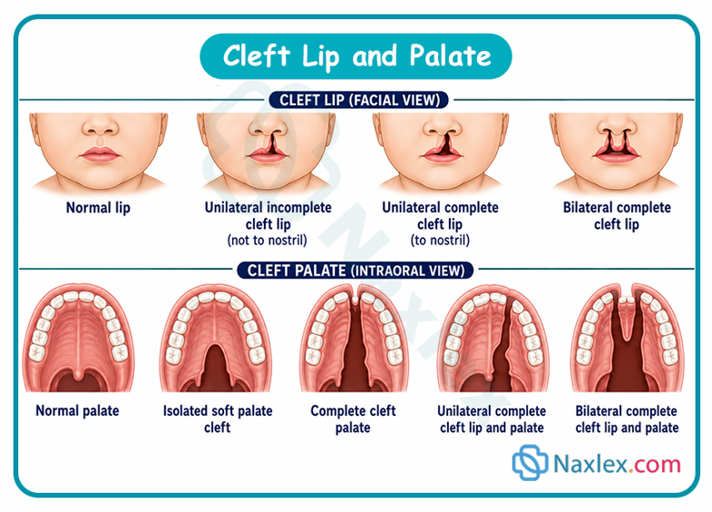
Epidemiology
- Incidence: Combined cleft lip with or without cleft palate occurs in approximately 1 in 700 to 1 in 1,000 live births globally. Isolated cleft palate occurs in roughly 1 in 2,500 live births.
- Demographics:
- Cleft lip (with or without cleft palate) is more prevalent in males.
- Isolated cleft palate is more common in females.
- Ethnic Variations: Highest incidence among Native American and Asian populations; lowest among African-descent populations.
Etiology & Risk Factors
Cleft malformations are primarily multifactorial in origin, stemming from a combination of genetic susceptibility and environmental exposures during early organogenesis (weeks 4 through 12 of gestation).
- Genetic Factors: Chromosomal abnormalities (e.g., Trisomy 13) and over 300 syndromic conditions (e.g., Pierre Robin sequence, Van der Woude syndrome).
- Maternal Environmental Exposures:
- Teratogenic medications (e.g., anticonvulsants like phenytoin, valproic acid, and retinoids).
- Maternal alcohol consumption and heavy cigarette smoking.
- Folate deficiency prior to and during early pregnancy.
- Maternal gestational diabetes or intra-uterine infections.
Pathophysiology
- Cleft Lip: Occurs during the 4th to 7th week of embryonic development. The failure of fusion between the medial nasal prominence and the maxillary prominence leads to a gap in the lip tissue and underlying orbicularis oris muscle.
- Cleft Palate: Occurs during the 7th to 12th week of embryonic development. The secondary palate fails to join when the lateral palatal shelves fail to elevate, approximate, and fuse at the midline due to abnormal cell migration, tongue interference, or structural restriction.
- Functional Impact: The absence of intact oral architecture prevents the creation of a negative pressure vacuum required for normal sucking, diverting oral liquids into the nasal cavity and placing the infant at high risk for aspiration and chronic middle ear effusion.
Clinical Manifestations
- Physical Findings:
- Visible separation of the upper lip (unilateral or bilateral).
- Direct visualization/palpation of a gap in the hard or soft palate.
- Bifid uvula (may indicate a submucous cleft palate).
- Nasal asymmetry or flattened ala.
- Functional Alterations:
- Inability to achieve a seal on a breast or standard bottle nipple.
- Nasal regurgitation of fluids during feeding.
- Excessive air swallowing during feedings (causes abdominal distension).
- Frequent middle ear infections (otitis media) due to Eustachian tube dysfunction.
- Hypernasal speech and articulation delays later in childhood.
Diagnostic Evaluation
- Prenatal Diagnosis: Routine high-resolution structural ultrasound (often detected as early as 18–20 weeks gestation). Cleft palate without cleft lip is difficult to visualize on prenatal ultrasound.
- Postnatal Physical Exam: Immediate visual evaluation of the lip and direct intraoral palpation using a gloved finger to inspect the hard and soft palates.
- Diagnostic Screening:
- Audiologic Testing: Auditory Brainstem Response (ABR) screening due to high risk of conductive hearing loss.
- Genetic Evaluation: Karyotyping or chromosomal microarray if associated congenital anomalies are suspected.
Therapeutic Management
Repair requires a coordinated, interprofessional team effort (pediatrician, plastic surgeon, pediatric dentist, speech-language pathologist, audiologist, pediatric nurse, otolaryngologist, social worker).
- Surgical Timing:
- Cheiloplasty (Cleft Lip Repair): Performed around 2 to 3 months of age. Often follows the "Rule of 10s": 10 weeks old, 10 pounds (4.5 kg), and hemoglobin of 10 g/dL.
- Palatoplasty (Cleft Palate Repair): Performed between 6 to 12 months of age (before significant speech development occurs) to optimize speech outcomes and anatomical growth.
Nursing Care Management
Feeding Strategies (Highest Priority Pre-Op)
- Positioning: Feed infant in an upright position (at least 60–90 degrees) to prevent fluid from flowing into the nasal cavity and Eustachian tubes.
- Specialized Equipment:
- Haberman Fe سف (SpecialNeeds Feeder): Utilizes a one-way valve and squeeze-controllable nipple.
- Mead Johnson Cleft Palate Nurser: Squeezable bottle to assist fluid delivery without requiring strong suction.
- Soft, wide-base nipples with cross-cut openings.
- Technique:
- Compress the bottle rhythmically to timed sucks.
- Burp frequently (every 0.5 to 1 oz or every 3–5 minutes) because these infants swallow large amounts of air.
- Feedings should be completed within 30 minutes to prevent calorie exhaustion.
Pre-Operative Care
- Educate caregivers on modified feeding techniques and ensure weight gain metrics are met.
- Acclimate the infant to post-operative positioning and restraint devices (e.g., arm elbow immobilizers).
- Ensure NPO status prior to surgery as ordered.
Post-Operative Care
- Airway & Respiratory Protection:
- Maintain clear airway; position to promote drainage (side-lying or back; never prone after cleft lip repair).
- Keep emergency suction and oxygen equipment at bedside. Suction gently only if critical, avoid deep or harsh oral suctioning.
- Suture Line Protection:
- Apply soft elbow immobilizers ("no-nos") to prevent the infant from touching the surgical site or placing objects/hands in the mouth. Remove per protocol (e.g., every 2 hours) to assess skin and perform range of motion.
- Avoid hard objects in the mouth (e.g., pacifiers, spoons, straws, thermometers, standard nipples).
- Apply prescribed topical antibiotic ointment or petroleum jelly to the lip repair line as ordered.
- Pain Management: Administer round-the-clock analgesics (e.g., acetaminophen, ibuprofen) to minimize crying, which places stress on suture lines.
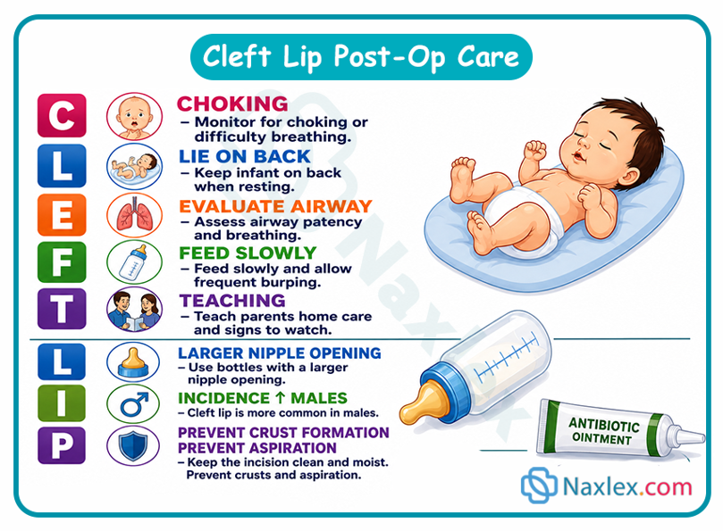
Caregiver Education
- Wound Care: Demonstrate gentle cleaning of the suture line with sterile water/saline using a cotton swab, followed by antibiotic ointment application.
- Feeding Progression: Instruct on open-cup or specialized syringe/dropper feeding during immediate post-op recovery as directed by the surgeon.
- Signs of Infection: Teach parents to monitor for redness, swelling, purulent drainage, or fever above 100.4°F (38°C).
- Restraint Compliance: Emphasize the vital need for elbow restraints at all times when the infant is unattended.
-
Long-Term Complications
- Hearing Impairment: Recurrent otitis media due to inefficient tensor veli palatini muscle function, causing Eustachian tube dysfunction; often requires tympanostomy tube placement.
- Speech Disorders: Hypernasality, compensatory articulation errors, and velopharyngeal insufficiency (VPI) requiring speech therapy or secondary surgical revision.
- Dental/Orthodontic Problems: Malocclusion, missing or supernumerary teeth, delayed tooth eruption, and alveolar arch misalignment.
- Psychosocial Impact: Body image concerns, self-esteem challenges, and potential peer-related socialization issues during school age and adolescence.
Esophageal Atresia And Tracheoesophageal Fistula
Definition
- Esophageal Atresia (EA): A congenital malformation in which the esophagus fails to develop as a continuous passage, ending in a blind pouch instead of connecting to the stomach.
- Tracheoesophageal Fistula (TEF): An abnormal fistulous connection between the esophagus and the trachea, allowing gastric contents or oral secretions to move directly into the respiratory tract.
Epidemiology
- Incidence: Occurs in approximately 1 in 2,500 to 1 in 4,500 live births globally.
- Associations: Over 50% of infants with EA/TEF have coexisting congenital anomalies, most commonly the VACTERL association:
- Vector/Vertebral defects
- Anal atresia
- Cardiac defects
- Tracheo-Esophageal fistula / Esophageal atresia
- Renal anomalies
- Limb defects (e.g., radial dysplasia)
Etiology
- Embryological Defect: Caused by defective lateral compartmentalization and incomplete separation of the primitive foregut into distinct anterior respiratory (trachea) and posterior digestive (esophagus) tubes during the 4th to 6th weeks of gestation.
- Genetics & Teratogens: Primarily sporadic, but can be linked to chromosomal abnormalities (e.g., Trisomy 18, 21, or 13) or maternal exposure to teratogens.
Pathophysiology
- Proximal Blind Pouch: Saliva and swallowings accumulate in the upper pouch, overflowing into the larynx and lungs, leading to acute choking, excessive drooling, and aspiration pneumonia.
- Distal Fistula: Air enters the distal esophagus from the trachea during inspiration, causing severe gastric distension and elevated diaphragm (compromising lung expansion). Reflux of acidic gastric juice into the lungs via the fistula leads to chemical pneumonitis.
Common Types (Vogt / Gross Classification)
|
Type |
Description |
Frequency |
|
Type C (EA with Distal TEF) |
Proximal esophagus ends in a blind pouch; distal segment connects to lower trachea/bronchus. |
~85% (Most Common) |
|
Type A (Isolated EA) |
Pure atresia; both upper and lower esophageal segments end in blind pouches with no fistula. |
~8% |
|
Type E (Isolated TEF / H-type) |
Continuous esophagus and trachea connected by an "H-shaped" fistulous tract (no atresia). |
~4% |
|
Type B (EA with Proximal TEF) |
Upper pouch connects to trachea; lower segment ends in a blind pouch. |
~1% |
|
Type D (Double TEF) |
Both proximal and distal esophageal segments connect independently to the trachea. |
~1% |
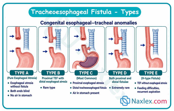
Clinical Manifestations
- The Classical "Three Cs": Coughing, Choking, and Cyanosis (especially during initial feeding attempts).
- Infant Presentation:
- Excessive fine, frothy bubbles of mucus/saliva at the mouth and nose.
- Recurrent cyanotic spells during feedings.
- Abdominal distension (due to air entering the stomach via a distal fistula).
- In isolated EA without TEF, the abdomen will appear abnormally flat/scaphoid.
Diagnostic Evaluation
- Prenatal: Maternal polyhydramnios on ultrasound (due to inability of fetus to swallow amniotic fluid) with a non-visualized fetal stomach bubble.
- Postnatal Bedside Test: Inability to pass a rigid 8–10 Fr radiopaque Replogle or nasogastric tube into the stomach (meets resistance/coils at 10–12 cm from the lips).
- Chest & Abdominal X-ray:
- Confirms tube coiling in the upper blind esophageal pouch.
- Gas in the GI tract indicates the presence of a distal TEF; a completely airless abdomen indicates isolated EA.
- Echo & Renal Ultrasound: Performed to Rule out associated VACTERL anomalies before surgery.
Therapeutic Management
- Pre-Operative Stabilization: Immediate cessation of oral intake (NPO), fluid hydration via IV, continuous suctioning of the upper pouch, and airway protection.
- Surgical Repair:
- Primary Repair: Right thoracotomy or thoracoscopic repair featuring fistulous tract ligation and primary end-to-end esophageal anastomosis.
- Staged Repair: Used for "long-gap" EA where segments are too far apart; involves temporary gastrostomy tube placement for nutrition, followed by delayed repair or esophageal replacement (e.g., colonic interposition).
Nursing Care Management
Immediate Pre-Operative Nursing Interventions
- Maintain NPO Status: Stop all oral feedings immediately.
- Airway Maintenance & Decompression:
- Insert a double-lumen Replogle tube into the upper pouch connected to low continuous suction (-20 to -30 mmHg) to clear mucus and prevent aspiration.
- Position infant Head-of-Bed (HOB) elevated 30–45 degrees to prevent gastric reflux into the lungs via the distal fistula.
- Oxygenation: Administer oxygen with caution. Avoid high-pressure positive pressure ventilation (can cause gastric rupture via fistula).
Post-Operative Nursing Care
- Airway & Respiratory Care:
- Maintain mechanical ventilation or supplemental oxygen as ordered.
- Suctioning Caution: Suction with extreme care using a pre-measured catheter; never pass a catheter past the suture line (anastomosis) to prevent disruption.
- Positioning: Maintain HOB elevated; avoid neck hyperextension to reduce tension on the esophageal suture line.
- Drain Management: Monitor and document chest tube or trans-anastomotic wound drain output (sudden increases in saliva-like output indicates an anastomotic leak).
- Nutrition:
- Maintain IV fluids / TPN initially.
- Initiate Gastrostomy / Trans-anastomotic tube (TAT) feedings slowly around post-op day 3–5 as ordered.
- Administer sham/comfort oral feeding (e.g., non-nutritive sucking on a pacifier) during G-tube feeds to promote normal sucking reflexes.
- Prior to oral feedings, an esophagogram (contrast study) is performed to verify absence of anastomotic leaks.
Complications
- Early Post-Op:
- Anastomotic Leak: Manifests as chest drainage, subcutaneous emphysema, or pneumothorax.
- Surgical Wound Infection or sepsis.
- Late/Long-Term:
- Esophageal Stricture: Dysphagia and feeding refusal due to scar tissue at the surgical site (requires balloon dilation).
- Severe Gastroesophageal Reflux (GERD): Impaired lower esophageal sphincter function.
- Tracheomalacia: Flaccid tracheal rings causing a characteristic "TEF cough" (barking cough), stridor, or recurrent respiratory infections.
Prognosis
- Overall Survival: Exceeds 90–95% in tertiary centers for infants with normal birth weight and no major cardiac anomalies.
- Primary Prognostic Factor: Mortality is primarily dictated by the severity of coexisting congenital heart defects or extreme prematurity/low birth weight rather than the esophageal repair itself.
Omphalocele
Definition
- Omphalocele: A midline congenital abdominal wall defect in which abdominal organs (bowel, liver, stomach, and occasionally spleen or gonads) herniate into the base of the umbilical cord, remaining enclosed within a protective sac.
- Distinction: Differs from gastroschisis, which is a full-thickness paraumbilical wall defect (typically to the right of the umbilicus) with no protective sac covering the exposed bowel.
Image Title: Omphalocele
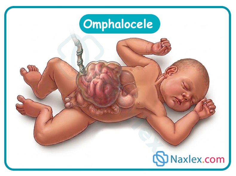
Epidemiology
- Incidence: Occurs in approximately 1 in 3,000 to 1 in 5,000 live births globally.
- Associated Anomalies: Over 50% to 70% of infants with an omphalocele have associated congenital, structural, or chromosomal abnormalities.
- Common Syndromes & Associations:
- Chromosomal Trisomies: Trisomy 13 (Patau), Trisomy 18 (Edwards), and Trisomy 21 (Down).
- Beckwith-Wiedemann Syndrome: Characterized by macrosomia, macroglossia, hypoglycemia, and omphalocele.
- Pentalogy of Cantrell: Midline abdominal wall defect, lower sternal defect, anterior diaphragmatic defect, pericardial defect, and intracardiac anomalies.
Etiology & Risk Factors
- Embryological Defect: Caused by the failure of the physiological midgut loop to return from the extraembryonic coelom (umbilical cord) to the abdominal cavity during the 10th to 12th week of embryonic development.
- Risk Factors: Advanced maternal age, maternal smoking, and exposure to certain medications (e.g., SSRIs) during early organogenesis.
Pathophysiology
- Sac Integrity: The protective sac prevents direct chemical irritation of the intestines from exposure to amniotic fluid in utero. However, if the sac ruptures during delivery, the risk of severe bacterial peritonitis, hypothermia, and fluid loss increases drastically.
- Abdominal Cavity Underdevelopment: Because the abdominal organs develop outside the abdominal cavity, the abdominal domain remains abnormally small (hypoplastic).
- Cardiorespiratory Impact: Rapid reduction of the herniated organs back into a small abdominal cavity can elevate intra-abdominal pressure, compressing the diaphragm, compromising pulmonary compliance, and obstructing vena cava blood return.
Clinical Manifestations
- Central Mass: A midline, variable-sized abdominal wall defect at the umbilical ring.
- Enclosing Membrane: A translucent, membranous sac covering the herniated viscera (umbilical cord inserts directly into the top of the sac).
- Associated Physical Findings:
- Macroglossia or umbilical enlargement (indicative of Beckwith-Wiedemann syndrome).
- Respiratory distress (secondary to associated pulmonary hypoplasia or elevated intra-abdominal pressure).
- Murmurs or signs of heart failure (due to coexisting congenital heart defects).
Diagnostic Evaluation
- Prenatal Diagnosis:
- Elevated Maternal Serum Alpha-Fetoprotein (MSAFP): Elevated in the second trimester.
- Prenatal Ultrasound: Visualizes a membrane-covered abdominal mass with umbilical cord insertion at the apex (detectable as early as 12–14 weeks).
- Fetal Echocardiogram & Karyotyping/Microarray: Strongly recommended prenatal tests to evaluate for cardiac defects and chromosomal anomalies.
- Postnatal Evaluation:
- Immediate physical inspection to confirm sac integrity (intact vs. ruptured).
- X-rays & Echocardiogram: Performed post-delivery to assess for associated cardiac, chest, and skeletal malformations.
Therapeutic Management
Management depends primarily on the size of the defect, sac integrity, and the presence of associated anomalies.
- Surgical Management:
- Primary Closure: For small-to-moderate omphaloceles where viscera can safely fit back into the abdominal cavity in a single operation.
- Staged Repair (Silo Placement): Used for large/giant omphaloceles. A prosthetic silo is suspended over the abdomen, and the viscera are gradually reduced daily into the abdominal cavity using gravity before final abdominal closure.
- Non-Surgical Management ("Paint and Wait"):
- Used for giant omphaloceles with pulmonary hypoplasia or severe cardiac defects. Topical escharotic agents (e.g., silver sulfadiazine or povidone-iodine) are applied to the sac to promote epithelialization, forming a stable ventral hernia that is repaired surgically years later.
Nursing Care Management
Immediate Pre-Operative / Neonatal Stabilization
- Sac Protection: Cover the sac immediately with sterile, warm, saline-soaked non-adherent dressings and wrap in a sterile bowel bag or plastic wrap to conserve heat and moisture.
- Positioning: Place the infant on their side or back; never prone to prevent mechanical rupture or ischemia of the sac.
- Thermoregulation: Place the infant under a radiant warmer immediately. Exposed sacs cause rapid radiant and evaporative heat loss.
- Fluid & Gastrointestinal Management:
- Place a low-intermittent suction Replogle or nasogastric tube to decompress the stomach and prevent bowel distension/aspiration.
- Keep the infant strict NPO and maintain IV fluid replacement (maintenance TPN/electrolytes).
Post-Operative Nursing Care
- Respiratory Monitoring: Monitor for signs of increased intra-abdominal pressure (elevated peak airway pressures, tachypnea, desaturations) as the abdominal domain adapts.
- Abdominal Compartment Syndrome Monitoring:
- Measure abdominal girth and monitor lower limb perfusion (capillary refill, femoral pulses).
- Assess urine output (oliguria < 1 mL/kg/hr indicates renal compression from elevated intra-abdominal pressure).
- Pain & Sedation: Administer continuous analgesics to minimize crying and intra-abdominal straining.
- Infection Control: Maintain aseptic technique during dressing changes and inspect the reduction site for erythema, drainage, or foul odor.
Complications
- Sac Rupture: Converts an omphalocele into an emergency equivalent to gastroschisis, drastically increasing risk of sepsis and hypothermia.
- Abdominal Compartment Syndrome: Elevated abdominal pressure causing decreased renal perfusion, mesenteric ischemia, and respiratory failure.
- Sepsis / Peritonitis: Wound infection or contamination of exposed bowel tissue.
- Long-Term Complications: Feeding intolerance, gastroesophageal reflux disease (GERD), prolonged ileus, and ventral hernias.
Prognosis
- Survival Rate: Approaches 90% for isolated omphaloceles treated in specialized surgical centers.
- Prognostic Determinants: Overall survival and long-term morbidity are overwhelmingly dictated by the presence and severity of associated anomalies (especially severe congenital heart disease and chromosomal trisomies) and the degree of pulmonary hypoplasia.
Gastroschisis
Definition
- Gastroschisis: A full-thickness anterior abdominal wall defect, typically located immediately to the right of an intact umbilical cord, resulting in the protrusion of uncovered abdominal organs (primarily small and large intestines, and occasionally the stomach or gonads).
- Key Characteristic: There is no protective sac or membrane covering the exposed bowel.
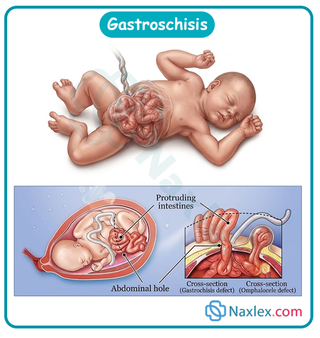
Epidemiology
- Incidence: Occurs in approximately 1 in 2,000 to 1 in 4,000 live births.
- Maternal Demographics: Strongly associated with young maternal age (mothers under 20 years old have a significantly higher risk).
- Associated Anomalies: Unlike omphalocele, extra-gastrointestinal or chromosomal anomalies are rare (<10%). Most coexisting defects are secondary intestinal malformations (e.g., intestinal atresia, stenosis, or malrotation).
-
Etiology & Risk Factors
- Embryological Defect: Vascular disruption of the right omphalomesenteric (vitelline) artery or abnormal involution of the right umbilical vein during early embryonic development, leading to ischemic weakness and rupture of the abdominal wall.
- Risk Factors: Young maternal age, low maternal body mass index (BMI), maternal cigarette smoking, alcohol use, recreational drug exposure (e.g., cocaine), and over-the-counter vasoconstrictive medications (e.g., pseudoephedrine).
-
Pathophysiology
- Chemical Peritonitis: Prolonged intrauterine exposure to acidic amniotic fluid and waste products causes severe inflammation, resulting in thickened, matted, edematous, and leathery bowel loops coated with a fibrous peel.
- Massive Evaporative Heat & Fluid Losses: Direct atmospheric exposure of the uncovered microvascular bowel mucosa leads to severe hypothermia, third-spacing of fluids, and hypovolemic shock if not immediately covered.
- Intestinal Dysmotility: Chronic inflammation damages the enteric nervous system and intestinal smooth muscle, causing prolonged post-operative paralytic ileus and delayed gastric emptying.
-
Clinical Manifestations
- Visual Presentation: Eviscerated bowel loops protruding through a small (<4 cm) abdominal wall defect to the right of the intact umbilical cord.
- Bowel Characteristics: Viscera appear edematous, hyperemic, thickened, shortened, and covered in a thick fibrous exudate.
- Systemic Signs: Dehydration, poor peripheral perfusion, hypothermia, and metabolic acidosis due to rapid fluid/heat loss.
-
Diagnostic Evaluation
- Prenatal Diagnosis:
- Elevated Maternal Serum Alpha-Fetoprotein (MSAFP): Significantly higher levels compared to omphalocele due to direct, uncovered contact between fetal serum/bowel and amniotic fluid.
- Prenatal Ultrasound: Detects free-floating loops of bowel in the amniotic cavity without an enclosing membrane (usually identified during the 18–20 week anomaly scan).
- Postnatal Evaluation:
- Direct physical inspection to evaluate bowel viability (inspect for dusky, ischemic, or necrotic segments indicating volvulus or constriction at the defect ring).
-
Therapeutic Management
- Surgical Management:
- Primary Closure: Eviscerated bowel is manually reduced back into the abdominal cavity in the operating room or at the bedside (sutureless closure) under analgesia, followed by abdominal wall closure.
- Staged Reduction (Silo Placement): Used when the abdominal cavity is too small or bowel edema is too severe. A transparent, pre-formed spring-loaded silastic silo is placed over the exposed bowel. The silo is gradually squeezed/reduced daily using gravity over 3–14 days until the viscera reside fully within the abdomen, followed by surgical closure.
-
Nursing Care Management
Immediate Pre-Operative / Resuscitation Priorities
- Bowel Protection & Positioning:
- Place the lower torso and exposed bowel inside a sterile bowel bag (clear plastic wrap/saline-soaked sterile gauze) up to the axillae to prevent evaporative water loss, heat loss, and contamination.
- Position the infant on their right side or supine with supportive rolls to prevent kinked mesenteric vessels and intestinal ischemia.
- Fluid & Hemodynamic Resuscitation:
- Establish IV access immediately; administer IV fluids (isotonic crystalloids) at 1.5 to 2 times maintenance rates (150–200 mL/kg/day) to offset massive third-spacing.
- Gastrointestinal Decompression:
- Insert a continuous low-suction Replogle or nasogastric tube (8–10 Fr) to decompress the stomach and bowel, preventing aspiration and further bowel distension.
- Thermoregulation:
- Place infant immediately under a pre-warmed radiant warmer. Maintain continuous core temperature monitoring.
-
Post-Operative Nursing Care
- Abdominal Compartment Syndrome Monitoring:
- Assess for elevated intra-abdominal pressure: monitor lower extremity perfusion (femoral pulses, capillary refill), urine output (maintain >1 mL/kg/hr), and peak airway pressures on mechanical ventilation.
- Nutritional Support:
- Maintain total parenteral nutrition (TPN) and lipids via a central line due to prolonged paralytic ileus (often lasting several weeks).
- Initiate trophic minimal enteral nutrition (breast milk preferred) only when bowel function returns (indicated by decreased bilious NG output, passage of stool, and presence of bowel sounds).
- Infection Control & Pain Management:
- Maintain strict aseptic technique with silo dressings and central lines.
- Administer continuous analgesia (e.g., morphine infusion) to prevent intra-abdominal pressure spikes from crying.
-
Complications
- Intestinal Atresia / Stenosis: Occurs in 10–15% of cases due to localized vascular compression at the abdominal wall defect ring.
- Necrotizing Enterocolitis (NEC) & Sepsis: Elevated risk during bowel reduction or initial feeding trials.
- Short Bowel Syndrome: Secondary to extensive surgical resection of necrotic or atretic bowel loops.
- Prolonged Paralytic Ileus: Dysmotility requiring months of parenteral nutritional support.
-
Prognosis
- Survival Rate: Exceeds 90–95% in tertiary pediatric surgical centers.
- Determinants: Long-term outcome depends on the presence of secondary bowel complications (e.g., short bowel syndrome, severe dysmotility, or sepsis) rather than associated structural organ anomalies.
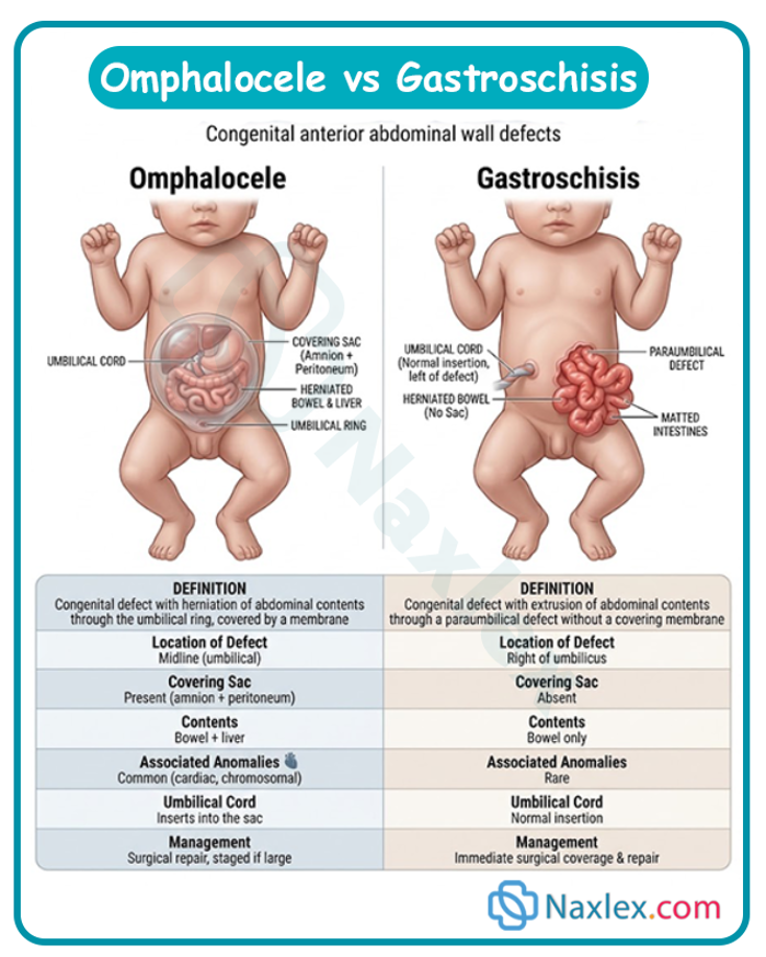
Anorectal Malformations
Definition
- Anorectal Malformations (ARMs): A congenital complex of structural defects characterized by the maldevelopment or absence of a normal anal opening, abnormal termination of the rectum, and frequent fistulous connections to the perineum or adjacent urogenital structures (imperforate anus).
Epidemiology
- Incidence: Occurs in approximately 1 in 2,500 to 1 in 5,000 live births globally.
- Gender Predilection: Slightly more common in males. Males present more frequently with rectourethral fistulas; females present more commonly with rectovestibular fistulas.
- Associated Anomalies: Found in 50% to 60% of cases. The most common is the VACTERL association (Vertebral, Anal, Cardiac, Tracheoesophageal, Renal, Limb defects) or sacral dysplasia.
Etiology
- Embryological Defect: Results from improper division of the primitive cloaca by the urorectal septum between the 4th and 7th weeks of embryonic development.
- Genetics: Primarily sporadic and multifactorial, though higher risks occur in specific chromosomal syndromes (e.g., Down syndrome, Townes-Brocks syndrome, Currarino triad).
Pathophysiology
- Structural Failure: Incomplete mesodermal development prevents the rectum from descending fully through the levator ani muscle complex to reach the perineal skin.
- Fistula Formation: The persistent embryonic cloaca leaves open abnormal fistulous tracts connecting the distal bowel to either the urinary tract (urethra/bladder in males) or reproductive tract (vestibule/vagina in females).
- Neuromuscular Impact: Hypoplasia of the internal and external anal sphincters and underlying sacral nerve roots directly influences lifelong fecal continence.
Classification (Krickenbeck System)
Anorectal malformations are categorized clinically by the presence and anatomical path of the fistulous tract:
- Major Clinical Groups:
- Perineal (Cutaneous) Fistula: Low defect where the rectum opens onto the perineum anterior to the center of the external sphincter.
- Rectourethral Fistula (Males): High/intermediate defect connecting the rectum to the prostatic or bulbar urethra.
- Rectovesical Fistula (Males): High defect connecting the rectum directly to the bladder neck.
- Rectovestibular Fistula (Females): The rectum opens into the vestibule of the female genitalia outside the hymen.
- Persistent Cloaca (Females): Complex defect where the rectum, vagina, and urinary tract converge into a single shared common channel opening on the perineum.
- ARM without Fistula: Imperforate anus with a blind-ending rectal pouch and no connection to adjacent tracts.
Clinical Manifestations
- Inability to Pass Meconium: Failure to pass meconium within the first 24 to 48 hours of life.
- Physical Inspection Findings:
- Absence of a normal anal opening or mislocated anal orifice.
- Presence of meconium discharging from the urethra, scrotum, or vaginal vestibule.
- Flat perineum or absence of a gluteal fold (indicates poor sacral development and poor sphincter muscle complex).
- Systemic Signs of GI Obstruction: Progressive abdominal distension, bilious vomiting, and irritability as bowel gas accumulates.
Diagnostic Evaluation
- Physical & Perineal Exam: Performed under good lighting 16–24 hours after birth (allows intra-abdominal pressure to build up gas and meconium, forcing it down to reveal a fistula).
- Cross-Table Invertogram / Prone Cross-Table Radiograph: Evaluates the distance between the distal gas-filled rectal pouch and a radio-opaque marker placed on the perineum (helps differentiate high vs. low defects).
- Pelvic & Abdominal Ultrasound: Evaluates distance to the perineum and screens for associated renal anomalies (hydronephrosis, renal agenesis).
- Spinal Ultrasound or MRI: Assesses for spinal cord tethering or sacral dysgenesis.
- Echocardiogram: Performed prior to surgery to Rule out coexisting VACTERL structural heart defects.
Therapeutic Management
Surgical Interventions
- Low Defects (e.g., Perineal Fistula): Managed with primary Anoplasty (repositioning the rectal opening within the sphincter complex) in the immediate newborn period without a colostomy.
- High/Complex Defects (e.g., Rectourethral, Cloaca): Managed via a staged surgical approach:
- Stage 1 (Neonatal Period): Creation of a divided diverting colostomy (usually high sigmoid) to decompress the bowel and protect the urinary tract.
- Stage 2 (3–6 Months of Age): Posterior Sagittal Anorectoplasty (PSARP / Peña Procedure) to pull the rectum down through the center of the sphincter muscle complex.
- Stage 3: Colostomy closure weeks to months after the PSARP site has healed and dilations are complete.
Nursing Care Management
Pre-Operative Care
- Keep the infant strict NPO and administer IV fluids.
- Maintain nasogastric or Replogle tube decompression to relieve abdominal distension and prevent aspiration.
- Avoid taking rectal temperatures.
- Monitor urine for meconium flakes (indicates a rectourinary fistula and high risk for ascending urinary tract infection).
Post-Operative Care (Post-PSARP & Anoplasty)
- Suture Line & Wound Care:
- Keep the surgical site clean and dry; perform gentle cleansing after voids/stools.
- Position the infant side-lying or prone (or elevated hips) to keep pressure off the operative site.
- Pain Management: Administer IV analgesics systematically to reduce crying and surgical site tension.
- Anal Dilations:
- Teach parents how to perform scheduled Hegar dilator routines (typically started 2 weeks post-op) to prevent scar stricture of the new anus.
- Dilator size is gradually increased according to a strict schedule provided by the surgeon.
Stoma Management (If Colostomy Created)
- Assess stoma color (should remain beefy red; pale/dusky indicates ischemia).
- Protect peri-stomal skin using skin barrier pastes and appropriately fitted pouching systems.
Complications
- Short-Term: Surgical site infection, wound dehiscence, urethral injury, or anastomotic stricture.
- Long-Term:
- Fecal Incontinence: Inability to control bowel movements due to underdeveloped sphincter muscles or altered nerve pathways.
- Severe Chronic Constipation / Megacolon: Secondary to impaired rectal motility and anterior muscle weakness.
- Neurogenic Bladder / Urinary Incontinence: Secondary to coexisting sacral nerve abnormalities or operative nerve injury.
Prognosis
- Low Defects: Excellent prognosis; over 80% to 90% achieve voluntary bowel control and full fecal continence.
- High / Complex Defects (e.g., Cloaca, Rectovesical): Variable long-term continence outcomes. Many children require lifelong bowel management protocols (e.g., daily enemas, specialized high-fiber diets, laxative regimens) and multidisciplinary follow-up.
Meckel Diverticulum
Definition
Meckel's Diverticulum: A true congenital pouch-like sacculation located on the antimesenteric border of the ileum, caused by the failure of the embryonic vitelline duct (omphalomesenteric duct) to fully atrophy and close.
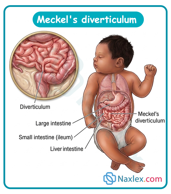
Epidemiology
Meckel's diverticulum classically follows the "Rule of 2s":
- Occurs in approximately 2% of the general population.
- Located within 2 feet (approx. 60 cm) of the ileocecal valve.
- Approximately 2 inches (approx. 5 cm) in length.
- Contains 2 types of common ectopic mucosa (gastric and pancreatic).
- Most commonly becomes symptomatic before 2 years of age.
- 2 times more common in males to be symptomatic than females.
Etiology
- Embryological Defect: Caused by the incomplete obliteration of the vitelline duct (which normally connects the yolk sac to the midgut in the early embryo) during the 5th to 7th week of gestation.
- Incomplete Regressional Variants: Partial persistence can result in a Meckel's diverticulum, a vitelline cyst, an umbilical-enteric fistula, or a fibrous band connecting the ileum to the umbilicus.
Pathophysiology
- Heterotopic/Ectopic Tissue: Up to 50% of symptomatic diverticula contain ectopic tissue, most commonly ectopic gastric mucosa (and less frequently heterotopic pancreatic tissue).
- Ulceration & Hemorrhage: The heterotopic gastric mucosa secretes hydrochloric acid and pepsin. Because the adjacent ileal mucosa lacks protective mechanisms against gastric acid, chronic irritation leads to peptic ulceration of the ileal wall, causing painless intestinal bleeding.
- Mechanical Obstruction: The diverticulum can act as a lead point for intussusception or cause bowel volvulus around a persistent fibrous vitelline cord.
Clinical Manifestations
- Painless Lower GI Bleeding: The hallmark presentation in young children is the sudden onset of painless, massive rectal bleeding ("brick-red," "currant jelly," or maroon-colored stools).
- Anemia & Hypovolemia: Pallor, fatigue, tachycardia, hypotension, and dizziness secondary to acute or chronic blood loss.
- Intestinal Obstruction Signs: Bilious vomiting, severe colicky abdominal pain, abdominal distension, and obstipation (if complicated by intussusception or volvulus).
- Diverticulitis: Symptoms closely mimicking acute appendicitis (periumbilical pain shifting to the right lower quadrant, fever, guarding, and rebound tenderness).
Diagnostic Evaluation
- Meckel's Scan (Technetium-99m Pertechnetate Scintigraphy): The diagnostic test of choice. Technetium-99m is selectively taken up by ectopic gastric mucosa, highlighting the diverticulum in the lower abdomen.
- Complete Blood Count (CBC): Evaluates severity of anemia (decreased Hemoglobin and Hematocrit) and leukocytosis (if diverticulitis or perforation is present).
- Stool for Occult Blood / Visual Inspection: Assesses stool color and presence of frank blood.
- Abdominal Ultrasound / CT Scan: Helps Rule out appendicitis, intussusception, or abscess formation.
- Wireless Capsule Endoscopy or Angiography: Used in obscure GI bleeding cases when nuclear medicine scans are equivocal.
Therapeutic Management
- Surgical Resection: The definitive treatment for a symptomatic Meckel's diverticulum:
- Meckel's Diverticulectomy: Excision of the pouch alone with transverse closure of the base.
- Segmental Ileal Resection: Excision of the diverticulum along with the adjacent ulcerated segment of ileum, followed by end-to-end anastomosis.
- Asymptomatic Incidental Findings: Incidental resection during unrelated abdominal procedures remains controversial and is determined based on patient age and diverticulum characteristics.
Nursing Care Management
Pre-Operative Care & Hemodynamic Stabilization
- Assess Hemodynamic Status: Closely monitor vital signs for signs of hypovolemic shock (tachycardia, hypotension, delayed capillary refill, weak peripheral pulses).
- Fluid & Blood Replacement: Establish large-bore IV access; administer isotonic IV fluids and blood products (Packed Red Blood Cells) as ordered.
- Gastrointestinal Decompression: Place the infant/child on strict NPO status and insert a nasogastric (NG) tube to continuous low intermittent suction if bowel obstruction or active vomiting is present.
- Stool Assessment: Document color, consistency, frequency, and volume of all bloody stools.
Post-Operative Care
- Airway & Respiratory Management: Maintain HOB elevated 30 degrees; monitor pulse oximetry and breath sounds.
- Gastrointestinal & Stoma/Wound Care:
- Maintain NG tube suction until bowel function returns.
- Monitor for signs of paralytic ileus: assess bowel sounds, abdominal girth, and passage of flatus or stool before initiating oral fluids.
- Inspect abdominal incision for signs of infection (erythema, swelling, purulent drainage) or wound dehiscence.
- Pain Management: Administer prescribed IV analgesics regularly to maintain comfort and facilitate deep breathing/movement.
- Nutrition: Slowly progress diet from clear liquids to regular foods once bowel sounds return and the NG tube is removed.
Complications
- Severe Hemorrhagic Shock: Rapid, massive blood loss from deep ileal ulcerations.
- Intestinal Obstruction / Intussusception: Invagination of the diverticulum into the intestinal lumen causing bowel ischemia.
- Diverticular Perforation & Peritonitis: Severe inflammation leading to rupture, causing acute abdomen, sepsis, and shock.
Prognosis
- Overall Prognosis: Excellent following surgical resection.
- Long-Term Outcomes: Full recovery is expected with zero long-term digestive deficits once the symptomatic diverticulum is successfully excised.
Inguinal Hernias
Definition
- Inguinal Hernia: The protrusion of abdominal contents (such as loops of small intestine, omentum, or occasionally an ovary or Meckel's diverticulum) through the internal inguinal ring into the inguinal canal.
- Indirect Inguinal Hernia: Herniation through the internal inguinal ring lateral to the inferior epigastric vessels (accounts for nearly all pediatric cases).
- Direct Inguinal Hernia: Herniation through a weakness in the abdominal floor (Hesselbach's triangle) medial to the inferior epigastric vessels (rare in children; more common in adults).
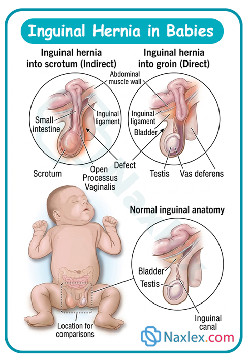
Epidemiology
- Incidence: Occurs in 1% to 5% of all full-term newborns, but rises significantly to 10% to 30% in premature infants.
- Gender Predilection: Approximately 3 to 4 times more common in males than in females.
- Laterality: More common on the right side (~60%), followed by the left side (~30%), and bilateral (~10%). This is due to the later descent of the right testicle and subsequent later closure of the right processes vaginalis.
-
Etiology & Risk Factors
- Congenital Defect: Caused by the persistence or incomplete obliteration of the processus vaginalis (an embryonic peritoneal pouch that accompanies the testis during its descent into the scrotum during the 7th to 8th month of gestation).
- Risk Factors:
- Prematurity and low birth weight.
- Male sex and undescended testes (cryptorchidism).
- Conditions causing increased intra-abdominal pressure (e.g., ascites, ventriculoperitoneal shunts, cystic fibrosis, chronic cough).
- Connective tissue disorders (e.g., Ehlers-Danlos syndrome, Marfan syndrome).
-
Pathophysiology
- Patent Processus Vaginalis (PPV): If the processus vaginalis remains open after birth, a potential tract is left open connecting the peritoneal cavity to the inguinal canal and scrotum.
- Herniation Mechanism: Increases in intra-abdominal pressure (crying, coughing, straining during bowel movements) force abdominal viscera down into the patent sac.
- Incarceration & Strangulation Sequence: If the herniated organ becomes trapped within the narrow internal ring, it becomes incarcerated (irreducible). Edema builds up, leading to venous stasis, arterial compromise, and ultimately strangulation (ischemia, necrosis, and bowel perforation).
-
Clinical Manifestations
- Painless Bulge: Smooth, soft, painless mass or swelling in the inguinal groin region, scrotum, or labia majora.
- Intermittent Nature: The mass typically appears or enlarges when the child cries, coughs, strains, or stands, and disappears or shrinks when the child is calm, lying down, or asleep.
- Silk Glove Sign: Palpation over the inguinal canal yields a sensation of two layers of silk rubbing together (representing the empty, thickened hernia sac).
- Signs of Incarceration / Strangulation (Medical Emergency):
- Inability to reduce the bulge back into the abdominal cavity.
- Irritability, inconsolable crying, and severe pain in the groin.
- Discoloration (erythema, purple, or blue tint) and extreme tenderness over the hernia sac.
- Systemic signs of GI obstruction: bilious vomiting, abdominal distension, obstipation, and fever.
-
Diagnostic Evaluation
- Clinical Physical Examination: Primary diagnostic method. Inspection and palpation of the inguinal region while the child is awake, upright, or crying.
- Transillumination: Differentiates a fluid-filled hydrocele (transilluminates brightly) from a solid tissue-filled hernia (does not transilluminate, or transilluminates poorly).
- Duplex Ultrasound: Used if physical examination is ambiguous or to differentiate between an inguinal hernia, hydrocele, retracted testicle, or undescended testicle.
-
Therapeutic Management
- Elective Surgical Repair (Hernioplasty / Herniorrhaphy):
- Definitive treatment for all diagnosed pediatric inguinal hernias due to the high risk of incarceration.
- Performed as an outpatient day surgery. The surgeon identifies the hernia sac, isolates it from the spermatic cord structures, ligates it high at the internal ring (high ligation), and excises the excess sac tissue.
- Management of Incarcerated Hernia:
- Manual Reduction: Attempted if the hernia is non-strangulated, the infant is hemodynamically stable, and symptoms have been present for less than 12 hours. The child is sedated and placed in the Trendelenburg position with ice applied to the groin to reduce edema before gentle manual pressure is applied.
- Emergency Surgery: Indicated if manual reduction fails, if signs of bowel ischemia or strangulation are present, or if the hernia has been incarcerated for a prolonged period.
-
Nursing Care Management
Pre-Operative Nursing Interventions
- Assess and document the characteristics of the hernia (location, size, ease of reduction, skin integrity).
- Monitor for signs of incarceration: report sudden pain, inconsolable crying, vomiting, or skin discoloration over the groin immediately.
- Maintain NPO status as ordered prior to surgery.
- Provide developmentally appropriate reassurance to the parents and child to reduce anxiety.
-
Post-Operative Nursing Care
- Incision & Wound Care:
- Keep the surgical incision clean and dry.
- In diapered infants, protect the groin incision from urine and fecal contamination (frequent diaper changes; apply protective waterproof barriers or collodion dressings if ordered).
- Pain Management: Administer mild oral analgesics (e.g., acetaminophen or ibuprofen) as prescribed to keep the infant/child comfortable.
- Activity Restrictions:
- Advise older children to avoid vigorous play, contact sports, or bicycle riding for approximately 2 to 3 weeks post-op.
- Monitoring Elimination: Ensure the child voids successfully before discharge and assess for normal bowel movements.
-
Complications
- Incarceration: Trapping of the herniated bowel segment within the inguinal canal.
- Strangulation & Bowel Infarction: Ischemic necrosis of the trapped bowel loop secondary to vascular compromise.
- Testicular Atrophy or Ovarian Torsion: Compression of testicular or ovarian blood vessels by an incarcerated hernia leading to gonadal ischemia and permanent damage.
- Recurrence: Occurs in <1% of elective repairs, but risk increases significantly following emergency repair of an incarcerated hernia or in premature infants.
- Iatrogenic Injury: Damage to the vas deferens or spermatic vessels during surgical repair.
Umbilical Hernias
Definition
- Umbilical Hernia: A ventral abdominal wall defect caused by the failure of the umbilical ring to completely close after birth, allowing abdominal viscera (most commonly omentum or small bowel) to bulge through the fascial defect beneath the umbilicus.
Epidemiology
- Incidence: Occurs in up to 10% to 20% of all full-term infants.
- Preterm Prevalence: Significantly higher in premature and low-birth-weight infants (up to 75% to 84% in infants weighing less than 1,500 grams).
- Demographics: Equal incidence in males and females. Up to 8 times more common in infants of African descent.
- Associated Conditions: High association with conditions that alter connective tissue or cause low muscle tone, such as Down syndrome (Trisomy 21), congenital hypothyroidism, Beckwith-Wiedemann syndrome, and mucopolysaccharidosis.
Etiology & Risk Factors
- Congenital Defect: Results from the incomplete fusion or defective closure of the umbilical ring (the fascial aperture through which the umbilical vessels passed during fetal life) after the umbilical cord sloughs off.
- Risk Factors:
- Prematurity and low birth weight.
- Increased intra-abdominal pressure (e.g., chronic coughing, crying, ascites, or constipation).
- African genetic ancestry.
- Congenital metabolic or chromosomal disorders.
Pathophysiology
- Fascial Defect: The umbilical ring is formed by the linea alba, rectus abdominis muscles, and rectus sheath. When this muscular ring fails to constrict fully after birth, a potential space remains behind the intact skin.
- Herniation Mechanism: Increases in intra-abdominal pressure push abdominal contents (usually omentum or small intestine loops) through the fascial defect, expanding the overlying skin and subcutaneous tissue.
- Spontaneous Closure: As the child grows, the rectus abdominis muscles enlarge, move toward the midline, and the fascial defect contracts. Most defects close spontaneously by 3 to 5 years of age as abdominal muscle tone improves.
Clinical Manifestations
- Soft Umbilical Bulge: A soft, skin-covered protrusion or swelling directly at the umbilicus.
- Fluctuant Mass: The protrusion easily enlarges when intra-abdominal pressure rises (e.g., during crying, coughing, straining, or defecating) and shrinks or flattens when the infant is calm or supine.
- Easily Reducible: The hernia mass can be easily pushed back into the abdominal cavity with gentle digit pressure, often yielding a palpable edge around the circular fascial defect.
- Lack of Pain: Uncomplicated umbilical hernias are typically painless and do not cause discomfort or crying.
Diagnostic Evaluation
- Clinical Physical Examination: Primary diagnostic approach. Inspection and palpation of the umbilicus while the child is supine and during maneuvers that increase abdominal pressure (e.g., crying or raising the head).
- Fascial Defect Measurement: The diameter of the underlying fascial ring (not the skin bulge) is measured to evaluate the likelihood of spontaneous closure (defects <1.5 cm are highly likely to close spontaneously).
- Abdominal Ultrasound: Rarely necessary; indicated only if there are signs of incarceration, acute severe pain, or an atypical presentation.
Therapeutic Management
- Observation ("Watchful Waiting"):
- Primary approach for the vast majority of cases. Spontaneous resolution occurs in >80% of children by age 3 to 5.
- Elective Surgical Repair (Umbilical Herniorrhaphy):
- Performed as an outpatient day procedure. A small infraumbilical curved skin incision is made, the hernia sac is isolated and excised, and the rectus fascia is closed in the midline using durable sutures.
- Indications for Surgical Repair:
- Fascial defect greater than 1.5 to 2 cm in children older than 2 years.
- Hernia that persists past 4 to 5 years of age.
- Evidence of incarceration, strangulation, or bowel obstruction (at any age).
- Progressive growth of the hernia after 1 to 2 years of age.
- Significant cosmetic concern or skin breakdown over the hernia sac.
Nursing Care Management
Caregiver Education & Reassurance (Primary Priority)
- Reassure Parents: Explain that umbilical hernias are usually painless, harmless, and resolve on their own as the child's abdominal muscles strengthen.
- Discourage Harmful Traditional Practices: Strongly advise parents NEVER to use belly bands, coins, tape, or strapping over the hernia. These methods do not speed up closure and significantly increase the risk of skin excoriation, tissue necrosis, and localized infection.
- Recognize Incarceration: Teach caregivers the warning signs that require immediate emergency evaluation:
- Inability to gently push the hernia bulge back in.
- Sudden appearance of severe abdominal pain, inconsolable crying, or extreme tenderness to touch over the umbilicus.
- Discoloration of the hernia skin (redness, purple, or dark blue).
- Onset of bilious (green) vomiting, abdominal distension, or inability to pass stool/gas.
Perioperative Nursing Care (If Surgery is Indicated)
- Pre-Operative: Maintain NPO status as ordered, perform baseline physical assessment, and address parental anxiety.
- Post-Operative:
- Keep the surgical dressing clean and dry. Use sponge baths for the first 2 to 3 days post-op to protect the incision from soaking.
- Manage post-operative discomfort with mild oral analgesics (e.g., acetaminophen or ibuprofen).
- Prevent contamination of the lower abdominal incision in diapered infants by performing frequent diaper changes.
- Restrict vigorous activities, contact sports, or climbing for 1 to 2 weeks post-surgery in older children.
Complications
- Incarceration & Strangulation: Extremely rare (<1% of all cases) due to the wide, flexible nature of the umbilical ring, but remains a surgical emergency when it occurs.
- Skin Integrity Breakdown: Rare skin erosion or ulceration over an exceptionally large hernia sac.
- Surgical Complications: Wound infection, hematoma formation, or umbilical deformity (rare following elective repair).
Summary
- Structural anomalies of the gastrointestinal (GI) tract represent a significant category of congenital malformations in the pediatric population.
- These conditions result from disruptions during crucial stages of embryonic development, specifically between the third and twelfth weeks of gestation, when processes such as tissue fusion, gut elongation, rotation, and septation take place.
- Disruptions in these embryonic sequences lead to anatomical defects that compromise the physiological integrity of the digestive system, ranging from craniofacial split malformations to incomplete closure of the anterior abdominal wall, foregut septation failures, and hindgut malformations.
- Management of pediatric GI structural defects requires a thorough understanding of their anatomical features, clinical manifestations, and emergency pre-operative priorities.
- Upper GI defects, such as cleft lip and cleft palate, primarily impair sucking mechanisms and predispose infants to otitis media and aspiration, requiring specialized feeding strategies and staged surgical reconstruction (cheiloplasty and palatoplasty).
- Foregut anomalies like esophageal atresia and tracheoesophageal fistula present as life-threatening neonate emergencies characterized by the classic "3 Cs" (coughing, choking, cyanosis) and frothy salivation, necessitating immediate airway protection, upper pouch continuous sump suction, and surgical division of fistulas.
- Abdominal wall defects present distinct management challenges: omphalocele involves herniation through the umbilical ring protected by a peritoneal sac, frequently associated with complex cardiac and chromosomal syndromes; gastroschisis involves exposed, sacless bowel lateral to the umbilicus requiring urgent protective wrapping, aggressive fluid resuscitation, and staged silo reduction to prevent compartment syndrome.
- Lower GI anomalies, including anorectal malformations and Meckel diverticulum, disrupt waste elimination or cause painless GI hemorrhage from ectopic gastric tissue, requiring precise surgical intervention.
- Pediatric hernias represent persistent embryonic pathways, where indirect inguinal hernias carry a high risk of incarceration requiring surgical repair, whereas umbilical hernias usually resolve spontaneously as abdominal muscles mature.
Naxlex
Videos
Login to View Video
Click here to loginTake Notes on Structural Anomalies Of The Gastrointestinal Tract
This filled cannot be empty
Join Naxlex Nursing for nursing questions & guides! Sign Up Now